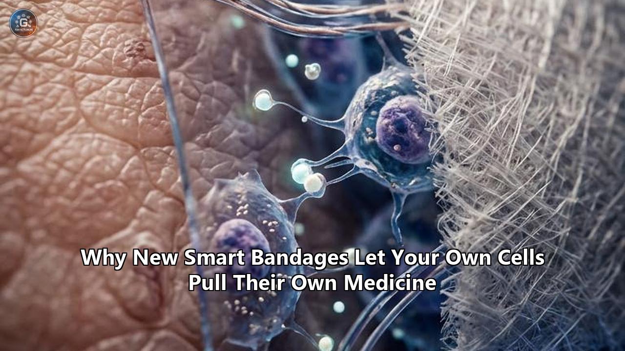
A bioengineering team at Imperial College London has developed a class of mechanically responsive wound dressings that allow migrating human cells to physically pull therapeutic proteins out of a bandage on demand. Detailed in a study published in Nature Materials, the platform—termed traction force-activated payloads, or TrAPs—dispenses with the microprocessors, batteries, and external sensors typical of electronic wound monitors. Instead, it uses the nanonewton-scale mechanical tension generated by living cells to trigger drug release at the exact point of tissue repair.
The implications of this mechanism alter the trajectory of smart bandage technology. Where previous smart dressings forced a binary choice—either passive cotton and silicone pads that merely soak up fluid, or battery-laden electronic patches that flood tissue with synthetic drugs based on algorithmic timers—the new system transforms the injured body into its own automated dispenser. By deploying folded DNA aptamers that tether directly to an underlying biomaterial scaffold, the dressing captures growth factors naturally present in blood and wound exudate. The proteins remain locked and shielded from tissue-degrading enzymes until a repairing fibroblast, keratinocyte, or endothelial cell grips a molecular handle on the dressing and tugs.
"What particularly stands out with this research is that the patient's own body becomes the pharmacy," said Dr. Ben Almquist, senior author of the study and associate professor in the Department of Bioengineering at Imperial College London. "We are not delivering a manufactured drug and hoping it survives long enough to work. We are capturing what the body is already making and giving it back to the cells that need it, by encoding intelligence directly into the material, activated by the one signal guaranteed to be present right where healing is happening: the physical force of a cell pulling on its surroundings".
Lead author Dr. Magdalene Ho and the research team demonstrated that this mechanism operates within living human skin. Across tests spanning rat bone defects, mouse cutaneous lesions, and explanted human tissue maintained ex vivo, the force-responsive platform accelerated cellular migration and capillary sprouting while reducing required therapeutic protein concentrations by up to three orders of magnitude compared to standard clinical biologics. The discovery initiates an impact chain that extends from patient survival rates in chronic diabetic clinics to the economic structures governing modern wound care.
The Chronic Wound Epidemic and the Failure of Flooding
To evaluate the operational impact of this technology, one must examine the clinical landscape of non-healing wounds. Chronic wounds—primarily diabetic foot ulcers, venous leg ulcers, arterial ulcers, and stage 3 or 4 pressure sores—represent a silent, continuous drain on global healthcare infrastructure. In the United States alone, chronic wounds afflict an estimated 4.5 million individuals each year, generating more than $28 billion in direct healthcare costs. Globally, roughly 18.6 million individuals live with diabetic foot ulcers annually, with approximately 20% of those cases progressing to partial or total lower-limb amputation. For patients undergoing amputation, the five-year mortality rate sits near 70%—a prognosis more lethal than several aggressive forms of cancer.
The primary barrier to resolving these wounds is biological desynchronization. In healthy cutaneous repair, the body conducts a precisely timed sequence of four distinct phases:
- Hemostasis, marked by platelet aggregation and fibrin clot formation.
- Inflammation, driven by neutrophils and macrophages clearing debris and pathogens.
- Proliferation, characterized by fibroblast migration, collagen deposition, and angiogenesis.
- Remodeling, where immature collagen is cross-linked and organized over months.
Chronic wounds stall indefinitely in the inflammatory phase. Stagnant wound beds host an overabundance of neutrophils that secrete matrix metalloproteinases (MMPs) and serine proteases. These enzymes degrade newly formed extracellular matrix proteins as quickly as cells assemble them. Worse, they systematically destroy the body's native signaling proteins—such as vascular endothelial growth factor (VEGF), basic fibroblast growth factor (FGF-2), and platelet-derived growth factor (PDGF)—before these factors can instruct adjacent cells to close the wound.
Standard Pharmacological Approach:
[High-Dose Recombinant Protein] ──> [Hostile Wound Bed / High Proteases] ──> 95%+ Enzymatic Degradation ──> Off-Target Toxicity / Failure

TrAP-Mediated Mechanosensitive Approach:
[Endogenous Growth Factors] ──> [Aptamer Pocket Shielding] ──> [Cell Traction Tug] ──> [Direct Receptor Delivery at Nanoscale]
Pharmaceutical attempts to overcome this hurdle have historically relied on a brute-force approach: flooding the wound bed with massive, supraphysiological doses of manufactured recombinant human growth factors. The clinical reality of this strategy is best exemplified by becaplermin (marketed as Regranex), a recombinant human PDGF-BB gel. While biochemically sound in principle, becaplermin requires high daily topical concentrations because the hostile microenvironment deactivates the drug within hours.
This excessive dosing carries steep consequences. Regranex received an FDA black-box warning after post-marketing surveillance identified an increased risk of systemic cancer mortality in patients treated with three or more tubes of the product. Supplying intense, uncoordinated growth signals over prolonged durations risks triggering uncontrolled cellular proliferation elsewhere in the body. Furthermore, these products cost thousands of dollars per treatment course, limiting adoption across resource-constrained health systems and rural clinical settings.
Active wound therapies have attempted to solve this distribution problem by building complex hardware. Over the past decade, academic and industrial teams have constructed patches that incorporate flexible silicone substrates, Bluetooth transmitters, microfluidic channels, and electromechanical pumps intended to squirt medications into tissue. While these devices advance diagnostic monitoring, their translational utility remains constrained by practical physics. Electronic dressings require onboard power supplies, risk circuit failure in the presence of heavy liquid exudates, complicate biological waste disposal, and create sheer mechanical rigidity that can shear fragile granulation tissue on a diabetic heel or ankle.
The Nanoscale Mechanics: How Cells Pull Their Own Payloads
The Imperial College London team bypassed the need for electronic circuitry by weaponizing mechanobiology. Cells do not traverse tissue passively; they rely on actomyosin networks to crawl, crawl-stride, and anchor themselves to their extracellular environment. As a fibroblast or microvascular endothelial cell migrates through injured tissue, it extends finger-like protrusions called filopodia and lamellipodia. Transmembrane integrin receptors within these protrusions lock onto extracellular ligands, such as the Arg-Gly-Asp (RGD) amino acid motifs abundant in native collagen and fibronectin.
Once integrins bind, intracellular actin filaments contract via non-muscle myosin II motor proteins. This generates physical traction forces ranging from several hundred piconewtons to tens of nanonewtons per cell. In natural physiology, these cellular tugs serve as mechanical probes: cells literally feel the stiffness of their substrate to determine whether they should divide, differentiate, or synthesize scar tissue.
Mechanosensitive Aptamer Architecture:
[Collagen Sponge Scaffold]
       │
       ▼ (Covalent Anchor)
[Folded DNA Aptamer] ── <Holds Endogenous Growth Factor Inactive / Shielded>
       │
       ▼ (Molecular "Leash")
[Cell-Adhesive Peptide (RGD Motif)]
       ▲
       │ (Mechanical Grip via Integrin Receptor)
[Migrating Repair Cell] ── [Actomyosin Contraction Forces: 100 pN to 10 nN]
The TrAP architecture mimics and hijacks this process through four engineered components:
- The Structural Backbone: The system uses standard, biocompatible collagen sponges or porous hydrogels already cleared for human surgical use. The core mechanics do not require an exotic or unproven matrix.
- The Base Anchor: Short synthetic oligonucleotides are covalently tethered directly to the collagen fibers.
- The Aptamer Payload Pocket: Attached to the anchor is a carefully folded, single-stranded DNA aptamer (typically 30 to 40 nucleotides long). Nucleic acid aptamers fold into stable three-dimensional architectures possessing distinct binding pockets that grasp specific growth factor proteins with high affinity and exquisite chemical specificity.
- The Cellular Handle: Extended from the terminus of the aptamer sequence is an integrin-targeting peptide handle, such as an RGD sequence or a customized peptide specific to distinct cellular subtypes.
The operational mechanics of this molecular trap mimic a shoelace knot. When a growth factor is captured by the aptamer, its active binding epitopes are physically occluded. This conformational cage shields the protein from proteolytic cleaving by circulating matrix metalloproteinases. The growth factor remains stable, inert, and anchored to the sponge.
When a migrating repair cell enters the wound scaffold, it encounters the synthetic handle. The cell's surface integrins bind to the peptide. As the cell moves forward, its cytoskeleton contracts, applying directional traction force through the integrin-handle connection. Because the aptamer’s folded secondary structure is engineered to have a mechanical unzipping threshold lower than the rupture force of the integrin-peptide bond, the cell's mechanical pull pulls the aptamer apart.
The aptamer uncoils like an untied knot. The structural deformation eliminates the binding pocket, releasing the encapsulated growth factor directly into the nanometer-scale cleft between the cellular membrane and the material. The liberated growth factor binds immediately to the pulling cell’s surface tyrosine kinase receptors, initiating downstream cascades for proliferation, migration, or vessel formation.
By systematically adjusting the GC-content (guanine-cytosine base pairs) and secondary hairpin loops within the aptamer, researchers can precisely program the exact mechanical force required to open the payload. A low-threshold aptamer can be built to release vascular endothelial growth factor (VEGF-A) when pulled by delicate endothelial cells. A higher-threshold sequence can be calibrated to release platelet-derived growth factor (PDGF-BB) or fibroblast growth factor (FGF-2) only when subjected to the forceful pulls of mature, contractile myofibroblasts.
This design shifts drug delivery from a spatial flood to a spatial lock-and-key. Quiescent, bystander cells do not trigger the release. Circulating bacteria cannot access the payload. Proteolytic enzymes cannot pre-degrade the protein. The drug is delivered exclusively to cells that are actively attempting to reconstruct the injured site, precisely at the moment they arrive.
Who Is Affected: Mapping the Stakeholders
The arrival of mechanically triggered therapeutic release introduces distinct shifts across multiple sectors of medicine, clinical practice, and healthcare administration.
Patients with Chronic and Complex Wounds
For patients with diabetes, peripheral vascular disease, or immobility-induced ulcers, this technology alters the biological odds of healing. In diabetic individuals, cellular signaling is chronically impaired; white blood cells produce deficient cytokine profiles, and peripheral neuropathy prevents patients from feeling localized necrosis. When applied to these wounds, mechanosensitive smart bandage technology bridges the biochemical communication breakdown. Because the platform can capture growth factors from the patient’s own wound fluids or autologous blood products (such as platelet lysates) applied at the point of care, it circumvents the need for patients to receive manufactured biologics that their bodies might neutralize or fail to process.
Crucially, the treatment format minimizes physical trauma. Conventional hydrogel and gauze changes must be performed every 24 to 72 hours, an agonizing process that frequently tears away newly formed, fragile epithelial tongues. Because TrAP scaffolds remain biologically viable as long as cells continue to enter and pull, the operational lifespan of a single dressing application extends considerably, sparing patients repetitive, painful dressing changes and lowering infection opportunities.
Plastic, Reconstructive, and Trauma Surgeons
Surgical teams handling severe burns, high-energy blast trauma, and large oncological resections face the persistent issue of graft failure. Autologous split-thickness skin grafts require rapid, robust neo-vascularization from the wound bed to survive—a process termed inosculation. If capillaries do not sprout through the wound bed within 72 to 96 hours, the graft dies.
Professor Shehan Hettiaratchy, a reconstructive plastic surgeon and clinical collaborator on the Imperial College project, noted that the mechanosensitive platform addresses an enduring clinical deficit in difficult wound reconstruction. By integrating TrAPs designed to harvest and display endogenous VEGF-A and hepatocyte growth factor (HGF), surgeons can prime a prepared wound bed prior to grafting. As endothelial cells crawl from healthy wound edges, they actively pull out the localized pro-angiogenic signals, multiplying microvascular density without flooding the delicate tissue with inflammatory exogenous proteins that could promote seroma or hematoma formation.
Outpatient Nurses and Wound Care Centers
Specialized wound care nurses spend hours weekly managing exudate levels, cleaning necrotic slough, and reapplying antimicrobial ointments across patient caseloads. Electronic smart bandages—though useful for monitoring metrics like pH, temperature, and moisture levels—introduce significant clinical friction. Staff must learn specialized software interfaces, manage battery charging cycles, and worry about electronic component disposal.
A mechanosensitive biomatrix, by contrast, handles like a familiar, non-electronic dressing. It requires no batteries, cables, or calibration routines. Nurses can cut the scaffold to size, pack it into an irregular wound cavity, and apply a standard secondary protective cover. The sophistication is molecular rather than digital, fitting within the fast-paced reality of home health visits and overloaded outpatient wound centers.
Comparative Workflow Burden:

[Electronic Wearable Bandage]
Application ──> Sensor Calibration ──> Bluetooth Pairing ──> Continuous Battery Management ──> Electronic Waste Protocols

[Mechanosensitive TrAP Matrix]
Application ──> Standard Packing into Wound Bed ──> Cellular Pulling Operates Autonomously ──> Standard Biodegradation / Disposal
Health Systems, Insurers, and Device Manufacturers
For institutional payers and national health systems like the NHS or Medicare, the financial calculus of chronic wound management is unsustainable. The primary cost drivers are hospital readmissions, emergency surgical interventions, and post-amputation long-term rehabilitation.
Recombinant growth factor therapies, while capable of driving healing in selected patient cohorts, have seen restricted clinical coverage due to cost-to-benefit ratios and oncological liabilities. If mechanically responsive materials can replace multi-thousand-dollar vials of synthetic human proteins by capturing endogenous cytokines, the drug component cost collapses. Medical device firms that currently manufacture passive collagen, alginate, and foam dressings gain a clear pathway to upgrade commodity materials into high-margin, biologically active therapies without manufacturing electronic hardware.
What Changes: Structural Shifts Across Wound Therapeutics
The transition from external, battery-driven smart bandages to cell-actuated matrices marks several fundamental shifts in how biotechnology approaches tissue repair.
| Performance Attribute | First-Generation Passive Dressings | Electronic Smart Bandages (Wearable IoT) | Cell-Traction Smart Bandage Technology |
|---|---|---|---|
| Release Trigger | Passive diffusion, swelling, erosion | Micro-pumps, electro-thermal signals | Cellular traction forces (actomyosin pulling) |
| Payload Source | Antimicrobials, silver ions, bulk hydrogels | Manufactured recombinant drugs, antibiotics | Native endogenous growth factors or ultra-low-dose pre-loaded biologics |
| Hardware Overhead | Zero | Microcontrollers, flexible batteries, wiring | Zero (entirely chemical and mechanical) |
| Dosing Accuracy | Uncontrolled; high burst release at onset | Programmed bolus or metered electronic pump | Nanoscale precision; strictly restricted to actively migrating cells |
| Local Enzyme Shielding | Minimal to none | None (dependent on reservoir protection) | High; folded aptamers protect active protein epitopes |
| Regulatory Category | Class I or II Medical Device | Combination Device (Electronics + Drug Delivery) | Regulated Biomaterial / Device-Biologic combination |
Shift 1: The End of "Bath-and-Wait" Pharmacology
Traditional medicine relies on systemic or topical concentration gradients: an agent is administered in bulk, diffusing down concentration paths until an adequate amount reaches target cell receptors. The mechanosensitive paradigm inverts this logic.
Because the drug release trigger is physical tension, the local effective concentration at the target receptor remains high while the surrounding concentration across the broader wound bed remains zero. In preclinical models published by Ho and Almquist, aptamer-functionalized dressings accelerated tissue repair using doses hundreds to thousands of times lower than conventional delivery systems. Cells do not sit in a biochemical bath; they pluck single molecules from the substrate as they build tissue.
Conventional Topical Delivery Gradient:
[Bulk Formulation Pool: 10,000 ng/mL] ──> [Hostile Wound Fluid Gradient: 100 ng/mL] ──> [Cell Surface Receptor: < 1 ng/mL]
                                                  ▲
                                                  │
                                       (Severe Systemic Leaching Risk)

TrAP Direct Mechanical Delivery:
[Matrix Anchor] ── [Aptamer] ── [Bound Factor] ── (Tug) ──> [Immediate Direct Transfer to Receptor]
Broad Wound Exudate Concentration = 0 ng/mL (Zero Off-Target Toxicity)
Shift 2: Passive Structural Materials Become Intelligent Systems
Historically, biomaterials used in surgery—collagen matrices, decellularized dermis, gelatin sponges—served merely as inert scaffolding. They occupied space, prevented cellular collapse, and waited for host cells to populate them.
Functionalizing these materials with traction-responsive aptamers bridges the gap between material science and synthetic biology. The scaffold ceases to be passive scaffolding; it becomes an interactive participant in cellular repair. The matrix continuously communicates with the cell collective, releasing vascular cues when endothelial cells demand them and structural cues when myofibroblasts pull.
Shift 3: De-Electrifying the Smart Dressing Field
Significant public and venture capital funding has poured into electronic smart bandage technology over the past decade. Researchers have produced impressive engineering accomplishments: dressings that stream real-time impedance readings to smartphones or deliver low-voltage electrical stimulation to orient cell migration. Yet, outside of clinical trials, real-world deployment of electronic bandages remains hampered by economics and logistical fragility.
The traction-activated platform matches the responsive functionality of a closed-loop electronic device without using a single milliampere of current. The sensing mechanism is the aptamer's mechanical stability; the processor is the cell's own biological signaling pathway; the actuator is the cell's actomyosin motor. By offloading logic and power to the patient’s own cells, the system eliminates the electronic failure points that have kept wearable wound-sensing patches confined to proof-of-concept stages.
Short-Term Consequences: Preclinical Validation and the Road to Trials
The validation of TrAPs in Nature Materials represents a major basic science advance, but several operational hurdles must be cleared within the next 24 to 36 months before human patients receive these dressings.
Translational Timeline & Milestones:

[2026: Nature Materials Publication]
      │
      ▼
[2027: Traxion Biotech In Vivo Diabetic Scaling]
   • Testing in hyperglycemic, low-perfusion porcine models
   • Validation of aptamer stability in heavy bacterial biofilm exudates
      │
      ▼
[2028: Early-Stage Clinical Trials (Phase I/IIa)]
   • Human safety trials on refractory diabetic foot ulcers
   • Evaluating cellular traction vigor in elderly/senescent patient cohorts
      │
      ▼
[2029+: Regulatory Approval & Commercial Integration]
   • Clearance pathway: De Novo Device vs. Biologic Combination
   • Scale-up of synthetic oligonucleotide-collagen conjugation
The In Vitro to In Vivo Gap
The data generated by Ho, Almquist, and colleagues verified that traction forces function reliably as an actuation mechanism in three distinct experimental settings:
- In rat bone defects, TrAPs loaded with pro-osteogenic and vascular growth factors elicited substantial vascular ingrowth.
- In mouse full-thickness dermal wounds, the force-responsive materials accelerated wound closure rates relative to non-responsive controls.
- In human skin explants harvested from surgical procedures and kept alive in culture, resident cells migrated directly into the collagen scaffolds, successfully pulling payloads free.
However, translating these findings to complex patient pathology requires overcoming distinct biological realities. The experimental animal models featured young, healthy rodents possessing robust microvascular architecture, clear cellular responsiveness, and absence of systemic disease. The explanted human skin tissues, while human, lacked dynamic blood flow, hydrostatic pressure, and functional systemic immune responses—elements that drive the hostile biochemistry of real-world diabetic ulcers.
In an elderly patient with long-standing type 2 diabetes, cells do not behave like cells in a laboratory culture. Fibroblasts harvested from chronic ulcers often suffer from cellular senescence. These "exhausted" cells express fewer surface integrins, demonstrate disrupted focal adhesion kinase (FAK) signaling, and generate demonstrably lower traction forces than healthy cells. If an ulcerated cell cannot muster the nanonewtons of force required to uncoil the aptamer, the dressing will remain locked.
The immediate next step for the research team—and their commercial spin-out vehicle, Traxion Biotech—is mapping the mechanical force profiles of senescent cells taken directly from diabetic patient margins. If necessary, aptamer sequence thresholds will need to be loosened to accommodate the weaker physical grip of compromised tissue.
Biochemical Longevity in Severe Biofilms
Chronic wounds are rarely sterile; they are colonized by polymicrobial biofilm communities dominated by Pseudomonas aeruginosa and Staphylococcus aureus. These bacteria churn out endogenous nucleases designed to chew up extracellular DNA as well as elastases that degrade structural proteins.
Because TrAPs rely on synthetic DNA oligonucleotides, researchers must address susceptibility to bacterial DNases. While chemical modifications to the oligonucleotide backbone—such as phosphorothioate linkages, 2'-O-methyl alterations, or locked nucleic acids (LNAs)—can render aptamers impervious to enzymatic degradation, these molecular edits must be integrated without blunting the aptamer’s binding affinity for the growth factor or altering its mechanical unfolding curve. Demonstrating structural durability within infected, enzyme-saturated porcine wound models remains the critical short-term milestone.
Long-Term Consequences: Systemic Ripples in Regenerative Medicine
Looking beyond cutaneous wound management, the ability to control biological payloads through mechanical pulling force opens new territory across several branches of clinical bioengineering.
Long-Term Cross-Disciplinary Applications of Cell-Traction Delivery:

                      ┌──> Orthopedic Non-Union Fractures
                      │    (Osteoblast-mediated BMP-2 extraction without heterotopic ossification)
                      │
                      ├──> Cardiovascular Myocardial Infarction Patches
[Traction-Responsive ─┼──> (Perfusion-guided VEGF/angiopoietin release driven by cardiac myofibroblasts)
 Biomaterial Matrix]  │
                      ├──> Peripheral Nerve Reconstruction Guides
                      │    (Axonal-growth-cone traction releasing neurotrophic factors: NGF/BDNF)
                      │
                      └──> Musculoskeletal Tendon-Bone Anchors
                           (Tension-calibrated TGF-beta release along mechanical stress lines)
Orthopedics and Non-Union Bone Repair
Bone healing is governed strictly by mechanotransduction. In orthopedic surgery, non-union fractures—breaks that fail to bridge after months of immobilization—are often treated with recombinant human Bone Morphogenetic Protein-2 (rhBMP-2, sold as Infuse). Like becaplermin in wound care, Infuse requires massive, unphysiological doses to stimulate bone formation. These high doses frequently trigger severe clinical complications, including painful heterotopic ossification (uncontrolled bone growth into surrounding muscle and nerves) and lifethreatening cervical swelling when used in spinal fusions.
Adapting mechanosensitive smart bandage technology to porous titanium or hydroxyapatite bone implants changes this dynamic. By designing TrAPs tuned to osteoblast and mesenchymal stem cell traction forces, bone-inductive proteins can remain anchored to the implant surface. Only osteoblasts crawling directly across the fracture gap can liberate the BMP-2 payload. The signal remains strictly locked to the structural scaffold, preventing the uncontrolled leakage into adjacent soft tissue that causes heterotopic bone complications.
Cardiovascular Regeneration Post-Infarction
Following a myocardial infarction, millions of cardiomyocytes die from ischemia, replaced by a non-contractile, rigid collagen scar. Attempts to regenerate heart tissue through cardiac hydrogel patches loaded with pro-survival factors have stalled because therapeutics wash away into the coronary bloodstream within minutes of ventricular implantation.
Equipping a cardiac patch with traction-sensitive aptamers creates an entirely different therapeutic interface. As migrating myofibroblasts and circulating angiogenic progenitor cells enter the damaged border zone of the infarction, the rhythmic physical stretching of the myocardium, combined with the cells' own intrinsic traction, can selectively release survival cues and angiogenic cytokines precisely along the mechanical stress vectors of the heart wall.
Peripheral Nerve Regeneration
When peripheral nerves are severed across extensive gaps, surgeons bridge the defect using hollow silicone or bioresorbable nerve guidance conduits (NGCs). Axons must regenerate from the proximal nerve stump, traversing several centimeters of empty matrix to reconnect with target muscle tissue.
By lining the internal lumen of a conduit with TrAPs carrying neurotrophic factors (such as nerve growth factor [NGF] or brain-derived neurotrophic factor [BDNF]), migrating Schwann cells and advancing axonal growth cones can pull down guidance cues sequentially as they advance. The nerve literally pulls its own chemical pathway into existence, avoiding the desensitization that occurs when an entire conduit is flooded with stagnant neurotrophic solutions.
The Regulatory and Commercial Landscape
Transforming a peer-reviewed biomaterial into a standard hospital dressing requires navigating complex regulatory classifications and developing reproducible, high-yield manufacturing supply chains.
Device or Biologic: Navigating the FDA's Combination Product Hurdle
The primary regulatory question surrounding cell-actuated dressings is jurisdiction. In modern medical device regulation, products are divided into three distinct classes:
- Medical Devices (CDRH): Cleared via 510(k) or Premarket Approval (PMA), requiring evidence of mechanical/physical efficacy and safety.
- Biologics and Drugs (CDER/CBER): Requiring full-scale, three-phase clinical trials that take years and cost hundreds of millions of dollars.
- Combination Products: Combining a structural matrix with an active pharmaceutical or biological agent.
Because TrAP materials can operate simply as "empty sponges" that catch and re-display the patient’s own circulating endogenous growth factors without containing factory-made recombinant drugs, Traxion Biotech and future manufacturers may pursue clearance as a 510(k) or de novo medical device. If the dressing contains zero active biological molecules in the sterile packaging—relying entirely on capturing the patient's own native factors at the bedside—the platform shifts away from the burdensome pharmaceutical category.
This represents an immense economic advantage. Producing a dressing cleared under device pathways allows it to reach the market years faster, at a fraction of the capital cost demanded by traditional recombinant protein drugs.
However, if clinical formulations require pre-loading with defined, low-dose recombinant proteins to ensure performance in severely depleted, malnourished, or vasculopathic patients, the product will instantly default to combination product oversight. Navigating this pathway will necessitate exhaustive Phase I through Phase III clinical trials, demonstrating that the mechanosensitive release mechanism does not lead to unwanted systemic leakage or aberrant immunological reactions to the synthetic DNA aptamers.
Manufacturing and Scalability
Producing aptamer-functionalized dressings at industrial volume presents technical challenges that traditional dressing manufacturers have never faced. Passive bandages are stamped out in automated roll-to-roll processes using high-heat vulcanization, ultraviolet sterilization, or gamma irradiation.
DNA aptamers are biologically delicate polymers. Exposing them to standard gamma irradiation during terminal packaging destroys the precise three-dimensional tertiary structures that create the binding pockets, rendering them non-functional.
Manufacturing plants will need to adapt sterile bio-conjugation processes:
- Synthesizing the oligonucleotide sequences using high-throughput phosphoramidite chemistry.
- Purifying the aptamers to pharmaceutical grade via high-performance liquid chromatography (HPLC).
- Covalently coupling them to porous collagen or polysaccharide sponges under sterile, temperature-controlled conditions.
- Utilizing ethylene oxide (EtO) sterilization or aseptic assembly lines to prevent structural denaturation of the aptamer loops.
While technically complex, the biopharmaceutical sector has developed the required manufacturing infrastructure over the past decade, driven by the global expansion of oligonucleotide and mRNA production platforms. Leveraging these established contract manufacturing networks will dictate how rapidly traction-responsive materials scale to clinical trial volumes.
Key Milestones and Unresolved Questions on the Path to Market
As research transitions from academic laboratories into translational pipelines, the coming years will establish whether mechanosensitive smart bandage technology can replace conventional wound modalities.
Several milestones will determine the trajectory of this platform:
- Porcine Chronic Wound Validation: Demonstrating efficacy in swine models that exhibit both hyperglycemia and impaired vascular perfusion. Pig skin provides the closest anatomical and physiological analogue to human dermis; proving that TrAPs drive faster wound closure without premature degradation by pig proteases is the necessary gateway to clinical trials.
- First-in-Human Phase I Safety Trials: Assessing patient tolerance to modified synthetic oligonucleotides in open wounds. Clinical teams will watch for local immunogenicity, foreign-body granuloma formation, or allergic contact dermatitis caused by the nucleic acid fragments.
- Mechanical Benchmarking Across Diverse Patient Age Groups: Quantifying how cellular traction forces vary across patients suffering from vascular insufficiency, renal failure, and advanced age. Determining if "one force threshold fits all" or if dressings must be customized for different degrees of patient cellular vigor.
- Point-of-Care Loading Protocols: Establishing clinical workflows for utilizing autologous blood concentrates. If clinics choose to charge the dressing using a tube of the patient’s own blood or platelet-rich plasma (PRP) drawn chairside, protocols must be simple enough for a clinic nurse to complete within minutes without specialized lab equipment.
Unresolved questions remain regarding long-term matrix remodeling. In natural wound healing, as cells infiltrate a scaffold, they steadily deposit nascent collagen and fibronectin around themselves. In prolonged chronic settings, this rapid deposition of fresh matrix proteins could bury the unactivated TrAP aptamers, physically blocking subsequent waves of repairing cells from reaching the handles. Determining the effective functional half-life of an un-pulled aptamer within an active cellular matrix remains a crucial area of investigation.
The achievement realized by Ho, Almquist, and their collaborators marks a definitive turning point in the philosophy of advanced wound repair. For decades, medical engineering approached smart wound care as an electronics problem—attempting to digitize the wound bed with sensors, circuits, and motorized pumps.
The TrAP platform demonstrates that true systemic intelligence does not require microchips. The human body already possesses a sophisticated diagnostic and regulatory network governed by the mechanics of its own cells. By engineering biomaterials that listen to and obey that biological pulling force, smart bandage technology moves away from forceful pharmacological intervention, enabling injured tissues to repair themselves on their own structural terms.
Reference:
- https://scientificinquirer.com/2026/08/21/this-smart-bandage-waits-for-cells-to-pull-before-it-releases-its-healing-signal/
- https://diabeticfootonline.com/2026/07/27/the-wound-pulls-the-trigger-force-responsive-biomaterials-put-cells-in-charge-of-repair/
- https://studyfinds.com/smart-material-uses-bodys-own-healing-proteins-to-help-repair-wounds/
- https://blog.wcei.net/smart-bandages-new-era-healing
- https://www.earth.com/science/smart-wound-dressing-growth-factors/
- https://www.researchgate.net/figure/TrAP-functionalized-sponges-accelerate-wound-healing-in-an-ex-vivo-human-skin-model-a_fig5_410852516
- https://pmc.ncbi.nlm.nih.gov/articles/PMC6511580/
- https://pmc.ncbi.nlm.nih.gov/articles/PMC8336080/
- https://keck.usc.edu/news/new-smart-bandages-hold-potential-for-revolutionizing-the-treatment-of-chronic-wounds/
- https://www.caltech.edu/about/news/smart-bandage-clears-new-hurdle-monitors-chronic-wounds-in-human-patients
- https://cellscale.com/blog/research-highlights/smart-bandage-technology/
- https://www.ijpsjournal.com/article/Smart+Bandage+Containing+Herbal+Hydro+Gel+for+Controlled+Drug+Release+Wound+Healing
- https://cheme.stanford.edu/wireless-smart-bandage-provides-new-insights-healing-chronic-wounds
- https://www.facebook.com/rockinscience/posts/a-new-way-to-treat-chronic-woundsa-spartan-led-team-is-developing-an-inexpensive/1632041680319459/
- https://www.imperial.ac.uk/news/189689/new-material-could-drive-wound-healing/
- https://www.nmlett.org/index.php/nml/article/view/1116
- https://www.researchgate.net/publication/331880828_Local_nascent_protein_deposition_and_remodelling_guide_mesenchymal_stromal_cell_mechanosensing_and_fate_in_three-dimensional_hydrogels
- https://physicsworld.com/a/traction-forces-release-growth-factors-from-cells-to-heal-wounds/
- https://www.the-scientist.com/smart-dressing-harnesses-the-body-s-own-proteins-to-accelerate-wound-healing-74801
- https://www.traxionbiotech.com/
- https://www.drugdiscoverytrends.com/smart-bandage-could-accelerate-healing-of-chronic-injuries-and-battlefield-wounds/
- https://www.forbes.com/sites/robertglatter/2018/07/18/smart-bandage-can-dispense-drugs-while-keeping-an-eye-on-your-wound/