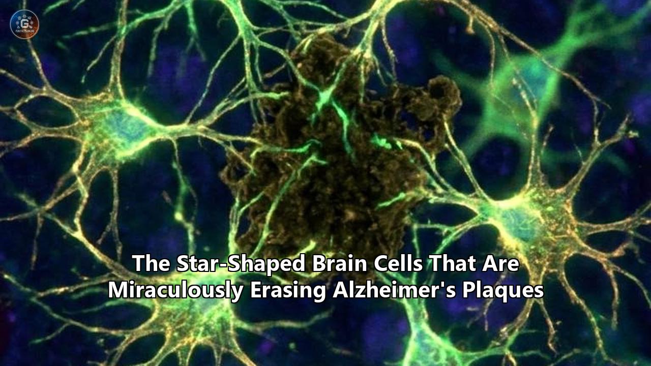
A pair of independent biological discoveries in the spring of 2026 has radically altered the trajectory of dementia research, demonstrating that the human brain possesses a dormant, built-in mechanism capable of erasing the toxic protein deposits associated with cognitive decline. Research teams at Baylor College of Medicine and Washington University School of Medicine have successfully activated astrocytes—star-shaped support cells long thought to be mere structural scaffolding—transforming them into highly efficient scavengers that consume and destroy amyloid beta plaques.
The findings, published recently in Nature Neuroscience and Science, represent a stark departure from the traditional pharmaceutical approach to dementia. Rather than relying on synthetic antibodies infused into the bloodstream to target amyloid accumulations, scientists are now genetically instructing the brain’s own native cells to execute the cleanup operation.
In mice exhibiting advanced memory deficits and heavy plaque burdens, this cellular activation not only cleared existing amyloid deposits by up to 50% but also halted the progression of cognitive decline over six months of testing.
The Baylor College of Medicine team, led by Dr. Benjamin Deneen and Dr. Dong-Joo Choi, achieved this by manipulating a single protein called Sox9, which governs the aging process of astrocytes. When Sox9 levels were artificially boosted, the star-shaped cells physically expanded, increased their structural complexity, and began ingesting amyloid plaques at an accelerated rate.
"We found that increasing Sox9 expression triggered astrocytes to ingest more amyloid plaques, clearing them from the brain like a vacuum cleaner," said Deneen, director of the Center for Cell and Gene Therapy at Baylor. "Most current treatments focus on neurons or try to prevent the formation of amyloid plaques. This study suggests that enhancing astrocytes' natural ability to clean up could be just as important."
Simultaneously, a team at Washington University in St. Louis borrowed a concept directly from oncology. Led by pathologist Dr. Marco Colonna, the researchers engineered a virus to deliver a Chimeric Antigen Receptor (CAR) specifically to astrocytes. Similar to the CAR-T cell therapies used to hunt down leukemia, these modified CAR-astrocytes were equipped with a genetic "homing device" that forced them to seek out and dismantle amyloid beta.
For the tens of millions of families globally affected by neurodegenerative diseases, these parallel breakthroughs signal a definitive shift in the scientific understanding of brain health. The search for a viable alzheimers plaques treatment has historically focused almost exclusively on neurons. By redirecting attention to the vast, largely untapped network of astrocytic support cells, researchers are opening an entirely new frontier in neuro-immunology.
The Biology of the Star-Shaped Cell
To understand how astrocytes can be mobilized against Alzheimer's disease, one must look at how heavily these cells have been historically underestimated.
When 19th-century German pathologist Rudolf Virchow first observed the non-neuronal cells of the central nervous system, he coined the term "neuroglia"—literally "nerve glue." For over a century, the scientific consensus held that neurons were the solitary drivers of cognition, memory, and disease, while glial cells merely held the neurons in place, provided them with nutrients, and maintained the blood-brain barrier.
Astrocytes, named for their distinctive, star-like branching appendages, are the most abundant type of glial cell in the central nervous system. Modern transcriptomic technologies have revealed that they are far from passive bystanders. Astrocytes actively regulate blood flow, recycle neurotransmitters, and manage the precise synaptic connections where memories are formed and stored.
As the brain ages, however, astrocytes undergo profound functional alterations. They enter a state known as reactive astrogliosis in response to the toxic buildup of amyloid beta—the sticky protein fragments that clump together between neurons, disrupting cellular function and triggering Alzheimer's disease. During this reactive phase, astrocytes often lose their complex branching structures, become inflamed, and abandon their homeostatic duties. Some distinct populations of reactive astrocytes even begin secreting neurotoxins that accelerate the death of nearby neurons.
The core objective for both the Baylor and Washington University research teams was to determine if this dysfunctional aging process could be intercepted and reversed. If reactive astrocytes naturally gather around amyloid plaques during the progression of Alzheimer's, the researchers theorized, perhaps they could be genetically coerced into consuming the plaques rather than simply reacting to them.
The Baylor Discovery: Unlocking the Sox9 Protein
The research published in Nature Neuroscience on May 2, 2026, provides the most detailed molecular map to date of how astrocytes can be biologically reprogrammed from passive observers into aggressive cleanup crews.
The Baylor team focused their attention on Sox9, a transcription factor—a protein that controls the rate of transcription of genetic information from DNA to messenger RNA. Sox9 is heavily involved in regulating the functional changes that occur in astrocytes as the mammalian brain grows older.
To test the specific impact of this protein, the researchers utilized a highly specific genetically engineered animal model known as the APP NLGF mouse. The design of the experiment was critical: rather than treating mice before they showed signs of disease, the team waited until the mice had already developed severe cognitive impairments and heavy amyloid plaque accumulation. This mirrors the clinical reality of most human patients, who typically do not receive an Alzheimer's diagnosis until significant neurological damage has already occurred.
The results yielded a stark biological contrast. When the researchers deliberately knocked out (removed) the Sox9 gene, the astrocytes simplified their structures, pulling back their star-like branches. Plaque formation accelerated rapidly, and the astrocytes demonstrated a near-total inability to clear the toxic deposits. Microglial cells—another type of immune cell in the brain—also lost their support network and failed to mitigate the damage.
Conversely, when the team utilized gene therapy vectors to overexpress (boost) Sox9 in the astrocytes, the trajectory of the disease reversed.
The Sox9-enriched astrocytes grew more complex, extending their branching architecture deep into the neural tissue. This structural expansion was paired with a massive increase in lysosomal activity. Lysosomes are the internal "stomachs" of a cell, filled with enzymes capable of breaking down cellular waste. The activated astrocytes began actively engulfing the amyloid plaques and degrading them internally.
Beyond the microscopic cellular changes, the physical behavior of the mice shifted dramatically. Over a six-month monitoring period, the mice with elevated Sox9 levels retained their performance in novel object recognition, place recognition, and working memory tasks. The degradation of their cognitive abilities was essentially frozen in place.
Dr. Dong-Joo Choi, the first author of the study and currently an assistant professor at the University of Texas Health Science Center at Houston, noted the clinical relevance of the timeline. Because the APP NLGF model develops plaques and memory impairment concurrently, the preservation of memory over half a year of testing strongly suggests that astrocytic clearance of amyloid is directly responsible for halting neurodegenerative cognitive decline.
Engineering Cellular Assassins: The CAR-Astrocyte Approach
While the Baylor team focused on accelerating a naturally occurring cellular process, the Washington University School of Medicine team opted for a synthetic engineering approach. Their findings, published in Science in early March 2026, outline the first successful attempt to equip brain cells with artificial biological radar.
Their method relies on Chimeric Antigen Receptor (CAR) technology, a bedrock of modern cellular immunotherapy for blood cancers. In oncology, doctors extract a patient's T-cells, engineer them to grow CARs on their surface that specifically recognize tumor cells, and infuse them back into the body to hunt down the cancer.
Dr. Marco Colonna and his colleagues hypothesized that a similar homing mechanism could be deployed inside the central nervous system. Rather than using T-cells—which struggle to penetrate the brain in large numbers and can trigger severe neurotoxicity—the team targeted astrocytes, which are already abundant and deeply integrated into the brain's architecture.
Using an adeno-associated virus (AAV) as a delivery vehicle, the researchers injected a genetic payload into the brains of Alzheimer's-model mice. The virus selectively infected the astrocytes, rewriting their DNA to force the expression of a CAR designed specifically to latch onto amyloid beta proteins.
Upon receiving this synthetic assignment, the astrocytes became singularly focused on amyloid eradication. The researchers tested the viral gene therapy on two distinct groups of subjects: young mice in their pre-plaque developmental years, and older mice that already exhibited heavy plaque burdens.
When the researchers analyzed the brains three to six months later, the results were definitive. In the young cohort, the presence of the CAR-astrocytes essentially prevented the formation of amyloid plaques altogether. In the older cohort with established disease, a single injection of the gene therapy resulted in a roughly 50% reduction in existing plaques.
"This study marks the first successful attempt at engineering astrocytes to specifically target and remove amyloid beta plaques in the brains of mice with Alzheimer's disease," Colonna reported.
The efficiency of this targeted astrocytic attack provides a striking alternative to the current pharmacological standards. Implementing this methodology as an alzheimers plaques treatment requires only a one-time viral vector injection, initiating a permanent genetic alteration in the brain's support cells. The modified astrocytes continuously patrol the neural tissue, aggressively clearing amyloid debris as soon as it begins to aggregate.
The Pathological Context and Flaws of Current Monoclonal Antibodies
To fully grasp the magnitude of these dual astrocytic breakthroughs, they must be contextualized against the current state of Alzheimer's pharmacology.
For decades, the pharmaceutical industry experienced a 99% failure rate in Alzheimer's drug development. The dominant theory driving the field was the "amyloid cascade hypothesis," which posits that the accumulation of amyloid beta initiates a chain reaction leading to the buildup of tau protein tangles, widespread inflammation, and eventual neuronal death.
It was not until the early 2020s that drugmakers successfully developed monoclonal antibodies capable of meaningfully removing amyloid from the human brain. Drugs like lecanemab (marketed as Leqembi) and donanemab (marketed as Kisunla) were approved by regulatory agencies after clinical trials proved they could slow cognitive decline by roughly 27% to 35% over 18 months. In practical terms, this preserves about 7 to 10 months of independent living for patients in the early stages of the disease.
Despite their clinical success, these antibody therapies are fraught with severe logistical and medical limitations.
First, monoclonal antibodies require massive, continuous dosing. Because only a tiny fraction of the antibodies infused into a patient's bloodstream successfully cross the highly restrictive blood-brain barrier, patients must undergo intravenous infusions every two to four weeks, indefinitely.
Second, the mechanism by which these antibodies clear plaques relies on recruiting the body's peripheral immune system and native microglia to attack the amyloid. This aggressive immune response frequently triggers a dangerous side effect known as ARIA (Amyloid-Related Imaging Abnormalities). ARIA manifests as brain swelling (ARIA-E) or micro-hemorrhages and bleeding in the brain (ARIA-H). In clinical trials, up to a quarter of patients receiving these drugs developed some form of ARIA, requiring constant, expensive monitoring via regular MRI scans. Several patient deaths have been directly linked to severe ARIA complications.
Utilizing enhanced astrocytes as an alzheimers plaques treatment circumvents many of the biological barriers that plague antibody infusions. Because astrocytes are already residents of the central nervous system, there is no need to force synthetic molecules across the blood-brain barrier. Furthermore, because astrocytes clear plaques through a localized process of cellular autophagy (self-eating) rather than by triggering a massive inflammatory immune cascade, researchers are optimistic that astrocyte-mediated clearance will not induce the widespread brain swelling associated with ARIA.
Autophagy and the Ecosystem of the Brain
The exact mechanical process by which astrocytes dismantle amyloid is rooted in the cell's natural waste-management system.
In a healthy brain, astrocytes are highly phagocytic—meaning they regularly ingest cellular debris, dead synapses, and metabolic waste. They encapsulate this waste in specialized vesicles and merge them with lysosomes, exposing the toxic material to highly acidic enzymes that dissolve it into harmless base components.
Previous international studies, including a landmark July 2024 paper in Molecular Neurodegeneration, highlighted how astrocytes deploy this self-recycling mechanism (autophagy) to break down amyloid beta naturally. However, as the brain ages, this internal machinery breaks down.
A critical component of this process is the APOE gene, which is responsible for lipid (fat) metabolism in the brain. The APOE4 variant of this gene is the single strongest genetic risk factor for late-onset Alzheimer's disease. Transcriptomic studies have revealed that astrocytes expressing the APOE4 variant are significantly less efficient at clearing amyloid beta than those expressing the neutral APOE3 variant. Furthermore, APOE4 astrocytes struggle to regulate the formation of lipid droplets, leading to a toxic buildup of fats inside the cell that impairs its ability to perform phagocytosis.
By artificially upregulating transcription factors like Sox9 or introducing synthetic CAR mechanisms, scientists are effectively overriding the genetic handicaps imposed by age and APOE4, forcibly reviving the astrocyte's dormant autophagic engines.
This cellular manipulation does not happen in a vacuum. The brain operates as an intricate, interacting cellular ecosystem. Insights generated in recent years, heavily discussed at the Society for Neuroscience (SfN) global conferences, have shifted the field away from a linear model of Alzheimer's toward an understanding of intense immune-brain crosstalk.
Astrocytes and microglia (the brain's primary resident immune cells) operate in tandem. A study published in May 2024 by researchers at the Icahn School of Medicine at Mount Sinai detailed exactly how reactive astrocytes control the physical architecture around amyloid plaques.
The Mount Sinai team discovered that a specific gene and its associated protein, Plexin-B1, are upregulated in reactive astrocytes surrounding plaques. Plexin-B1 controls the spacing and physical barrier that astrocytes form around the toxic deposits. By manipulating Plexin-B1, the researchers found they could alter how tightly the astrocytes cordoned off the plaques, effectively opening up pathways for both microglia and other astrocytes to access and clear the harmful material more efficiently.
"Our findings offer a promising path for developing new treatments by improving how cells interact with these harmful plaques," noted Dr. Roland Friedel, a senior author of the Mount Sinai study. This highlights a growing consensus that the physical, spatial relationship between glial cells and plaques is just as vital as the chemical mechanisms of degradation.
The Synaptic Engulfment Dilemma
While transforming astrocytes into hyper-aggressive scavengers offers a compelling path toward disease modification, researchers are moving forward with intense caution. The primary risk of engineering glial cells to "eat" brain matter is the potential for collateral damage—specifically, the accidental consumption of healthy synapses.
Synapse loss is the most accurate physical correlate to cognitive decline in Alzheimer's disease. Long before neurons actually die, they lose the synaptic connections that allow them to communicate with one another.
Historically, scientists believed amyloid plaques physically crushed or poisoned these synapses. But advanced microscopy and postmortem human tissue analyses have revealed a more disturbing reality: the brain's own glial cells are actively severing and eating the synapses.
During normal fetal and childhood development, astrocytes and microglia perform a vital function called "synaptic pruning." They engulf and eliminate weak or unnecessary neural connections to streamline the brain's circuits. This process is heavily mediated by specific phagocytic receptors, particularly one known as MEGF10, found on the surface of astrocytes.
In the adult brain, this pruning system is supposed to power down. However, the presence of amyloid beta appears to trigger a catastrophic reactivation of this developmental process. In APP/PS1 mouse models of Alzheimer's, researchers have observed a massive upregulation of the MEGF10 receptor in vivo. The astrocytic lysosomes in these mice are heavily loaded with engulfed synaptic proteins, proving that the astrocytes are actively eating the synapses of viable neurons.
This dynamic presents a massive biological puzzle for researchers designing a new alzheimers plaques treatment. Are the astrocytes inappropriately attacking healthy synapses due to amyloid-induced confusion? Or are the synapses already damaged by amyloid toxicity, making the astrocytes' clearance of them a beneficial, protective response to remove dead tissue?
"Evidence from both human tissue and animal models suggests that glia remove synapses from viable neurons, not only from apoptotic or dying neurons, supporting the idea of direct synaptic attrition," noted a major 2025 review of Alzheimer's pathology.
Consequently, any gene therapy designed to increase astrocytic phagocytosis—like the Sox9 overexpression or the CAR-astrocyte intervention—must be meticulously calibrated. The engineered cells must be aggressive enough to seek out and destroy amyloid beta, but precise enough to leave the fragile synaptic bridges between neurons entirely untouched. The Washington University team addressed this by specifically tuning their CAR receptor to bind exclusively to amyloid beta markers, theoretically preventing the astrocytes from mistakenly targeting healthy synaptic proteins.
Economic and Healthcare Infrastructure Implications
The transition from chronic pharmaceutical management to acute genetic interventions carries massive implications for global healthcare economics.
Neurodegenerative diseases currently represent one of the heaviest financial burdens on modern medical systems. As populations across the globe rapidly age, the sheer volume of dementia patients is projected to break existing infrastructure. Current monoclonal antibody treatments cost upwards of $26,000 to $32,000 per year, per patient, strictly for the drug itself.
However, the hidden costs of current therapies are vastly higher. Because drugs like Leqembi require bi-weekly intravenous infusions, patients—who are frequently cognitively impaired and unable to drive—must be transported to specialized infusion centers twice a month. The strict monitoring protocols for ARIA require baseline PET scans to confirm amyloid presence, followed by three to four high-resolution MRI scans in the first year of treatment to monitor for brain bleeding.
The clinical infrastructure required to deliver this care simply does not exist at the scale required to treat the millions of individuals currently harboring amyloid plaques.
By contrast, an astrocyte-targeted viral gene therapy operates on a radically different economic model. A single intracranial or intravenous injection of an AAV viral vector delivering a CAR or a Sox9 booster would hypothetically represent a one-time, definitive procedure.
While single-dose gene therapies are exceptionally expensive to manufacture upfront—often costing millions of dollars per dose in the context of rare diseases like spinal muscular atrophy—the long-term economic calculus heavily favors one-time interventions. Eradicating the need for continuous bi-weekly infusions, specialized nursing staff, and perpetual MRI monitoring would eliminate billions in downstream healthcare costs while drastically improving the quality of life for both patients and their full-time caregivers.
However, scaling AAV manufacturing to meet the demands of a disease as prevalent as Alzheimer's presents an unprecedented industrial challenge. Current global bioreactor capacity is optimized for rare genetic disorders affecting mere thousands of patients. Adapting this supply chain to produce viral vectors for millions of dementia patients will require a multi-billion-dollar expansion of the pharmaceutical manufacturing sector over the next decade.
Translating to Human Clinical Trials
The timeline for bringing astrocyte-reprogramming therapies to the public is dictated by the immense difficulty of translating rodent data into human clinical success.
While the APP NLGF and 5xFAD transgenic mouse models are excellent tools for studying amyloid accumulation and memory deficits, the mouse brain differs fundamentally from the human brain. Human astrocytes are vastly larger, structurally more complex, and biologically more diverse than their murine counterparts. A genetic intervention that works flawlessly in a mouse astrocyte may fail to take hold in a human cell, or worse, may trigger unexpected toxicity.
To bridge this gap, researchers are increasingly relying on induced pluripotent stem cells (iPSCs). By taking skin or blood cells from human Alzheimer's patients and reversing them into a stem-cell state, scientists can grow patient-specific human astrocytes in a laboratory dish. These iPSC-derived human astrocytes are currently being used to test the safety and efficacy of the CAR and Sox9 modifications before they ever touch a living human patient.
Furthermore, human brain organoids—three-dimensional, miniaturized clusters of neural tissue grown in vitro—are allowing researchers to observe how engineered astrocytes interact with human neurons and microglia in real-time. This intermediate testing phase is critical for ensuring that hyper-activated astrocytes do not trigger runaway neuroinflammation or engage in rogue synaptic pruning.
Regulatory agencies require exhaustive safety data before approving genetic modifications to the human central nervous system. The next phase of research for the Washington University and Baylor teams will likely involve large animal models, primarily non-human primates, to test the distribution of the viral vectors and to monitor for long-term immune reactions.
If the primate data replicates the staggering 50% plaque reduction and cognitive preservation seen in mice, Phase 1 human safety trials could commence before the end of the decade. These initial human trials will likely enroll a small cohort of patients with genetically driven, early-onset Alzheimer's disease, as their predictable disease progression makes them ideal candidates for experimental interventions.
Expanding the Target: Brain Tumors and Beyond
The implications of engineering the brain's support cells extend far beyond the realm of Alzheimer's disease. The foundational technology demonstrated by these recent breakthroughs establishes astrocytes as programmable biological drones capable of operating deep within the central nervous system.
Dr. Colonna and the Washington University research team have already signaled that their CAR-astrocyte platform could be adapted for neuro-oncology. Glioblastoma, the most aggressive and lethal form of brain cancer, is notoriously difficult to treat because the blood-brain barrier blocks most traditional chemotherapies, and the tumors rapidly mutate to evade standard immune attacks.
By modifying the CAR homing device to recognize surface markers specific to glioblastoma cells rather than amyloid beta, researchers believe astrocytes could be redirected from clearing protein debris to directly assassinating tumor cells. Because astrocytes are highly migratory and naturally integrated into the brain tissue, they could potentially track down microscopic cancer cells that have migrated away from the main tumor mass—the primary cause of cancer recurrence.
Similarly, the concept of boosting astrocytic autophagy via Sox9 manipulation could provide a universal treatment pathway for other protein-misfolding neurodegenerative diseases. Parkinson's disease is driven by the toxic accumulation of alpha-synuclein proteins, while Huntington's disease is caused by mutant huntingtin proteins. If astrocytes can be triggered to engulf and digest amyloid plaques "like a vacuum cleaner," they could theoretically be stimulated to clear out the toxic protein aggregates driving these other fatal conditions.
The New Paradigm of Dementia Research
The dual discoveries emerging from Texas and Missouri in 2026 have fundamentally reshaped the landscape of neurodegeneration research. For decades, the field was trapped in a cycle of attacking amyloid beta with synthetic antibodies that struggled to enter the brain, yielding incremental clinical benefits at the cost of severe side effects and immense logistical burdens.
The realization that the brain already possesses the biological hardware necessary to clean itself—and that this hardware simply requires a genetic software update to combat the effects of aging and disease—shifts the scientific focus away from chemical suppression and toward cellular enhancement.
As researchers prepare to move these therapies out of the laboratory and into primate testing, the scientific community is watching closely. The upcoming milestones will rely heavily on biomarker readouts—specifically tracking levels of tau protein and neurofilament light chain (NfL) in spinal fluid—to confirm whether astrocytic plaque clearance actively halts the downstream death of neurons.
By prioritizing the complex, interacting cellular systems of the brain over a singular focus on neurons, science is closing in on a mechanism that addresses the root physical pathology of Alzheimer's disease from the inside out. The star-shaped cells that researchers ignored for more than a century have emerged as the most formidable weapons in the fight against cognitive decline.
Reference:
- https://www.sciencedaily.com/releases/2026/05/260502013550.htm
- https://scienceblog.com/brains-star-cells-rise-up-against-alzheimers-damage/
- https://www.sciencealert.com/scientists-cut-amyloid-plaques-by-50-in-mice-with-engineered-cells
- https://www.sciencedaily.com/releases/2026/03/260311004720.htm
- https://www.futura-sciences.com/en/revolutionary-discovery-on-alzheimers-a-new-treatment-path-identified_21291/
- https://pmc.ncbi.nlm.nih.gov/articles/PMC7999452/
- https://beacon-intelligence.com/blog/the-future-of-alzheimers-disease-drug-development/
- https://www.mountsinai.org/about/newsroom/2024/altering-cellular-interactions-around-amyloid-plaques-may-offer-novel-alzheimers-treatment-strategies
- https://pmc.ncbi.nlm.nih.gov/articles/PMC10816735/