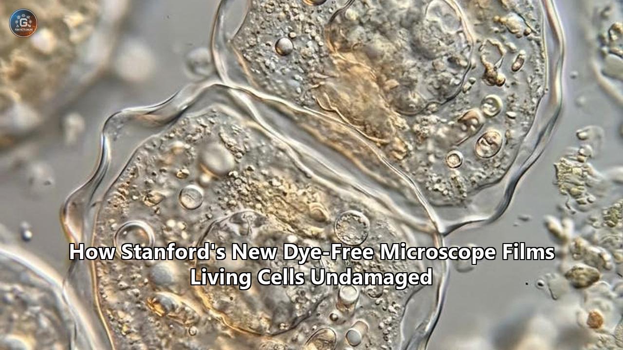
Stanford University optical physicists have filmed the sub-cellular machinery of living cells in real time at a resolution of 120 nanometers without using chemical stains, fluorescent tags, or destructive laser intensity. The development, detailed in Light: Science & Applications, introduces Interferometric Image Scanning Microscopy (iISM), an imaging technique that captures dynamic intracellular architecture while reducing the specimen's light exposure by tenfold.
Led by postdoctoral researcher Michelle Küppers and Nobel laureate W. E. Moerner, the Stanford team solved an optical contradiction that has constrained live-cell biology for more than three decades: the trade-off between spatial resolution and biological survival. By merging interferometric scattering physics with a multi-channel pixel-reassignment algorithm adapted for coherent light, the researchers filmed delicate organelles—such as endoplasmic reticulum tubules and trafficking vesicles—for hours on end without inducing heat stress, photobleaching, or cell death.
The instrument achieves this view using just 0.5 microwatts of incident laser power at the focal spot. At this power level, fragile eukaryotic structures can be recorded in their native states indefinitely. The technique is already being deployed across Stanford laboratories to observe how cancer therapeutics penetrate cell membranes, how plant roots exchange molecular signals with soil microbes, and how malaria parasites deform human red blood cells.
The path to this moment was defined by a series of physical barriers, experimental dead ends, and computational adjustments.
CHRONOLOGICAL ESCALATION OF LIVE-CELL OPTICAL IMAGING

1990s–2000s: The Fluorescence Gold Standard & Phototoxicity Trap
│  • Fluorophores enable targeted molecular imaging.
│  • Escalating Issue: Photobleaching limits observation times; reactive oxygen species (ROS) destroy living samples.
▼
2000s–2010s: The Rise of Label-Free Scattering (iSCAT)
│  • Sandoghdar and colleagues detect single nano-particles and viruses on clean glass substrates.
│  • Escalating Issue: Dense eukaryotic cytoplasm generates an impenetrable background "fog" of speckle.
▼
Early 2020s: The Confocal Impasse & The 0.15 Airy Dilemma
│  • Confocal iSCAT (C-iSCAT) rejects out-of-focus background using mechanical pinholes.
│  • Escalating Issue: A 0.15 AU pinhole discards >90% of photons; compensating with higher laser power fries cell structures.
▼
2023–2024: The Algorithmic Collision
│  • Moerner and Küppers pair Image Scanning Microscopy (ISM) arrays with coherent interferometry.
│  • Escalating Issue: Classical pixel reassignment assumes incoherent light; coherent wave interference causes phase artifacts.
▼
BREAKTHROUGH: Adaptive Pixel-Reassignment via Radial Variance Transform
   • iISM combines 9x9 virtual pinhole arrays with phase-aware reassignment algorithms.
   • Outcome: 120 nm resolution, 0.5 μW illumination, zero dyes, unlimited live recording.
1994–2014: The Fluorescence Boom and the Phototoxicity Trap
Modern cell biology was built on the back of fluorescence microscopy. When Martin Chalfie, Osamu Shimomura, and Roger Tsien developed green fluorescent protein (GFP) as a genetically encoded beacon in the 1990s, they gave researchers a way to locate individual proteins inside intact organisms. Over the subsequent two decades, fluorescence microscopy pushed beyond the classical diffraction limit described by Ernst Abbe in 1873.
Abbe established that light microscopy could not resolve two points closer together than roughly half the wavelength of light—about 200 to 250 nanometers for visible light. In the late 2000s, super-resolution methods broke this limit:
- Stimulated Emission Depletion (STED): Stefan Hell used engineered doughnut beams to switch off fluorophores at the periphery of the focal spot.
- Single-Molecule Localization (PALM/STORM): Eric Betzig and W. E. Moerner isolated individual glowing molecules across time to pinpoint their coordinates down to tens of nanometers.
These methods earned the 2014 Nobel Prize in Chemistry. Yet as biological researchers leaned into super-resolution fluorescence, an operational compromise hardened into an inescapable ceiling. Fluorescent molecules are delicate, short-lived light emitters. A fluorophore typically absorbs and re-emits a stream of photons before undergoing irreversible photochemical destruction, a process known as photobleaching. Most fluorescent dyes survive only tens of thousands to a few million excitation cycles before decaying, capping observation windows at minutes or even seconds.
THE FLUORESCENCE EXCITATION CYCLE & DAMAGE CASCADE

   Laser Photon (e.g., 488 nm)
            │
            ▼
   ┌───────────────────┐
   │ Fluorophore (S₀)  │ ──(Excitation)──► Fluorophore Excited State (S₁)
   └───────────────────┘                             │
            ▲                                        │ (Intersystem Crossing)
            │ (Fluorescence Emission)                ▼
            └────────────────────────── Fluorophore Triplet State (T₁)
                                                     │
                                                     ▼ Reacts with dissolved O₂
                                        ┌─────────────────────────┐
                                        │ Reactive Oxygen Species │
                                        │  (¹O₂, ·OH, H₂O₂, O₂·⁻) │
                                        └─────────────────────────┘
                                                     │
               ┌─────────────────────────────────────┴─────────────────────────────────────┐
               ▼                                     ▼                                     ▼
        Lipid Peroxidation                  Protein Crosslinking                   DNA Strand Breaks
    (Mitochondrial/ER lysis)            (Enzymatic function stalls)            (TK6 comet assay lesions)
The physical consequences for the host cell are severe. When a fluorophore is excited into higher energy states, it frequently crosses over to long-lived triplet states, which react directly with molecular oxygen dissolved in the cytoplasm. This interaction generates reactive oxygen species (ROS), including singlet oxygen ($^1\text{O}_2$), superoxide anions ($\text{O}_2^{\bullet-}$), and hydrogen peroxide ($\text{H}_2\text{O}_2$).
These free radicals oxidize cell membranes, cross-link enzymatic proteins, break structural cytoskeletal assemblies, and slice through genomic DNA. Single-cell electrophoresis studies (comet assays) conducted on TK6 human lymphoblast cells demonstrated that even standard live-cell fluorescent imaging routines with blue-wavelength laser exposure induce significant base lesions and single-strand breaks. The resulting oxidative cascade triggers mitochondrial depolarization, membrane blebbing, and apoptosis.
To overcome the low photon yields of individual dyes, scientists had to blast cells with illumination intensities ranging from hundreds of watts to kilowatts per square centimeter. Biologists were trapped: the higher the spatial resolution they achieved, the faster they killed the specimens they were trying to observe.
Beyond phototoxicity, fluorescent labeling alters cellular mechanics. A typical tag like GFP weighs approximately 27 kilodaltons. When fused to smaller biological proteins, the tag can equal or exceed the size of the protein it is tracking. This synthetic bulk can:
- Distort protein folding pathways.
- Sterically hinder multi-protein binding complexes.
- Alter localization within tight intracellular spaces.
- Distort membrane trafficking kinetics.
Furthermore, spectral overlap limits multi-target tracking. Because fluorescent emission spectra are broad, observing more than three or four targets simultaneously requires complex filter arrays and unmixing algorithms, shutting researchers out from viewing the whole, crowded cellular environment at once.
2004–2018: The Scattering Alternative and the Intracellular Fog
Faced with the phototoxicity trap, biophysicists searched for ways to image living specimens without exogenous dyes. Rather than coaxing engineered molecules into emitting fluorescence, they turned to light scattering—the elastic process where photons bounce off structures of varying refractive index.
Every biological component possesses an intrinsic polarizability governed by its local refractive index ($n$). Water in the cytoplasm has an index of around 1.33, while protein-dense ribosomes, lipid bilayers, and nucleic acid assemblies range from 1.45 to 1.60. When an incident laser beam hits these nanoscale variations, the structures scatter photons via Rayleigh scattering without any fluorescent absorption or molecular destruction.
In the mid-2000s, Vahid Sandoghdar and his research group developed Interferometric Scattering Microscopy (iSCAT). The technique transformed optical physics by amplifying weak scattered light through coherent optical interference.
THE INTERFEROMETRIC SCATTERING (iSCAT) SIGNAL MECHANISM

  Incoming Laser Field (E_inc)
             │
             ├─────────────────────────────────────────────────┐
             ▼                                                 ▼
   Coverslip-Water Interface                         Nanoscale Cellular Scatterer
  ┌─────────────────────────┐                       ┌─────────────────────────┐
  │  Reference Field (E_ref)│                       │ Scattered Field (E_scat)│
  │     (Strong, ~0.5%)     │                       │      (Extremely weak)   │
  └─────────────────────────┘                       └─────────────────────────┘
             │                                                 │
             └───────────────────────┬─────────────────────────┘
                                     ▼
                          Total Intensity at Detector
                                     │
     I_det = |E_ref + E_scat|² = |E_ref|² + |E_scat|² + 2|E_ref||E_scat|cos(θ)
                                   ▲           ▲               ▲
                                   │           │               │
                              Constant      Negligible     INTERFERENCE TERM
                             Background     Pure Scatter   Amplified by E_ref!
The mathematical foundation of iSCAT explains its sensitivity. If a tiny organelle or single protein scatters an electric field $E_{\text{scat}}$, the pure scattered intensity $I_{\text{scat}} = |E_{\text{scat}}|^2$ is minuscule. For a particle smaller than the wavelength of light, scattering cross-sections drop with the sixth power of the particle's diameter ($d^6$). Detecting that pure scatter against detector noise is functionally impossible for biological nanoparticles.
However, iSCAT does not measure pure scatter. It interferes the scattered wave with a strong reference beam—typically the reflection off the glass coverslip supporting the cell ($E_{\text{ref}}$). The total intensity arriving at the detector is:
$$I_{\text{det}} = |E_{\text{ref}} + E_{\text{scat}}|^2 = |E_{\text{ref}}|^2 + |E_{\text{scat}}|^2 + 2 |E_{\text{ref}}| |E_{\text{scat}}| \cos \theta$$
Because $|E_{\text{scat}}|^2$ is negligible, the signal simplifies to:
$$I_{\text{det}} \approx |E_{\text{ref}}|^2 + 2 |E_{\text{ref}}| |E_{\text{scat}}| \cos \theta$$
The third term—the interference term—is the key. The faint biological signal ($E_{\text{scat}}$) is multiplied directly by the strong reference field ($E_{\text{ref}}$). This cross-term scales with the volume of the particle ($d^3$) rather than its surface area squared ($d^6$), amplifying the signal far above detector read noise.
Using iSCAT, researchers in the early 2010s demonstrated the detection of individual unlabeled proteins, tracking myosin motors walking on actin filaments, and sizing single viral particles on bare glass slides.
THE CELLULAR NOISE BARRIER IN WIDE-FIELD iSCAT

  Clean Coverslip (Isolated Particle):
  [Cover Glass] ────────► E_ref
  [Nano-target] ────────► E_scat
  Result: Clean, shot-noise limited interference dip. Target easily detected.

  Dense Cytoplasmic Environment (Living Eukaryotic Cell):
  [Mitochondria]   ──► E_scat1 ┐
  [Actin Network]  ──► E_scat2 │
  [Ribosomes]      ──► E_scat3 ┼─► Chaotic, out-of-focus speckle pattern
  [Cell Membrane]  ──► E_scat4 │   Total background drowns individual nanostructures!
  [Target Organelle] ─► E_target┘
The moment biophysicists shifted iSCAT from clean glass slides to intact living cells, the physics turned against them. A eukaryotic cell is not an empty glass chamber; it is a crowded, 10-micrometer-thick suspension of thousands of organelles, vesicles, and macromolecular complexes.
Every single organelle scatters light. In wide-field iSCAT, the scattered waves from out-of-focus organelles above and below the focal plane propagate to the camera, interfering chaotically with the reference beam and with each other. This creates an impenetrable speckle pattern—an optical "fog" that obscures the delicate structures within the cell.
By 2018, the biophysics community realized that wide-field scattering could not resolve deep, nanoscale structures inside living cells. The background interference overwhelmed the detectors, preventing label-free scattering from challenging super-resolution fluorescence in real biological environments.
2019–2022: The Confocal Impasse and the 0.15 Airy Dilemma
To clear away this out-of-focus background, researchers turned to confocal geometry, creating Confocal Interferometric Scattering Microscopy (C-iSCAT).
Confocal architectures reject out-of-focus light by placing a physical aperture—a pinhole—in front of the photodetector, conjugate to the focal plane of the specimen. Light originating from the focal point passes cleanly through the pinhole, while scattered waves originating from out-of-focus planes strike the pinhole edges and are blocked.
THE CONFOCAL PINHOLE TRADE-OFF IN C-iSCAT

              A: Open Pinhole (e.g., 1.0 to 2.0 AU)
   Light from Focus ══════════════════════════► [ Detector ] (High Photon Flux)
   Out-of-Focus Light ─────────────────────────► [ Detector ] (Background Fog Retained)
   Result: High signal, but ruined contrast and low spatial resolution.

              B: Closed Pinhole (e.g., 0.15 AU)
   Light from Focus ═══════════[ Pinhole ]═════► [ Detector ] (Diffraction Sharpened)
   Out-of-Focus Light ─────────[ BLOCKED ]
   Photons Discarded: >90% of signal rejected!
   Result: Excellent background rejection, but signal collapses.
   Compensatory Action: Crank laser power up by 10x–50x.
   Consequence: Photothermal stress, local heating, specimen damage.
In C-iSCAT, focusing both the illumination beam and the detection channel to a diffraction-limited spot suppressed the out-of-focus background. But optical physics immediately imposed another penalty: The Airy Dilemma.
An optical pinhole’s diameter is measured in Airy Units (AU), where 1.0 AU corresponds to the diameter of the central disk of the Airy pattern formed by the microscope’s objective.
- If researchers opened the pinhole to 1.0 or 1.5 AU to capture enough scattered light, out-of-focus cellular fog leaked through, washing out low-contrast nanostructures.
- If researchers closed the pinhole down to 0.15 AU to maximize spatial resolution and reject out-of-focus light, diffraction mechanics stripped away more than 90% of the scattered photons.
To recover an adequate signal-to-noise ratio through a closed 0.15 AU pinhole, microscopists had to turn up the illumination laser. Powers at the focal plane climbed to tens of microwatts, and in some setups to milliwatts.
Although scattering does not involve fluorophore triplet-state cascades, extreme coherent laser intensity produces photothermal heating. When concentrated into a tiny 200-nanometer focal volume, high laser power induces thermal gradients, drives micro-cavitation, and damages the cell's delicate lipid membranes.
The confocal approach had reached an impasse. If the pinhole was open, the cell was safe, but the image was blurred and flooded with background noise. If the pinhole was closed, the image was crisp, but the laser power required to see anything cooked the specimen. The promise of an unperturbed look into cellular activity remained unfulfilled.
2023–2024: The Algorithmic Collision — Blending Array Detectors with Coherence
In 2023, the Moerner laboratory at Stanford began targeting this bottleneck. W. E. Moerner, having spent decades analyzing the photophysical limits of single-molecule imaging, saw that the physical pinhole was the critical point of failure in confocal scattering setups. He recruited Michelle Küppers, an optical scientist who had specialized in interferometric scattering during her doctoral studies, to tackle the problem.
The team looked at a parallel development in fluorescence imaging: Image Scanning Microscopy (ISM).
ISM was first conceptualized theoretically by British physicist Colin Sheppard in 1988 and later operationalized for fluorescence by Jörg Enderlein and colleagues in 2010. Sheppard realized that if you replace the single confocal pinhole and single photodetector with a detector array—such as a small camera or a matrix of avalanche photodiodes—you no longer have to throw away displaced photons.
SHEPPARD'S IMAGE SCANNING MICROSCOPY (ISM) GEOMETRY

  Point Illumination Spot ────► Scans Sample Coordinate (r_scan)
                                          │
                                          ▼
                                Objective Lens
                                          │
                                          ▼
                      Camera Detector Array (e.g., 9x9 pixels)
                      ┌──────┬──────┬──────┬──────┬──────┐
                      │ p₁₁  │ p₁₂  │ p₁₃  │ p₁₄  │ p₁₅  │
                      ├──────┼──────┼──────┼──────┼──────┤
                      │ p₂₁  │ p₂₂  │ p₂₃  │ p₂₄  │ p₂₅  │  Central pixel = on-axis pinhole
                      ├──────┼──────┼──────┼──────┼──────┤  Off-axis pixels = off-axis pinholes
                      │ p₃₁  │ p₃₂  │ Central p₃₃ │ p₃₄  │
                      ├──────┼──────┼──────┼──────┼──────┤
                      │ p₄₁  │ p₄₂  │ p₄₃  │ p₄₄  │ p₄₅  │
                      └──────┴──────┴──────┴──────┴──────┘
                                          │
                        Computational Pixel Reassignment
                         r_reassigned = r_scan + (d_pixel / 2)
                                          │
                                          ▼
                       Reconstructed Super-Resolved Image:
                       • Resolution boosted by factor of √2
                       • 100% of collected light preserved!
In standard fluorescence ISM, each pixel in the array acts as an independent, closed, off-axis pinhole. An off-axis detector pixel records light efficiently, but its point-spread function (PSF) is spatially shifted relative to the optical axis.
Because fluorescent light is incoherent—photons emitted by one dye molecule have no fixed phase relationship to photons emitted by another—the signals are additive intensities. Sheppard proved that if you computationally shift each off-axis pixel’s signal back toward the optical center by half the displacement distance (a factor of $M/2$, where $M$ is magnification) and sum them up, a clean mathematical result emerges:
- Every photon striking the detector is collected, matching the light efficiency of a wide-open pinhole.
- The effective optical point-spread function is narrowed by a factor of $\sqrt{2}$, boosting resolution beyond the classical diffraction limit.
Küppers and Moerner sought to port this mechanism over to interferometric scattering microscopy. If they could build an Interferometric Image Scanning Microscope (iISM), they could capture the scattered light across a detector array, synthesize the background-rejection performance of a sub-0.15 AU pinhole, collect every scattered photon, and lower the laser power into a safe regime.
However, when they tested this concept on scattering fields, the standard pixel-reassignment algorithms failed completely.
THE INCOHERENT VS. COHERENT PIXEL-REASSIGNMENT FAILURE

  A: Incoherent Fluorescence (Standard ISM)
     Total Signal = I₁ + I₂ + I₃ + ... + Iₙ
     • No phase terms.
     • Direct scalar addition of shifted pixel signals.
     • Reassignment yields a clean, sharpened Gaussian PSF.

  B: Coherent Scattering (The iSCAT Problem)
     Total Signal = |E_ref + E_scat1 + E_scat2|²
     • Retains phase cross-terms: 2·E_ref·E_scat·cos[φ(r)]
     • Scattered waves have spatially varying phases, sign flips, and interference rings.
     • Applying standard ISM scalar reassignment causes destructive interference,
       false-edge ringing, and phase cancellation artifacts!
The physical breakdown stemmed from the coherence of the laser light. In fluorescence ISM, individual molecules emit incoherent light waves, meaning their intensities add as positive real numbers ($I = \sum I_i$). In iSCAT, the scattered field and the reference field are mutually coherent.
The detected signal is governed by the phase difference $\phi(r)$ between the biological feature's scattered wave and the reference reflection:
$$S(r) \propto 2 |E_{\text{ref}}| |E_{\text{scat}}(r)| \cos[\phi(r)]$$
Depending on the axial position, local refractive index, and geometry of an organelle, $\cos[\phi(r)]$ swings between positive and negative values. An off-axis detector pixel does not merely see a laterally shifted intensity profile; it sees a shifted, phase-distorted interference pattern with dark and bright fringes.
Subjecting these coherent fringes to standard, intensity-based pixel reassignment caused destructive numerical interference. The algorithmic summing cancelled out valid biological signals, introduced edge artifacts, and blurred out fine detail. To make image scanning work with scattering, the Stanford team had to invent a computational method designed specifically for coherent point-spread functions.
2024–2025: Cracking the Coherent Code — The Birth of iISM
Between late 2024 and mid-2025, Küppers and Moerner restructured both the hardware detection pipeline and the underlying mathematics.
On the hardware bench, the researchers coupled a laser-scanning confocal microscope to an array detector positioned conjugate to the sample's focal plane. For every scan position across the biological specimen, the instrument recorded the full interferometric point-spread function (iPSF) across an array of detector elements, generating a multidimensional data hypercube representing multiple parallel, virtual off-axis pinholes.
SCHEMATIC ARCHITECTURE OF THE STANFORD iISM SYSTEM

  [Laser Source (Low Power, 0.5 μW)]
                 │
                 ▼
       [Galvo Scanning Mirrors]
                 │
                 ▼
      [High-NA Objective Lens]
                 │
                 ▼
  ═════════ Living Cell ═════════  ◄── Native intracellular organelles scatter light
  ───────────────────────────────
  Cover Glass / Aqueous Interface  ◄── Generates coherent reference reflection (E_ref)
                 │
                 ▼
      [High-NA Objective Lens]
                 │
                 ▼
       [Descanning Optics]
                 │
                 ▼
     [Camera Array Detector]       ◄── Records full iPSF matrix (9x9 virtual pinholes)
                 │
                 ▼
  [Adaptive Pixel Reassignment (APR)]
  [ via Radial Variance Transform  ] ◄── Corrects phase shifts & shifts photons
                 │
                 ▼
   Reconstructed Super-Resolved
   Label-Free Live Video Feed      ◄── 120 nm Lateral Resolution at 0.5 μW!
To reconstruct this data without destructive phase cancellation, Küppers developed a modified Adaptive Pixel-Reassignment (APR) algorithm tailored for coherent detection.
Instead of treating the off-axis pinhole signals as scalar intensity values, the algorithm applies a radial variance transform (RVT). The RVT acts as a phase-sensitive operator that analyzes the geometric curvature and intensity gradients of the coherent interference fringes captured across the virtual pinholes.
THE ADAPTIVE PIXEL-REASSIGNMENT (APR) WORKFLOW

   Raw 9x9 Array Detector Frames at each scan position (x, y)
                     │
                     ▼
   [ Radial Variance Transform (RVT) Processing ]
   • Maps spatial symmetry of coherent fringes across the virtual array.
   • Calculates the local phase offset φ_local and true displacement vectors.
                     │
                     ▼
   [ Coherent Shift-and-Sum Vector Reassignment ]
   • Off-axis signals are phase-corrected: S_corrected = S_measured · sign[cos(φ)]
   • Photons are shifted by the reassignment vector (M/2) to the true physical center.
                     │
                     ▼
   [ Super-Resolved Reconstruction ]
   • Resolution sharpened from ~200 nm down to ~120 nm.
   • Signal-to-noise ratio boosted by parallel photon collection.
   • Retains 100% of out-of-focus background rejection.
By calculating the radial variance across the detector array, the algorithm maps the precise spatial offset and phase orientation of the scattered signal at each pixel. It then shifts each signal back toward the central optical coordinate while adjusting for phase, preventing destructive numerical cancellation.
The system was first calibrated using 60-nanometer polystyrene calibration nanoparticles deposited on a glass substrate. The experimental data confirmed the theoretical models:
- The lateral point-spread function shrank, delivering a lateral resolution of approximately 120 nanometers—surpassing the diffraction limit for visible light without using fluorescent stains.
- By collecting scattered light across the entire array detector rather than rejecting it with a metal pinhole, the system captured the vast majority of available signal photons.
- This optical efficiency allowed the team to drop the illumination power to 0.5 microwatts at the specimen plane—roughly ten times lower than prior confocal scattering microscopes, while matching their background-rejection performance.
The development of dye-free microscope technology had repeatedly run up against physical compromises between laser damage, background noise, and spatial blur. The combination of array detection and phase-aware computational reassignment broke through that bottleneck.
2026: The Unperturbed Living Cell Revealed
The decisive validation of iISM came when Küppers and Moerner shifted their optics from synthetic nanoparticles to living eukaryotic cells. The resulting data, published in Light: Science & Applications, provided real-time, label-free movies of intracellular dynamics with exceptional clarity.
COMPARISON OF LIVE-CELL OPTICAL MODALITIES

Feature / Metric            Wide-Field Fluorescence     Super-Resolution (STED)   Confocal iSCAT (C-iSCAT)   Stanford iISM (Küppers & Moerner)
──────────────────────────────────────────────────────────────────────────────────────────────────────────────────────────────────────────
Fluorescent Dyes Needed?    Yes (GFP, DAPI, Alexa)      Yes (High-stability dyes) No (Label-Free)            No (Label-Free)
Lateral Resolution          ~200–250 nm                 ~30–60 nm                 ~180–200 nm                ~120 nm
Laser Power at Specimen     1–10 mW/cm²                 MW to GW/cm²              5–50 μW                    0.5 μW (Tenfold reduction)
Observation Lifespan        Seconds to Minutes          Seconds (Rapid bleach)    Minutes (Thermal stress)   Hours (Essentially unlimited)
Intracellular Target Scope  1–4 Tagged Proteins         1–2 Tagged Proteins       All native scatterers      All native scatterers simultaneously
Specimen Phototoxicity      High (Free Radicals / ROS)  Severe (Extreme damage)   Moderate (Photothermal)    Negligible / Unperturbed native state
The microscope resolved structures that were previously invisible without chemical staining:
- The Endoplasmic Reticulum (ER): The ER forms a sprawling, highly dynamic network of lipid tubules and sheets that extends throughout the cytoplasm. Because ER tubules are between 50 and 100 nanometers in diameter, diffraction-limited label-free microscopes see only a fuzzy, diffuse blur. With iISM, individual ER tubules emerged with sharp contrast, allowing researchers to track them elongating, retracting, and forming three-way junctions in real time.
- Intracellular Vesicles: Metabolic cargo vesicles ranging from 80 to 150 nanometers in diameter were tracked traversing the cytosol along cytoskeletal tracks.
- Mitochondrial Cristae Remodeling: The internal convolutions of mitochondrial membranes were imaged shifting during native metabolic cycles without the swelling and fragmentation caused by laser toxicity.
MICROSCOPIC VIEW: RETICULAR REMODELING RECORDED VIA iISM

   Time: t = 0 min                           Time: t = 15 min                         Time: t = 60 min
   ┌───────────────────────────────┐         ┌───────────────────────────────┐        ┌───────────────────────────────┐
   │    \     /         ○ (Vesicle)│         │      \   /          ○         │        │       \ /          ○          │
   │     \___/                     │         │       \_/                     │        │        X                      │
   │      / \                      │  ────►  │       / \                     │ ────►  │       / \                     │
   │     /   \-----[ER Tubule]     │         │      /   \----------○         │        │      /   \---------○          │
   │    /                          │         │     /                         │        │     /                       │
   └───────────────────────────────┘         └───────────────────────────────┘        └───────────────────────────────┘
    Laser Power: 0.5 μW                       Laser Power: 0.5 μW                      Laser Power: 0.5 μW
    Cell State: Active, dynamic, unperturbed  Cell State: Continual, steady transport   Cell State: Zero blebbing, zero stress
The most consequential result was temporal endurance. In super-resolution fluorescence experiments, viewing dynamic organellar remodeling at high frame rates typically leads to photobleaching and cell detachment within several minutes. With iISM operating at 0.5 microwatts, the Stanford team recorded continuous intracellular movies for extended experimental durations.
The cells exhibited zero signs of light-induced distress:
- No cytoplasmic vacuolization.
- No mitochondrial rounding or swelling.
- No membrane retraction or apoptosis.
The platform logged these nanoscale processes while preserving the native physiological state of the cell, marking a decisive advance for dye-free microscope technology.
The Next Phase: Three Biological Fronts Under Immediate Investigation
The development of iISM has rapidly expanded beyond optical physics into active biological research. Moerner and Küppers have initiated three interdisciplinary collaborations across Stanford University to deploy the microscope against long-standing questions in pharmacology, botany, and parasitology.
ACTIVE RESEARCH COLLABORATIONS AT STANFORD

    ┌────────────────────────────────────────────────────────┐
    │  Stanford University School of Humanities & Sciences   │
    │             Moerner Laboratory (iISM System)           │
    └───────────────────────────┬────────────────────────────┘
                                │
        ┌───────────────────────┼───────────────────────┐
        ▼                       ▼                       ▼
  Front 1: Oncology       Front 2: Botany        Front 3: Parasitology
  Cancer Drug Internal-   Plant-Microbe Inter-   Malaria Erythrocyte
  ization & Nanocarrier   actions in Soil        Deformation Dynamics
  Membrane Crossing       Ecosystems             (P. falciparum)
Front 1: The Pharmacology of Drug Uptake in Cancer
The transport of pharmaceutical compounds across cellular barriers remains an elusive stage in drug development. Small-molecule drugs and nanoparticle-based therapeutic carriers are typically tested using fluorescent tags to verify their delivery into target cells.
Yet adding a fluorescent tag alters a drug carrier's surface charge, molecular weight, hydrophobicity, and lipid-binding affinity. As a result, researchers often end up measuring how the fluorescent tag enters the cell, rather than how the therapeutic molecule behaves on its own.
By providing an unperturbed window into native cellular processes, dye-free microscope technology allows biophysicists to track untagged, pristine nanoparticles and drug-carrying liposomes as they touch the plasma membrane, become enclosed within endocytic vesicles, and travel through the cytoplasm. Oncologists can now quantify the direct internalization kinetics of candidate molecules in cancer cells under true physiological conditions, eliminating labeling artifacts from pre-clinical drug evaluations.
Front 2: Plant-Microbe Symbiotic and Pathogenic Interfaces
Plant roots exist within a dynamic ecological interface, exchanging nutrients with mycorrhizal fungi while fighting off pathogenic bacteria. Observing these multi-organism interactions under a microscope has long been hindered by plant biology:
- Plant tissues are naturally autofluorescent due to lignin, chlorophyll, and polyphenolic compounds, creating background noise that swamps standard fluorescent markers.
- Root hairs and fungal hyphae are vulnerable to light stress; laser exposure disrupts their calcium signaling cascades and stops growth entirely.
The Stanford team is using iISM to track the real-time physical interactions between plant roots, fungal hyphae, and soil bacteria simultaneously. Because iISM does not rely on fluorescent emission spectra, it bypasses native autofluorescence entirely.
Researchers can watch the physical contact zones where fungal filaments adhere to plant cell walls, observing cytoskeletal rearrangements and vesicle accumulation at the infection front without light-induced growth arrest.
MALARIA INFECTION: RED BLOOD CELL (RBC) NANOMECHANICAL ATTACK

  Normal Healthy Erythrocyte:
  [ Highly Flexible, Biconcave Disk ] ──► Squeezes smoothly through 2 μm splenic capillaries.

  Plasmodium falciparum Invasion (Observed via iISM):
  1. Merozoite binds to erythrocyte membrane.
  2. Parasite exports protein complexes (e.g., RESA, KAHRP) to the cell membrane.
  3. Cytoskeletal actin-spectrin network is crosslinked and stiffened.
  4. iISM maps the emergence of nanoscale membrane "knobs" (~100 nm) in real time!
     • No fluorescent antibody tags needed to perturb fragile RBC membrane mechanics.
     • Researchers observe the exact mechanics of vascular occlusion and cell destruction.
Front 3: Mechanical Remodeling in Malaria-Infected Red Blood Cells
A planned clinical research project applies iISM to human parasitology, focusing on the malaria pathogen Plasmodium falciparum.
When P. falciparum merozoites invade human erythrocytes, they radically restructure the host cell over a 48-hour cycle. The parasite exports specialized proteins that form nanoscale structures called "knobs" on the red blood cell membrane. These knobs anchor the cell to blood vessel walls, preventing the spleen from filtering out infected cells and driving severe complications such as cerebral malaria.
Studying this process in living red blood cells has challenged biophysicists because erythrocyte membranes are extremely fragile. Fluorescent antibody labels alter membrane stiffness, while the high illumination power used in super-resolution setups disrupts the spectrin-actin cytoskeleton.
Using iISM, researchers can study how erythrocytes deform when attacked by Plasmodium parasites at high spatial resolution. By recording changes in membrane topography and stiffness over hours without chemical tags, the team aims to identify how the parasite alters red blood cell elasticity, pinpointing mechanical vulnerabilities that can be targeted with small-molecule antimalarial drugs.
Resolving the Optical Trade-Off: Complementarity and Technological Horizons
The introduction of iISM does not make fluorescence microscopy obsolete, a point emphasized by both Küppers and Moerner. Rather, it establishes a complementary imaging framework that balances molecular specificity with holistic structural context.
Fluorescence microscopy remains unsurpassed in its ability to pinpoint a single specific protein out of thousands within a cell. If a biochemist needs to know whether Protein A binds exclusively to Protein B, fluorescent tagging provides an unmistakable, high-contrast answer.
However, fluorescence operates with narrow vision: it reveals only the tagged molecules, leaving the surrounding cellular environment invisible.
CORRELATIVE iISM AND FLUORESCENCE: COMBINED DUAL-CHANNEL SYSTEM

                     Illumination Source
                     (Dual-Line Laser: e.g., 488 nm / 635 nm)
                                │
                                ▼
                       Living Cell Specimen
                                │
                 ┌──────────────┴──────────────┐
                 ▼                             ▼
       Coherent Scattered Photons     Incoherent Fluorescent Photons
                 │                             │
                 ▼                             ▼
       [ Array Camera (iISM) ]         [ Confocal PMT / APD ]
                 │                             │
                 ▼                             ▼
        Structural Context:            Molecular Specificity:
        • ER Tubules (120 nm)          • Single Protein Complex
        • Vesicle Trafficking          • Transcription Factor
        • Mitochondrial Cristae        • Receptor Localization
                 │                             │
                 └──────────────┬──────────────┘
                                ▼
                   Fused Correlative Image:
                   Specific molecular actors viewed within
                   the dynamic, unperturbed cellular context!
By operating as a multi-modal system, the Stanford architecture enables correlative imaging. A researcher can use a minimal, low-intensity fluorescent channel to mark a single gene product, while running iISM simultaneously to visualize the entire cytoplasmic context—the surrounding organelles, membranes, and cytoskeletal structures—moving at 120-nanometer resolution.
This allows researchers to see both the target protein and its unperturbed cellular environment without destroying the living system.
The future implementation of dye-free microscope technology depends on overcoming several clear technical hurdles:
- Axial Resolution Limits ($Z$-Axis Sectioning):
While iISM achieves roughly 120-nanometer resolution in the lateral ($X$-$Y$) plane, its axial resolution remains constrained by standard confocal physics to approximately 400 to 500 nanometers. Out-of-focus background rejection is strong, but true isotropic three-dimensional resolution will require combining iISM with opposing dual-objective geometries, such as 4Pi optical architectures, or pairing it with light-sheet illumination.
- Optical Penetration in Thick Tissues:
The initial breakthroughs with iISM were accomplished in flat cell monolayers and cultured mammalian cells. As the microscope focuses deeper into multi-layered multicellular spheroids or thick living tissue slices (beyond 20 to 50 micrometers), multiple scattering events scramble the spatial phase of both the scattered wave and the reference beam. Overcoming this will require integrating adaptive optics—using deformable mirrors to measure and cancel out tissue-induced wavefront aberrations in real time.
- High-Speed Computational Processing Demands:
Recording the full interferometric point-spread function across a camera array for every pixel of a high-resolution scan generates massive data streams. A single high-speed video recording can produce hundreds of gigabytes of raw interferometric frames. Processing these arrays through the radial variance transform and executing adaptive pixel-reassignment requires dedicated GPU clusters.
To make iISM a routine laboratory tool, software teams are writing streamlined machine-learning algorithms and field-programmable gate array (FPGA) firmware to run coherent pixel reassignment in real time, delivering live, super-resolved video feeds directly to microscope monitors.
- Integration with Existing Commercial Microscopes:
Because iISM uses a laser-scanning framework and standard high-numerical-aperture objectives, it does not require an entirely exotic hardware footprint. The Moerner laboratory designed the system around an architecture that can be retrofitted onto commercial confocal systems.
Adding an array detector module and replacing scalar pixel-reassignment software with the team's coherent APR code allows standard confocal platforms to be converted into gentle, super-resolution scattering instruments.
DEVELOPMENT MILESTONES: THE PATH FORWARD FOR iISM

[ Current Achievement (2026) ]
  • 120 nm lateral resolution in cultured cell monolayers.
  • 0.5 μW illumination power; 10x lower light dose than prior C-iSCAT.
  • Demonstration of unperturbed ER tubule and vesicle dynamics.
         │
         ▼
[ Phase 1: Commercial Modular Upgrades (2026–2027) ]
  • Drop-in detector array units for existing commercial confocal stands.
  • Open-source FPGA algorithms for real-time coherent APR reconstruction.
  • Correlative dual-channel imaging (iISM + low-dose single-molecule tracking).
         │
         ▼
[ Phase 2: Deep-Tissue & 3D Implementations (2027–2029) ]
  • Adaptive optics integration to correct tissue aberrations deeper than 50 μm.
  • 4Pi / dual-objective configurations to push axial (Z) resolution below 150 nm.
  • In vivo imaging of intact model organisms (e.g., C. elegans, Zebrafish larvae).
A New Window into the Native Machinery of Life
The history of microscopy has been defined by a constant battle between illumination and survival. For centuries, the only way to see the microscopic world clearly was to fix it, slice it, dry it, or blast it with light. When super-resolution fluorescence broke through Abbe’s diffraction limit at the turn of the 21st century, it opened an extraordinary view into molecular biology, but at the cost of high phototoxicity and fleeting observation times.
The realization of Interferometric Image Scanning Microscopy at Stanford shows that the dilemma between spatial resolution and biological survival is not an absolute law of physics. By using coherent interference to amplify the faint signals of unstained cellular structures, and by applying adaptive pixel reassignment to collect every available photon, Küppers and Moerner turned discarded scattered light into a practical imaging tool.
Operating at an illumination power of just 0.5 microwatts, the instrument gives researchers a way to film the delicate mechanics of the cell—from moving transport vesicles to reshaping organelle networks—without cooking the sample or masking it behind synthetic dyes.
As this dye-free microscope technology moves into oncology, infectious disease, and plant biology laboratories, it establishes a new standard for biological imaging: the ability to observe the complex machinery of living cells in real time, with high clarity, completely undisturbed.
Reference:
- https://scitechdaily.com/stanfords-revolutionary-new-microscope-reveals-living-cells-in-stunning-detail/
- https://www.youtube.com/watch?v=7_fb1MaebZc
- https://news.stanford.edu/stories/2026/03/high-resolution-miscroscope-living-cells-interferometric-image-scanning-microscopy
- https://analyticalscience.wiley.com/content/news-do/high-resolution-label-free-microscopy-maps-structures-inside-living-cells
- https://www.photonics.com/Articles/Interferometric-Microscopy-Opens-Pathways-for/a72071
- https://www.eurekalert.org/news-releases/1118713
- https://scitechdaily.com/stanfords-revolutionary-new-microscope-reveals-living-cells-in-stunning-detail/
- https://www.biorxiv.org/content/10.1101/2025.09.19.677416v1.full
- https://pmc.ncbi.nlm.nih.gov/articles/PMC3677558/
- https://chemistry.stanford.edu/news/moerner-lab-new-microscope-reveals-living-cells-unprecedented-detail
- https://techfinder.stanford.edu/technology/interferometric-image-scanning-microscopy-label-free-high-resolution-imaging-live-cells