For decades, the standard medical explanation for the profound exhaustion that accompanies menopause has followed a predictable, surface-level script. Women are told that fluctuating estrogen levels disrupt sleep, that hot flashes and night sweats cause chronic night-time waking, and that the psychological stress of midlife transitions naturally saps daily energy.
While these factors certainly play a role, they fail to explain a critical clinical reality: the bone-deep, heavy-limbed exhaustion that persists even when a woman sleeps for eight hours, optimizes her diet, and eliminates external stressors. This is not merely a lack of sleep; it is a systemic cellular brownout.
The ultimate root of menopause fatigue causes lies not in the mind, nor even primarily in the sleep centers of the brain. Instead, it is located within the microscopic, double-membraned energy factories of your cells: the mitochondria.
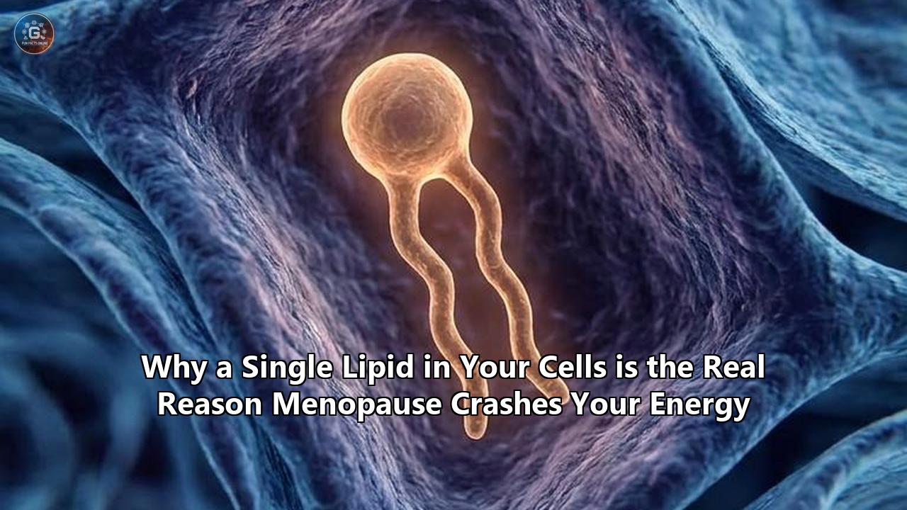
At the very heart of this mitochondrial crisis is a highly specialized, ancient lipid that constitutes the physical foundation of cellular power generation. This lipid is cardiolipin.
When estrogen levels plummet during the menopausal transition, this single, fragile lipid undergoes a catastrophic degradation process. Without it, the machinery that generates cellular energy collapses.
To truly understand why menopause crashes your energy, we must look beyond the standard endocrine model and venture into the highly technical, often overlooked field of mitochondrial lipidomics.
What is Cardiolipin? The Ancient Scaffold of Your Cell's Power Grid
To understand how menopause halts energy production, one must first look at the unique architecture of the mitochondrion. Unlike most organelles, the mitochondrion is surrounded by two distinct lipid membranes: an outer membrane and a highly folded inner mitochondrial membrane (IMM).
The IMM is where the magic of life happens. It is the site of oxidative phosphorylation—the process by which your cells convert the oxygen you breathe and the food you eat into adenosine triphosphate (ATP), the universal chemical currency of cellular energy.
[Outer Mitochondrial Membrane]
       │ (Intermembrane Space)
[Inner Mitochondrial Membrane (IMM)] ── Contains Cardiolipin (CL) & ETC Supercomplexes
       │ (Mitochondrial Matrix)
Cardiolipin (CL) is a unique phospholipid localized almost exclusively within this inner mitochondrial membrane, representing roughly 15% to 20% of its total lipid composition.
Unlike typical structural phospholipids (such as phosphatidylcholine or phosphatidylethanolamine) which possess a head group and two fatty acid tails, cardiolipin has a dimer-like structure. It consists of two phosphatidyl groups linked by a central glycerol bridge, giving it a total of four fatty acid (acyl) chains and two negative charges.
This specific molecular geometry gives cardiolipin a conical shape. When packed tightly together in the IMM, these conical lipids force the membrane to bend and fold inward, creating the deep, finger-like invaginations known as cristae.
The cristae are not passive structures; they are highly specialized subcompartments designed to maximize the surface area available for energy generation. By driving this curvature, cardiolipin literally builds the physical space where your body makes energy.
Beyond its structural role, cardiolipin functions as an indispensable physical scaffold. The generation of ATP relies on the Electron Transport Chain (ETC), a series of five protein complexes (Complexes I through V) embedded within the IMM.
To transfer electrons efficiently, these complexes cannot drift aimlessly through the membrane. Instead, they must assemble into tightly bound physical macro-structures called supercomplexes or "respirasomes".
Cardiolipin acts as the molecular glue that anchors these complexes together. It establishes highly specific, non-covalent bonds with Complex I, Complex III, Complex IV, and ATP Synthase (Complex V).
Additionally, cardiolipin holds Cytochrome C—a critical electron-shuttling protein—in close physical proximity to the IMM. When cardiolipin is abundant and structurally intact, electrons flow seamlessly down the chain, protons are pumped efficiently into the intermembrane space, and the ATP Synthase rotor spins at maximum capacity, flooding your body with energy.
The Estrogen Shield: How 17β-Estradiol Guards Mitochondrial Lipids
For a woman's entire reproductive life, this delicate, high-energy machinery is heavily protected by 17β-estradiol (E2), the dominant and most potent form of estrogen. Estrogen is far more than a reproductive hormone; it is a master metabolic survival factor.
At the cellular level, estrogen acts as a primary guardian of mitochondrial health. It does this through two main pathways: genomic signaling in the nucleus and direct, non-genomic actions inside the mitochondria themselves.
                        17β-Estradiol (Estrogen)
                                   │
         ┌─────────────────────────┴─────────────────────────┐
         ▼                                                   ▼
[Nuclear Receptors: ERα]                           [Mitochondrial Receptors: ERβ]
         │                                                   │
   Upregulates PGC-1α                               Upregulates MnSOD & GPX1
         │                                                   │
  Increases CL Synthesis & Remodeling                Suppresses ROS & Lipid Peroxidation
         │                                                   │
         └─────────────────────────┬─────────────────────────┘
                                   ▼
                   [Stable, Intact Cardiolipin]
                                   │
                   [Efficient ATP & High Energy]
1. Genomic Regulation via PGC-1α
In the cell nucleus, estrogen binds to Estrogen Receptor Alpha (ERα) to upregulate the expression of PGC-1α (Peroxisome proliferator-activated receptor gamma coactivator 1-alpha).
PGC-1α is the master regulator of mitochondrial biogenesis—the process by which cells grow and multiply their mitochondria.
Crucially, PGC-1α controls the transcription of the enzymes responsible for synthesizing and remodeling cardiolipin. This includes Cardiolipin Synthase 1 (CLS1), the enzyme that builds the cardiolipin molecule, and Tafazzin, the transacylase enzyme that remodels cardiolipin's fatty acid tails to ensure they contain the precise ratio of polyunsaturated fatty acids (PUFAs) needed for optimal membrane fluidity.
2. Direct Protection via Mitochondrial ERβ
Mitochondria contain their own local pool of estrogen receptors, with Estrogen Receptor Beta (ERβ) localized directly inside the mitochondrial matrix.
When E2 binds to mitochondrial ERβ, it directly stimulates the expression and activity of critical antioxidant defense enzymes:
- Manganese Superoxide Dismutase (MnSOD)
- Glutathione Peroxidase 1 (GPX1)
These enzymes act as an elite defense force patrolling the IMM, neutralising the reactive oxygen species (ROS) that are naturally generated as byproducts of ATP production.
By continuously suppressing ROS, estrogen prevents oxidative damage to the highly sensitive lipids in the mitochondrial membrane. Under this protective estrogen shield, cardiolipin remains highly stable, keeping your cellular power grid operating at peak efficiency.
The Peroxidation Cascade: The Biochemistry of Estrogen Depletion
When a woman enters perimenopause and transitions into postmenopause, this protective shield is abruptly dismantled. Estrogen levels do not merely decline; they crash.
Circulating E2 drops from premenopausal levels of 432–1,893 pmol/L to less than 100 pmol/L. This sudden hormone depletion triggers a devastating biochemical domino effect that directly targets cardiolipin.
Without estrogen to drive PGC-1α expression, the synthesis and remodeling of cardiolipin slows down significantly. Concurrently, the loss of mitochondrial ERβ activation leads to a rapid decline in the levels of MnSOD and GPX1.
As these antioxidant enzymes vanish, the mitochondria lose their ability to scavenge free radicals. The concentration of reactive oxygen species within the inner mitochondrial membrane surges exponentially.
This surge in ROS is highly problematic for cardiolipin due to its unique chemical composition. To maintain the high membrane fluidity required for the ETC, cardiolipin is heavily enriched with polyunsaturated fatty acids (such as linoleic acid).
PUFAs contain multiple carbon-carbon double bonds, which possess highly reactive allylic hydrogens. This makes cardiolipin one of the most chemically vulnerable targets for lipid peroxidation in the entire human body.
When a hydroxyl radical or superoxide anion attacks cardiolipin's unsaturated acyl chains, it steals a hydrogen atom, initiating a self-propagating chain reaction of lipid peroxidation.
This process transforms the structurally vital cardiolipin into oxidized cardiolipin.
   [Estrogen Drops]
          │
          ▼
   [Loss of MnSOD/GPX1]
          │
          ▼
   [ROS Surge in IMM]
          │
          ▼
   [Free Radical Attack on Cardiolipin's PUFA Tails]
          │
          ▼
   [Lipid Peroxidation]
          │
          ▼
   [Cardiolipin Oxidized & Degraded]
Once oxidized, cardiolipin undergoes a dramatic conformational change. It loses its conical shape and its negative charge, rendering it unable to maintain the structural curvature of the IMM cristae.
Furthermore, oxidized cardiolipin is rapidly targeted by phospholipases and degraded.
This biochemical devastation has been thoroughly documented in preclinical models of menopause. In seminal research led by scientists Sandra Zárate, Mariana Astiz, and Natalia D. Magnani, the effects of long-term ovarian hormone deprivation were evaluated in female rats.
The researchers discovered that chronic ovariectomy (the surgical removal of ovaries to mimic surgical menopause) led to a severe reduction in total cardiolipin content alongside a highly altered cardiolipin fatty acid profile.
This lipid depletion occurred alongside a significant decline in mitochondrial oxygen uptake and ATP production, even though the physical proteins of the respiratory complexes were still present.
The structural lipid milieu had deteriorated, and with it, the cell’s capacity to generate energy.
Why Cardiolipin Loss Causes Sleep-Resistant Menopause Fatigue
To understand why this molecular damage results in the clinical reality of menopause fatigue causes, we have to examine the physiological consequences of cardiolipin depletion.
When cardiolipin is oxidized and degraded, the physical structure of the mitochondrial inner membrane unravels. This structural failure triggers three distinct, energy-crashing mechanisms.
1. The Supercomplex Unraveling and Electron Leakage
Without cardiolipin acting as the structural scaffold, the individual respiratory complexes of the ETC lose their physical connections and drift apart within the liquid lipid bilayer.
This separation drastically increases the physical distance that electrons must travel to complete the chain. As a result, electron transfer slows to a crawl, and the efficiency of ATP generation plummets.
Crucially, these unanchored, struggling complexes begin to "leak" electrons directly into the mitochondrial matrix. These stray electrons react with oxygen to form massive quantities of superoxide radicals, further accelerating the lipid peroxidation of whatever healthy cardiolipin remains.
This is the classic mitochondrial "vicious cycle": cardiolipin loss causes electron leakage, which generates more ROS, which destroys more cardiolipin.
               [Supercomplexes Disassembled]
                             │
                             ▼
                     [Electron Leakage]
                             │
            ┌────────────────┴────────────────┐
            ▼                                 ▼
   [Decreased ATP Production]      [Superoxide Radical (ROS) Generation]
            │                                 │
            ▼                                 ▼
   [Systemic Fatigue]              [Further Cardiolipin Peroxidation]
2. Inner Membrane Depolarization and Proton Uncoupling
To make ATP, the mitochondria must maintain a massive electrochemical gradient across the inner membrane. As electrons move down the ETC, Complexes I, III, and IV pump protons ($H^+$) out of the matrix and into the intermembrane space.
This creates a high concentration of protons outside the matrix, generating a membrane potential of roughly -150 to -200 mV—an electrical field strength equivalent to 30 million volts per meter.
ATP Synthase (Complex V) acts like a hydroelectric turbine. Protons flow back down their concentration gradient through the turbine, causing it to spin and physically squeeze adenosine diphosphate (ADP) and inorganic phosphate together to form ATP.
However, for this turbine to work, the inner membrane must be completely impermeable to protons. Protons must only pass through the turbine.
When cardiolipin is depleted, the structural integrity of the IMM is compromised. The membrane becomes "leaky" or uncoupled.
Protons begin to slip directly through the damaged lipid bilayer, bypassing ATP Synthase entirely. This is known as proton leak or mitochondrial uncoupling.
Even though you are breathing and eating, the energy stored in your proton gradient is dissipated as wasted heat rather than being converted into ATP. Your cells are running, but your engine is slipping.
3. The Tissue-Specific Impact on High-Energy Organs
Because mitochondria are present in virtually every cell, this cardiolipin-driven energy deficit affects the entire body. However, the symptoms of menopause fatigue are highly concentrated in the organs that possess the highest density of mitochondria:
The Brain (The Cognitive Crash)
The human brain represents only 2% of total body weight but consumes over 20% of the body's total glucose and oxygen—most of which is used to power the mitochondrial ATP engines required for synaptic transmission.
As documented in the research by Sandra Zárate and her colleagues, long-term ovarian hormone deprivation causes an early and severe drop in cardiolipin levels within the hippocampus—the brain's primary center for learning, memory, and spatial orientation.
When hippocampal cardiolipin is depleted, local ATP levels crash, leading directly to the classic menopausal cognitive triad: brain fog, memory recall lapses, and severe mental fatigue.
The Heart (The Cardiovascular Shift)
The heart is the most mitochondria-dense organ in the body, requiring a continuous, massive supply of ATP to sustain roughly 100,000 beats per day.
In a landmark study published by Dr. Tami Martino, director of the Centre for Cardiovascular Investigations at the University of Guelph, researchers discovered that female hearts naturally maintain a highly specialized, protective cardiolipin profile that ensures superior energy production compared to male hearts.
However, this cardioprotective advantage was completely lost when the ovaries were removed.
The resulting cardiolipin degradation and metabolic shift away from efficient fatty acid oxidation not only contributes to the extreme physical exhaustion felt during menopause, but also explains why a woman’s risk of cardiovascular disease spikes dramatically after the menopausal transition.
Skeletal Muscle (The Physical Heavy-Limb Sensation)
Skeletal muscle fibers rely heavily on cardiolipin to keep their intermyofibrillar mitochondria functioning during physical exertion.
When cardiolipin degrades, the threshold for physical exhaustion drops significantly. Women find that walking up stairs, lifting weights, or recovering from exercise requires far more effort and time.
This is not a loss of motivation; it is a structural failure of the muscle mitochondria to generate the ATP necessary for muscle contraction.
Technical Comparison of Menopause Fatigue Pathways
To understand how cardiolipin degradation compares to more commonly discussed menopause fatigue causes, review this breakdown of cellular versus systemic drivers:
| Diagnostic Metric | Cardiolipin Degradation (Mitochondrial) | Sleep-Disruption Pathway (Endocrine) | Vasomotor-Induced Fatigue (Autonomic) | HPA-Axis Dysregulation (Adrenal) |
|---|---|---|---|---|
| Primary Location | Inner Mitochondrial Membrane (IMM) | Hypothalamus & Pineal Gland | Brainstem & Sympathetic Nervous System | Adrenal Cortex & Hypothalamus |
| Molecular Mechanism | Lipid peroxidation of PUFA chains; supercomplex disassembly | Decline in melatonin synthesis; frequent waking due to progesterone drop | Sudden surges in noradrenaline triggering hot flashes | Dysregulated cortisol rhythms and depleted DHEA levels |
| Primary Biomarker | Oxidized cardiolipin; decreased blood-plasma ATP potential | Reduced slow-wave sleep duration on polysomnography | Fluctuating skin conductance response (SCR) | Flattened salivary cortisol curve (4-point test) |
| Nature of Fatigue | Bone-deep, physical, and cognitive; completely resistant to rest | Drowsiness, morning grogginess, and daytime sleepiness | Episoic exhaustion following acute hot flash events | Late-afternoon crashes; "wired but tired" sensation at night |
| Response to Caffeine | Negligible; often worsens fatigue by depleting cellular NAD+ | Temporary improvement in alertness via adenosine receptor blockade | Worsens symptoms by triggering additional hot flashes | Exacerbates cortisol depletion and adrenal strain |
Behind-the-Scenes Clinical Reality: Why This Science Has Been Missed
If cardiolipin is so central to mitochondrial energy production and menopause-induced fatigue, why is this cellular lipid rarely mentioned in standard gynecological care?
The answer lies in the deep, historic division between clinical endocrinology and mitochondrial lipidomics.
Standard clinical medicine is built around a "hormone-centric" paradigm. When a patient presents with menopausal symptoms, clinicians measure serum hormone levels (such as FSH, LH, and estradiol) and treat the condition by attempting to replace those hormones.
However, standard blood tests cannot measure the biochemical state of internal cell membranes. Measuring cardiolipin levels or assessing inner mitochondrial membrane potential ($\Delta\Psi_m$) requires tissue biopsies or specialized, high-resolution mass spectrometry lipidomics—tools that are strictly confined to research laboratories and are entirely unavailable in standard clinical practice.
Furthermore, the pharmaceutical pipelines have historically favored broad-spectrum hormone therapies rather than targeted mitochondrial therapeutics.
Standard hormone replacement therapy (HRT) can help protect cardiolipin by restoring systemic estradiol levels, but it is often prescribed late in the menopausal transition—long after the initial wave of lipid peroxidation has already damaged the local mitochondrial architecture.
Because clinical guidelines are slow to adapt to subcellular research, millions of women continue to receive treatments that address systemic symptoms while leaving the underlying cellular battery damage completely untouched.
Emerging Therapies: Recharging the Cellular Battery
Fortunately, the field of mitochondrial medicine is advancing rapidly. In 2026, researchers and clinicians are developing and utilizing novel therapeutic strategies specifically designed to bypass the hormone pathway and directly target, stabilize, and rebuild mitochondrial cardiolipin.
               [Therapeutic Targets for Cardiolipin Recovery]
                                     │
         ┌───────────────────────────┼───────────────────────────┐
         ▼                           ▼                           ▼
 [Mito-Stabilizing Peptides]  [Membrane Lipid Replacement]  [Mito-Targeted Nutrients]
         │                           │                           │
  e.g., Elamipretide (SS-31)    e.g., Glycerophospholipids   e.g., CoQ10, ALCAR, Omega-3s
         │                           │                           │
         ▼                           ▼                           ▼
[Binds & Protects Cardiolipin] [Replaces Damaged IMM Lipids] [Enhances ETC Flow & Remodeling]
         │                           │                           │
         └───────────────────────────┬───────────────────────────┘
                                     ▼
                      [Restored Mitochondrial Power]
1. Mitochondria-Targeted Peptides (Elamipretide / SS-31)
The most sophisticated frontier in cardiolipin restoration is the development of mitochondria-targeted peptides, specifically Elamipretide (also known as SS-31).
SS-31 is a small, water-soluble, cell-permeable tetrapeptide that selectively targets the inner mitochondrial membrane.
Once inside the IMM, SS-31 binds directly to cardiolipin via electrostatic and hydrophobic interactions. This binding prevents cardiolipin from interacting with Cytochrome C to form a peroxidase complex, thereby halting the self-propagating cycle of cardiolipin oxidation.
By physically stabilizing cardiolipin, SS-31 helps preserve cristae structure, keeps the respiratory supercomplexes assembled, increases ATP production, and dramatically reduces ROS leakage.
While originally researched for severe mitochondrial diseases like Barth Syndrome (a genetic disorder of cardiolipin synthesis), SS-31 is now being actively studied as a therapy for age-related and menopause-induced mitochondrial decline.
2. Membrane Lipid Replacement (MLR)
Another highly effective clinical approach is Membrane Lipid Replacement (MLR).
MLR involves the oral administration of specific, protected mixtures of glycerophospholipids (GPLs), often derived from soy or egg lecithin and formulated to survive the harsh environment of the gastrointestinal tract.
Once absorbed, these exogenous phospholipids are transported via the lymphatic system and blood directly to cells.
Through natural lipid exchange processes, these healthy, unoxidized phospholipids insert themselves into damaged cellular and mitochondrial membranes, physically replacing the oxidized, degraded lipids.
In a series of clinical trials evaluating patients suffering from chronic, profound fatigue—including menopausal women—the administration of oral GPLs resulted in a significant and measurable reduction in fatigue levels, alongside a notable increase in cellular membrane potential and mitochondrial function.
3. Exogenous Cardiolipin Therapy (The Laplante & Bergdahl Findings)
A remarkable preclinical study published in April 2026 by researchers A. Laplante and A. Bergdahl provided direct evidence of cardiolipin's therapeutic potential.
The researchers evaluated the effects of exogenously administered cardiolipin in a rodent model of metabolic and physical performance.
They discovered that the administration of exogenous cardiolipin was associated with a direct and significant enhancement of skeletal muscle mitochondrial respiratory function.
Crucially, this improvement occurred without any change in mitochondrial density.
The study demonstrated that exogenously supplied lipids can integrate into the IMM, directly optimizing mitochondrial efficiency and aerobic capacity.
This research opens up exciting new avenues for lipid-based therapies specifically designed to combat muscle weakness and physical exhaustion in postmenopausal women.
4. Advanced Nutrient and Co-Factor Support
To synthesize and remodel cardiolipin, the mitochondria require a continuous supply of highly specific raw materials and enzyme co-factors. Clinicians specializing in mitochondrial medicine utilize targeted protocols that include:
- Acetyl-L-Carnitine (ALCAR): An amino acid derivative that plays a dual role: it shuttles long-chain fatty acids into the mitochondria to be used as fuel, and it acts as an acetyl donor during the synthesis and remodeling of mitochondrial lipids, including cardiolipin.
- Coenzyme Q10 (CoQ10): A vital component of the ETC that shuttles electrons from Complexes I and II to Complex III. CoQ10 acts as a powerful fat-soluble antioxidant within the IMM, protecting cardiolipin from local lipid peroxidation.
- High-Purity Omega-3 Fatty Acids (EPA and DHA): These specific polyunsaturated fatty acids serve as essential structural building blocks during the remodeling of cardiolipin. Ensuring an abundant supply of unoxidized omega-3s allows the Tafazzin enzyme to rebuild cardiolipin's delicate tails with the ideal structural geometry.
- Targeted Antioxidant Support (Vitamins E, C, and Selenium): These nutrients act synergistically to neutralize free radicals within both the lipid and aqueous phases of the mitochondrial membrane, halting the peroxidation cascade before it can destroy the cardiolipin scaffold.
Actionable Strategies: Reclaiming Your Vitality
If you are navigating perimenopause or postmenopause and struggling with profound, sleep-resistant fatigue, you do not have to wait for advanced peptide therapies to become mainstream.
You can begin supporting your mitochondrial lipid membrane and addressing these specific menopause fatigue causes immediately using targeted, science-backed lifestyle and dietary interventions.
               [Daily Protocol for Cardiolipin Protection]
                                    │
       ┌────────────────────────────┼────────────────────────────┐
       ▼                            ▼                            ▼
[Mitochondrial Nutrients]     [Strength Training]          [Glucose Management]
• CoQ10: 200mg/day            • Lift weights 3x/week       • Limit processed sugars
• ALCAR: 1000mg/day           • Stimulates mitophagy       • Enhances insulin sensitivity
• Omega-3: 2-3g/day           • Replaces old membranes     • Reduces mitochondrial stress
1. Deploy the Cardiolipin Protection Nutrient Stack
To protect and rebuild your mitochondrial membranes from the inside out, consider incorporating a highly specific combination of lipid-active supplements:
- Coenzyme Q10 (as Ubiquinol): 200 mg daily. This form is highly active and serves as a direct antioxidant shield for your inner mitochondrial lipids.
- Acetyl-L-Carnitine (ALCAR): 1,000 mg daily. Take this in the morning to optimize fatty acid transport and support cardiolipin remodeling.
- High-Dose, Low-Oxidation Omega-3 Fatty Acids: 2 to 3 grams of combined EPA/DHA daily. Ensure your supplement is third-party tested for low peroxide values to avoid introducing oxidized lipids into your body.
- Active B-Complex and Zinc: 15 mg of zinc and a methylated B-complex daily. These act as essential co-factors for the enzymes involved in cellular energy metabolism.
2. Leverage Strength Training to Force Mitochondrial Cleansing
While cardiovascular exercise is highly beneficial, progressive resistance training (lifting weights) is the ultimate stimulus for mitochondrial quality control.
When muscle tissue is subjected to mechanical load, it triggers a cellular clean-up process called mitophagy—the selective destruction and recycling of old, damaged mitochondria.
By forcing your body to clear out mitochondria with degraded, oxidized cardiolipin, strength training stimulates the rapid biogenesis of brand-new, healthy mitochondria packed with freshly synthesized cardiolipin.
Aim for 3 sessions of full-body resistance training per week, focusing on compound movements like squats, rows, and presses.
3. Implement Strict Blood Glucose Control
Surges in blood glucose and subsequent insulin spikes place a massive metabolic burden on the mitochondrial ETC.
When cells are flooded with excess glucose, the electron transport chain is forced to process an overwhelming number of electrons, leading to a massive increase in electron leakage and a catastrophic spike in ROS.
This localized oxidative stress directly damages your cardiolipin.
To protect your lipids, prioritize a diet built around whole, nutrient-dense foods, clean proteins, and healthy fats. Avoid processed sugars, refined carbohydrates, and continuous snacking, which keep your mitochondria in a state of constant, damaging overdrive.
Future Frontiers: Shift to Mitochondria-Centric Menopause Medicine
As research in mitochondrial lipidomics continues to expand, the clinical understanding of menopause is undergoing a dramatic shift. We are moving away from an era that viewed menopause simply as a "glandular decay" or a localized reproductive event.
Instead, the scientific consensus is beginning to recognize menopause as a profound bioenergetic remodeling process that affects every cell, tissue, and organ in the female body.
In the coming years, we can expect to see:
- The development of advanced, blood-based lipidomic panels capable of measuring oxidized cardiolipin and general mitochondrial membrane damage in a standard clinical setting.
- The clinical approval and widespread availability of mitochondria-targeted peptide therapies (like Elamipretide/SS-31) specifically tailored to treat age-related and menopause-induced fatigue.
- A major shift in gynecological guidelines that incorporates mitochondrial protective therapies—including targeted nutrition, membrane lipid replacement, and metabolic optimization—alongside traditional hormone replacement therapy.
For the millions of women experiencing the debilitating effects of menopause-related exhaustion, this scientific evolution offers a powerful message of validation and hope.
Your fatigue is not in your head, and it is not a personal failure of willpower or lifestyle. It is a physical, structural challenge occurring within the ancient lipid membranes of your cells.
By understanding the unique role of cardiolipin and taking proactive, targeted steps to protect your cellular power plants, you can reclaim your energy, restore your cognitive clarity, and step into the next chapter of your life with power, strength, and vitality.
Reference:
- http://denverholisticmedicine.com/blog/2025/12/20/mitochondrial-health-the-missing-link-in-perimenopause-amp-menopause-hormone-balance
- https://www.menopausenaturalsolutions.com/blog/mitochondria
- https://agerejuvenation.com/wellness-center/peptide-therapy/ss-31/
- https://pmc.ncbi.nlm.nih.gov/articles/PMC6678812/
- https://joe.bioscientifica.com/view/journals/joe/233/1/1.xml
- https://pmc.ncbi.nlm.nih.gov/articles/PMC3306973/
- https://pmc.ncbi.nlm.nih.gov/articles/PMC6702264/
- https://joe.bioscientifica.com/view/journals/joe/233/1/1.xml
- https://cellnatsci.com/wp-content/uploads/2025/08/10-61474-ncs-2024-00038.pdf
- https://www.mdpi.com/2073-4425/13/10/1889
- https://www.dovepress.com/intraperitoneal-administration-of-cardiolipin-enhances-mitochondrial-r-peer-reviewed-fulltext-article-JEP
- https://pmc.ncbi.nlm.nih.gov/articles/PMC5626828/
- https://www.dovepress.com/intraperitoneal-administration-of-cardiolipin-enhances-mitochondrial-r-peer-reviewed-fulltext-article-JEP
- https://pmc.ncbi.nlm.nih.gov/articles/PMC2709391/
- https://www.researchgate.net/figure/Cardiolipin-CL-function-in-the-mitochondria-A-CL-location-is-restricted-to-the-inner_fig2_334579599
- https://pmc.ncbi.nlm.nih.gov/articles/PMC10494270/
- https://www.mitoq.com/blogs/journal/menopause-and-heart-health-understanding-mitochondria-s-role
- https://pmc.ncbi.nlm.nih.gov/articles/PMC12254118/
- https://www.mdpi.com/1422-0067/23/3/1118
- https://www.frontiersin.org/journals/aging-neuroscience/articles/10.3389/fnagi.2018.00124/full
- https://drmalcolmkendrick.org/2017/11/12/what-causes-cvd-part-xl1-part-forty-one/
- https://www.mdpi.com/2039-4713/15/5/170
- https://www.mdpi.com/2077-0375/11/12/944
- https://joe.bioscientifica.com/view/journals/joe/233/1/1.xml
- https://www.mdpi.com/2072-6643/12/1/202
- https://www.researchgate.net/publication/377802955_Unsaturated_fatty_acids_aggravate_mitochondrial_dysfunction_in_senescent_primary_hepatocytes
- https://www.researchgate.net/scientific-contributions/Analia-Reines-39885513
- https://www.sciencedaily.com/releases/2017/10/171004120444.htm
- https://pmc.ncbi.nlm.nih.gov/articles/PMC3306973/
- https://www.medicalnewstoday.com/articles/menopause-fatigue
- https://pmc.ncbi.nlm.nih.gov/articles/PMC8704126/
- https://www.hopkinsmedicine.org/health/conditions-and-diseases/menopause-and-the-cardiovascular-system
- https://www.frontiersin.org/journals/neuroscience/articles/10.3389/fnins.2018.00128/full
- https://drbrighten.com/menopause-fatigue/