For decades, clinical oncology has operated under a relatively straightforward Darwinian premise: when subjected to the harsh selective pressure of chemotherapy, targeted small molecules, or immunotherapy, lung cancer cells survive by acquiring new genetic mutations. A single-nucleotide change here or a gene duplication there allows the cell to bypass the drug’s target, rendering the treatment obsolete.
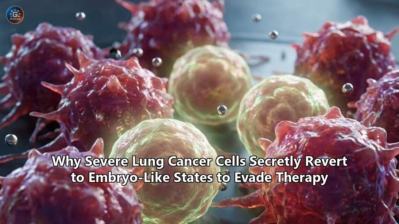
However, a pair of paradigm-shifting studies published in early 2026 has exposed a far more insidious, non-mutational escape mechanism. Rather than waiting for a random genetic mutation to rescue them, aggressive lung cancer cells possess an innate, highly dynamic ability to rewrite their own identity. By secretly reverting to primitive, embryo-like states, these cells establish a biological safe haven that is entirely invisible to modern drugs, driving lung cancer treatment resistance to unprecedented heights.
This revelation of developmental regression represents a fundamental shift in how scientists view cellular plasticity in oncology. Two competing yet complementary research papers have laid bare this phenomenon.
On May 27, 2026, researchers at the University of Southampton published a study in Molecular Oncology demonstrating that severe lung adenocarcinoma (LUAD) cells systematically deactivate their mature, gas-exchanging alveolar programs. Instead, they reactivate primitive branching morphogenesis—the highly invasive, migratory developmental program that sculpts the embryonic respiratory tree.
Meanwhile, a landmark paper published in Nature by researchers at the Memorial Sloan Kettering Cancer Center (MSK) identified a transient, injury-responsive "High-Plasticity Cell State" (HPCS). This state behaves like a temporary, embryonic-like transition hub, allowing tumor cells to seamlessly morph between highly sensitive and completely drug-tolerant identities.
These findings challenge the traditional, mutation-centric view of tumor evolution. They also force a clinical reckoning. If lung cancer treatment resistance is driven by fluid, epigenetic-mediated state changes rather than fixed genetic mutations, then traditional targeted therapies are merely pruning the branches of a tree while ignoring its deeply adaptive, developmental roots.
To overcome this clinical impasse, the scientific community is split between competing therapeutic strategies:
- blocking the developmental pathways that facilitate this regression,
- physically ablating these plastic "hubs" via cellular engineering, or
- resetting the chromatin architecture that allows these embryonic programs to be unmasked in the first place.
The Biological Reversion: Alveogenesis vs. Branching Morphogenesis
To comprehend why a lung cancer cell would revert to an embryonic state, one must first understand how the human lung is built during gestation. Lung development is a tightly orchestrated, temporal sequence that progresses through distinct phases:
- The Embryonic and Pseudoglandular Stages (Branching Morphogenesis): The lung bud undergoes rapid, iterative branching to generate the complex, tree-like structure of the conducting airways. This phase is defined by extreme cellular motility, proliferation, tissue remodeling, and invasiveness. Cells in this state must push through the extracellular matrix, ignore the boundary signals of surrounding tissues, and maintain a highly plastic, undifferentiated phenotype to expand the respiratory tree.
- The Canalicular, Saccular, and Alveolar Stages (Alveogenesis): The terminal ends of these branches thin out and differentiate into specialized gas-exchanging units called alveoli. This process relies heavily on Alveolar Type II (AT2) epithelial cells, which act as local progenitors, producing surfactant to keep alveoli open and differentiating into ultra-thin Alveolar Type I (AT1) cells. Alveogenesis is a state of physiological specialization, structural stability, and restricted plasticity. Master lineage transcription factors, most notably NKX2-1, act as molecular anchors, keeping these cells locked in their mature, specialized identities.
DEVELOPMENTAL TIMELINE
[Embryonic Branching Morphogenesis] ------------> [Mature Alveogenesis]
(High motility, proliferation, plasticity)         (Specialization, structural stability, NKX2-1+)

CANCER REVERSION (Lineage Plasticity)
[Mature Alveolar Tumor Cells] ------------------> [Branching Morphogenic Phenotype]
(Vulnerable to TKIs & Immunotherapy)             (Highly invasive, stem-like, drug-tolerant)
The Southampton study, led by senior researcher Dr. Chris Hanley and first author Kamila J. Bienkowska, revealed that aggressive lung adenocarcinomas systematically reverse this developmental timeline. By analyzing bulk and single-cell transcriptomic datasets from over 1,500 patients, the Southampton team discovered that the most severe, therapy-resistant tumors had systematically shut down their alveogenesis (ALV) genetic programs and reactivated branching morphogenesis (BM).
"In healthy lung development, the lungs initially grow through a branching process before later forming alveoli," Hanley explained. "Aggressive lung tumours appear to reverse this process. Cells revert from their alveoli-forming state back to a branching state. This allows the cancer to grow faster and become more resistant to treatment."
The Double-Whammy: TP53 Loss and Type-I Interferon Signaling
This reversion is not a random biological accident. The Southampton team identified a specific molecular "perfect storm" that drives this regression: the co-occurrence of tumor suppressor TP53 loss-of-function and the chronic activation of Type-I Interferon (IFN) signaling.
Normally, Type-I Interferon is an antiviral cytokine produced by the immune system to restrict cellular growth, induce apoptosis, and recruit immune cells to a threat. It is a cornerstone of the body’s natural defense mechanism. However, when a tumor cell loses its TP53 pathway—a genomic event present in over 50% of advanced non-small cell lung cancers (NSCLCs)—the cellular response to interferon is fundamentally warped.
Instead of triggering cell death or senescence, chronic Type-I IFN signaling in a TP53-deficient background acts as a powerful driver of transdifferentiation. It forces the mature AT2-like cancer cells to shed their alveolar lineage markers (such as surfactant proteins and NKX2-1) and adopt a basal-like, highly inflamed, and exceptionally plastic embryonic branching state.
This transdifferentiation creates an evolutionary escape hatch. Because targeted small molecules like epidermal growth factor receptor (EGFR) tyrosine kinase inhibitors (TKIs) and ALK inhibitors are biochemically designed to exploit the dependencies of mature, lineage-committed lung adenocarcinoma cells, they are completely ineffective against cells that have abandoned this identity. The drug is still present, and its genomic target may still be mutated, but the cell has shifted its entire biological architecture to an embryonic lineage that simply does not care about EGFR or ALK signaling.
Furthermore, this basal-like, inflamed embryonic state is equally adept at evading the immune system. By expressing fetal-specific surface antigens and secreting immunosuppressive cytokines, these reverted cells actively shut down infiltrating cytotoxic T lymphocytes, explaining why patients with high branching morphogenesis (BM) gene signatures show profound resistance to immune checkpoint blockade (ICB) therapies like pembrolizumab and nivolumab.
Competing Paradigms: Southampton’s Developmental Reversion vs. MSK’s High-Plasticity Cell State (HPCS)
While the Southampton team focused on a directional, developmental reversion from alveoli back to embryonic branching, researchers at Memorial Sloan Kettering Cancer Center, led by Tuomas Tammela, Scott Lowe, and Jason E. Chan, approached cellular plasticity from an entirely different angle.
Published in Nature, the MSK study bypassed the traditional focus on stable, long-term lineage transitions—such as the classic histological transformation of lung adenocarcinoma into small-cell lung cancer (SCLC). Instead, they focused on a highly transient, incredibly fluid state that they coined the High-Plasticity Cell State (HPCS).
The conceptual differences between these two models represent a major debate within translational oncology.
| Feature | Southampton’s Developmental Reversion (BM) | MSK’s High-Plasticity Cell State (HPCS) |
|---|---|---|
| Primary Paper | Bienkowska et al., Molecular Oncology (May 2026) | Chan et al., Nature (Jan/March 2026) |
| Underlying Metaphor | Developmental Time-Travel (Alveogenesis $\rightarrow$ Branching Morphogenesis) | Injury-Induced Regenerative Program (Wound Healing/Emergency State) |
| Cellular Longevity | Stable, persistent, lineage-reprogrammed states | Highly transient, dynamic, and reversible transition hub |
| Key Molecular Drivers | TP53 loss-of-function + Type-I Interferon (IFN) signaling | Localized, microenvironmental, injury-like stromal cues |
| Proportion in Tumor | Varies widely; highly enriched in aggressive, late-stage, and recurrent disease | ~3% in precancerous lesions, ~15% in established tumors, up to ~30% in metastases |
| Therapeutic Target | Developmental pathways (Interferon, BM-associated gene networks) | Direct physical ablation of the plastic "hub" (via CAR T-cells or suicide genes) |
HPCS: The Transitional Hub of the Tumor
The classic model of tumor hierarchy dictates that tumors are driven by a rare, stable subpopulation of "Cancer Stem Cells" (CSCs). These CSCs are thought to sit permanently at the top of the developmental pyramid, self-renewing while slowly shedding progeny that differentiate into the bulk, non-tumorigenic cells that make up the mass of the cancer.
The MSK study completely upends this static model. Using sophisticated mouse models (specifically the Kras$^{G12D/+}$; Trp53}$^{-/-}$ or "KP" model) equipped with multiplexed, spectral cell-sorting surface markers and longitudinal lineage tracing, Tammela’s team demonstrated that plasticity is not a permanent property of a select group of stem cells. Rather, it is an inducible, transient state that any cancer cell can slide into when exposed to stress.
These highly plastic cells do not resemble steady-state stem cells. Instead, they biologically mirror the temporary, emergency regenerative programs that normal lung tissues activate following a severe injury. When the lung is damaged (e.g., by toxic inhalation or severe infection), differentiated cells temporarily cast off their specialized identities to become highly plastic, migrating to the site of injury and proliferating rapidly to rebuild the tissue, before returning to their mature states once healing is complete.
Lung cancer cells hijack this physiological emergency response. Early in tumor development, only about 3% of the cells in precancerous lesions reside in this high-plasticity state. However, as the tumor expands, experiences hypoxia, and is bombarded by inflammatory signals from the stroma, the proportion of HPCS cells swells to 15% in primary tumors and up to 30% in metastatic sites.
Crucially, when a patient is treated with targeted therapies—such as a KRAS$^G12D$ inhibitor or chemotherapy—the bulk, lineage-committed tumor cells are killed off. But the HPCS cells, sensing this existential threat as an extreme cellular "injury," rapidly adapt. They transition into drug-tolerant, slow-cycling developmental states, acting as a biological seed bank. Once the therapeutic pressure is removed or the cell acquires additional survival adaptations, these HPCS cells "re-differentiate" into a diverse array of aggressive cancer cell types, driving tumor recurrence.
"Rather than being akin to steady-state stem cells, these highly plastic cells more closely resemble the temporary, regenerative program that normal tissues activate in response to an injury," Dr. Tammela explained.
The MSK study proved this functional dependency by genetically ablating the HPCS cells in vivo. When they systematically destroyed this tiny fraction of highly plastic cells, the transition from benign adenoma to malignant adenocarcinoma was completely blocked. In established tumors, ablating the HPCS population destroyed the tumor's transition hub, causing the remaining tumor cells to succumb to standard therapies and robustly reducing overall tumor burden.
Epigenetic Erasure: The Chromatin Landscape of Lineage Plasticity
To understand how a cancer cell can physically execute these dramatic identity shifts—whether transitioning from alveoli to embryonic branching or sliding in and out of an HPCS state—one must look beyond the genomic sequence to the epigenome. These rapid transitions happen too quickly to be driven by classic genetic mutation and selection; they are instead driven by sweeping, coordinated changes in chromatin accessibility and histone modifications.
Under normal physiological conditions, adult cells are kept in their differentiated states by "epigenetic locks". These locks consist of chemical modifications to DNA and histone proteins that keep embryonic genes tightly wound around histones (in closed, transcriptionally silent heterochromatin), while keeping adult, lineage-specific genes open and accessible (in active euchromatin).
In severe lung cancer, these epigenetic locks are systematically broken. When master lineage regulators like NKX2-1 are downregulated, and chromatin-remodeling complexes like Polycomb Repressive Complex 2 (PRC2) are dysregulated, the chromatin landscape becomes highly fluid. Closed embryonic loci are suddenly unmasked, allowing developmental transcription factors like SOX2, SOX9, and SALL4 to bind and reactivate long-dormant fetal programs.
EPIGENETIC LANDSCAPE OF LINEAGE PLASTICITY

Differentiated Adult State (Lineage-Locked):
[NKX2-1 bound] ---> Euchromatin (Adult Genes: Surfactants, AT2 markers) [OPEN]
[PRC2 / Histone Methylation] ---> Heterochromatin (Embryonic/Branching Genes) [LOCKED]

Reprogrammed Embryonic State (Lineage-Plastic):
[NKX2-1 lost] ---> Heterochromatin (Adult Genes shut down) [CLOSED]
[Histone Deacetylation / Crotonylation Depletion] ---> Euchromatin (Embryonic/Branching Genes) [UNMASKED]
The Crotonylation Conundrum: A Newly Uncovered Epigenetic Driver
Adding another layer of complexity to this epigenetic remodeling is a third major discovery published in late 2025 in the Proceedings of the National Academy of Sciences (PNAS). The study, focused on post-translational modifications (PTMs) on histones, uncovered a highly specific link between a novel histone modification—histone lysine crotonylation (Kcr)—and lung cancer treatment resistance.
Histone crotonylation is a highly dynamic epigenetic mark that is closely tied to cellular metabolism. It relies on the availability of crotonyl-CoA, which is regulated by the metabolic enzyme acyl-CoA synthetase short-chain family member 2 (ACSS2). Under normal conditions, high levels of histone crotonylation keep active metabolic and lineage-maintaining genes highly expressed.
The PNAS study established that in diverse preclinical lung tumor models resistant to EGFR TKIs, there is a profound, resistance-associated drop in histone crotonylation levels across the genome. This depletion is driven directly by the metabolic shutdown of ACSS2. When histone crotonylation is erased, the cell's chromatin architecture undergoes a dramatic structural contraction, silencing the genes that maintain mature epithelial lineage fidelity.
Remarkably, when the researchers treated these resistant cells with a histone decrotonylase inhibitor, they were able to selectively restore histone crotonylation levels across these silenced loci. This epigenetic "reset" forced the cells to abandon their primitive, drug-tolerant states and re-adopt their mature, lineage-committed identities, completely restoring their sensitivity to EGFR TKIs in vitro and in vivo.
This metabolic-epigenetic axis represents a third, competing paradigm for how lineage plasticity is regulated. While the Southampton study views plasticity as a response to immunological/genetic cues (Type-I IFN and TP53 loss) and MSK views it as a response to microenvironmental injury cues, the PNAS study frames it as a metabolic adaptation, where a drop in key metabolites allows the cell to slide down the developmental landscape into a drug-resistant state.
Compare and Contrast: Competing Clinical Strategies to Overcome Plasticity
The emergence of these diverse biological frameworks has triggered an intense debate over how to translate these discoveries into clinical benefit. If oncology is to finally conquer lung cancer treatment resistance, it must move beyond simply developing "fourth-generation" TKIs that target yet more single-nucleotide mutations. Instead, clinicians must target the structural plasticity of the cell itself.
Currently, there are three competing, biochemically distinct therapeutic strategies vying for clinical development:
- Developmental Pathway Interdiction (The Southampton Approach)
- Cellular Ablation of the Plastic State (The MSK Approach)
- Epigenetic Re-Anchoring / Chromatin Resetting (The PNAS/Chromatin Approach)
1. Developmental Pathway Interdiction: Freezing the State Transition
The strategy favored by the Southampton researchers is to surgically disable the developmental programs that the cancer cell is trying to co-opt. By identifying the specific molecular channels that facilitate the reversion from alveogenesis to branching morphogenesis, clinicians can potentially "freeze" the cancer cells in their mature, drug-sensitive states.
The key therapeutic node in the Southampton model is the combination of Type-I Interferon (IFN) signaling and TP53 pathway deficiency. Because chronic Type-I IFN drives transdifferentiation in TP53-mutant cells, blocking this interferon cascade is an obvious clinical target.
- The Therapeutic Agents: Monoclonal antibodies targeting the Type-I Interferon receptor (IFNAR1), such as anifrolumab (currently approved for systemic lupus erythematosus), or small-molecule JAK1/2 inhibitors (like ruxolitinib) that block downstream interferon-stimulated gene (ISG) transcription.
- The Mechanism: By blocking IFNAR1, the chronic "inflamed" stimulus is removed. Without this signal, the TP53-deficient cell cannot execute the transdifferentiation program required to transition from an alveolar type II cell to a basal-like branching morphogenic cell.
- The Trade-Offs: The major risk of this approach is systemic immunosuppression. Type-I Interferon is a critical component of host viral defense and plays a major role in natural anti-tumor immunosurveillance. Systemically blocking IFNAR1 could severely compromise a patient’s ability to fight off opportunistic infections and, paradoxically, could prevent the immune system from recognizing other non-plastic clones within the same tumor. Furthermore, this approach relies on "freezing" the cells, meaning it does not actually kill the plastic cells; it merely prevents them from shifting states, requiring long-term, continuous combination therapy with TKIs or chemotherapy.
2. Cellular Ablation of the Plastic State: Direct Physical Destruction
The MSK group proposes a far more aggressive, definitive strategy: instead of trying to manipulate the fluid signaling pathways that control plasticity, simply identify the plastic cells by their unique surface markers and physically eliminate them from the body.
Using their advanced spectral cell-sorting platform, Tammela’s team identified a highly specific multiplexed cell-surface marker signature that is uniquely expressed on the surface of High-Plasticity Cell State (HPCS) cells, but is absent on mature, healthy lung epithelial cells.
- The Therapeutic Agents: Chimeric Antigen Receptor (CAR) T-cells engineered to target these HPCS-specific surface markers, or antibody-drug conjugates (ADCs) designed to deliver a lethal cytotoxic payload directly to cells expressing this plastic signature.
- The Mechanism: In mouse models, when HPCS cells were selectively destroyed using a suicide gene or HPCS-targeted CAR T-cells, the tumor's entire "evolutionary reservoir" was wiped out. Even when the remaining, non-plastic tumor cells were challenged with chemotherapy or KRAS inhibitors, they were unable to develop drug-tolerant states because the transition "hub" (the HPCS) no longer existed.
- The Trade-Offs: This approach represents a massive technological leap, but its clinical translation is fraught with challenges. The primary concern is on-target, off-tumor toxicity. The HPCS program is almost identical to the transient regenerative state that healthy tissues use to heal from injury. If a patient receiving HPCS-targeted CAR T-cells experiences a physical injury, a severe lung infection, or even standard radiation-induced tissue damage, the CAR T-cells could aggressively attack the healthy, regenerating tissues, leading to catastrophic organ failure or impaired wound healing. Additionally, producing patient-specific CAR T-cells remains an incredibly expensive, logistically complex process that may be difficult to scale for the massive population of patients with advanced lung cancer.
3. Epigenetic Re-Anchoring: Resetting the Chromatin Landscape
The third approach is to target the enzymatic machinery that governs chromatin packaging. Rather than targeting a single surface marker or cytokine receptor, this strategy aims to rewrite the epigenetic "code" of the cell, closing the embryonic loci and permanently re-locking the cells into their mature, differentiated lineages.
This strategy utilizes a growing arsenal of chromatin-modifying small molecules.
- The Therapeutic Agents: Histone deacetylase (HDAC) inhibitors, histone decrotonylase inhibitors, EZH2 (PRC2 core enzyme) inhibitors, and BET bromodomain inhibitors (which block the reading of active histone marks).
- The Mechanism: In the case of the PNAS crotonylation study, utilizing a histone decrotonylase inhibitor successfully restored crotonylation (Kcr) marks on histones, forcing the open, plastic chromatin of TKI-resistant cells to condense and silencing the embryonic escape programs. Similarly, in EGFR-mutant lung cancers undergoing lineage transition to squamous or small-cell states, EZH2 inhibitors can prevent the epigenetic silencing of NKX2-1, keeping the cells anchored in their TKI-sensitive adenocarcinomal lineage.
- The Trade-Offs: The primary drawback of epigenetic therapies is their lack of specificity. Chromatin-modifying enzymes regulate gene expression across every single tissue in the human body. Systemically inhibiting EZH2 or HDACs can cause severe side effects, including profound bone marrow suppression (thrombocytopenia, anemia), gastrointestinal toxicity, and fatigue. Moreover, the epigenome is notoriously dynamic; while an epigenetic drug might successfully close one embryonic locus, the cell may rapidly find alternative chromatin-remodeling pathways to bypass the blockade, leading to secondary epigenetic resistance.
Comparative Analysis of Anti-Plasticity Interventions
To evaluate these competing strategies, it is helpful to contrast their key features, mechanisms, and translational hurdles:
| Metric | Developmental Pathway Interdiction (Southampton) | Cellular Ablation of HPCS (MSK) | Epigenetic Re-Anchoring (PNAS / Chromatin) |
|---|---|---|---|
| Primary Target | IFNAR1 / JAK-STAT / BM-gene networks | Surface markers of the high-plasticity state (HPCS) | Histone modifications (Kcr, acetylation) / EZH2 / PRC2 |
| Biochemical Strategy | Block the stimulus driving the cell down the developmental slope | Physically destroy the cell that resides in the plastic hub | Rewrite the cellular code to lock the chromatin in an adult state |
| Modality | Small molecule inhibitors (JAKi) or Monoclonal Antibodies (anti-IFNAR1) | CAR T-cells or Antibody-Drug Conjugates (ADCs) | Systemic small molecule epigenetic modifiers |
| Therapeutic Phase | Preventative; must be given concurrently with primary therapy | Ablative; can be given to clear residual disease and prevent relapse | Restorative; can be used to re-sensitize already resistant tumors |
| Translational Status | High; several drugs are already FDA-approved for other indications | Low-Moderate; preclinical mouse validation, clinical-grade CAR T-cells in development | Moderate; multiple HDAC/EZH2 inhibitors in early-phase oncology trials |
| Primary Safety Concern | Systemic immunosuppression and vulnerability to viral infections | Severe auto-inflammatory damage to regenerating healthy tissues | Off-target systemic toxicity (bone marrow suppression, gastrointestinal issues) |
| Resistance Escape Route | Bypass pathways (e.g., activation of Wnt or Hedgehog developmental signaling) | Antigen loss / mutation of targeted HPCS surface markers | Alternative chromatin remodeling, histone modifications, or metabolic shifts |
The Microenvironmental Niche: How the Stroma Anchors the Embryonic State
The cell autonomous changes—genetic mutations, transcription factor shifts, and histone modifications—do not occur in a vacuum. A lung cancer cell’s ability to regress to an embryonic state is heavily dependent on constant, bi-directional communication with the surrounding Tumor Microenvironment (TME).
During embryonic development, the extracellular matrix (ECM) and surrounding mesenchymal cells provide a tightly regulated physical and chemical scaffold that guides branching morphogenesis. In advanced lung cancer, the tumor stroma is remodeled to mimic this embryonic niche, actively encouraging cancer cells to shed their mature epithelial identities.
THE ONCOFETAL MICROENVIRONMENTAL LOOP

[Tumor-Associated Macrophages (TAMs)] --------> Iron Release & Pro-inflammatory Cytokines
                                                    │
                                                    ▼
[Cancer Epithelial Cells] <------------------- Chronic Tissue Injury & Hypoxia Cues
  │  (TP53-mutant / ACSS2-depleted)
  │
  ▼ (Reversion to Embryonic State)
[Branching Morphogenesis / HPCS Activated] ---> Secretion of Oncofetal Fibronectin & LncRNA H19
                                                    │
                                                    ▼
                                               Immunosuppressive Niche (T-Cell Exclusion)
1. Tumor-Associated Macrophages (TAMs) and Oncofetal Reprogramming
A key component of this microenvironmental support system is the presence of specialized immune cells. As noted in recent single-cell analyses of treatment-naive lung cancer patients, tumor-associated macrophages (TAMs) within these aggressive niches express a distinct transcriptional signature that is highly similar to the macrophages found in human fetal lungs during gestation.
These "oncofetal TAMs" (specifically defined by the expression of the scavenger receptor STAB1) show a dramatic increase in gene expression that promotes iron release into the surrounding extracellular space. In a developing embryo, this localized iron release is crucial for fueling the rapid metabolic demands of branching morphogenesis. In a tumor, this fetal-like iron release fuels the rapid metabolic demands and proliferative capacity of the reverted branching cancer cells, while simultaneously suppressing the metabolic fitness of infiltrating cytotoxic T-cells.
2. The Extracellular Matrix (ECM) and Embryonic Fibronectin
Mature adult lungs have a stable, rigid ECM dominated by collagen and mature laminins that keep alveoli physically anchored. However, tumors characterized by high levels of branching morphogenesis actively remodel this matrix, secreting oncofetal fibronectin and other embryonic ECM variants.
These embryonic matrix components bind to integrins on the surface of cancer cells, triggering a powerful mechanotransductive signaling cascade. This cascade activates the FAK/Src kinase axis, which in turn inactivates the Hippo tumor-suppressor pathway, allowing the transcriptional co-activators YAP1 and TAZ to rush into the nucleus. Once in the nucleus, YAP1/TAZ complexes with AP-1 to act as a master transcriptional engine of oncofetal reprogramming, opening up embryonic loci and driving the cell-state transition.
3. Fetal Non-Coding RNAs: The H19 Sponge
The microenvironment is also heavily influenced by non-coding RNAs secreted by both the plastic tumor cells and the stroma. The long non-coding RNA lncRNA H19 is exceptionally abundant during early mammalian embryogenesis but is robustly silenced in adult tissues.
In advanced lung cancers, particularly those showing high branching morphogenesis signatures, lncRNA H19 is highly upregulated and secreted in extracellular vesicles. Within the tumor cell, H19 acts as a molecular "sponge" for adult-specific microRNAs (such as let-7), preventing them from silencing embryonic genes. In the microenvironment, H19-containing vesicles are taken up by endothelial cells and fibroblasts, reprogramming them to build highly vascularized, embryonic-like stromal networks that shield the plastic cancer cells from therapeutic penetration.
Why Traditional Clinical Trials Fail: The RECIST Blindspot
The realization that lung cancer cells can evade therapies by sliding into embryonic or high-plasticity states exposes a fundamental flaw in how the oncology community designs and evaluates clinical trials.
For decades, the gold standard for determining whether a new cancer drug is effective has been the RECIST (Response Evaluation Criteria in Solid Tumors) guidelines. RECIST relies entirely on physical tumor measurements—using CT or MRI scans to calculate whether the sum of the diameters of target lesions has shrunk by a certain percentage (e.g., a 30% reduction indicates a partial response).
While RECIST is highly effective at measuring the destruction of the bulk, rapidly proliferating, lineage-committed tumor cells, it is completely blind to the highly dynamic dynamics of cell-state transitions.
THE RECIST BLINDSPOT IN TARGETED THERAPY

Pre-Treatment Tumor:
[Bulk TKI-Sensitive Cells: 85%] + [HPCS / Embryonic Cells: 15%]
Total Tumor Volume: Large

Post-Treatment (Clinical "Response" under RECIST):
[Bulk TKI-Sensitive Cells: 0%]  + [HPCS / Embryonic Cells: 30%] (Doubled in proportion!)
Total Tumor Volume: Small (Shrunk by 70%)
Result: Declared a "success" in Phase II trials.

Relapse (Agressive Recurrence):
[HPCS / Embryonic Cells] differentiate back into highly aggressive, drug-tolerant heterogeneous tumor cells.
Total Tumor Volume: Massive, metastatic, and completely resistant to all subsequent lines of therapy.
When a patient is treated with a potent targeted therapy (such as a third-generation EGFR TKI), the therapy successfully kills off the 85% of the tumor that consists of mature, lineage-committed cells. Under RECIST criteria, this is classified as a dramatic clinical response. The oncologist celebrates, and the drug is heralded as a success.
However, this therapeutic onslaught acts as an extreme physical stressor. The remaining 15% of the tumor cells—the HPCS or embryonic branching morphogenic cells—survive the treatment. In fact, the physical destruction of the surrounding cells relieves mechanical pressure and releases massive amounts of inflammatory "injury" cytokines, actively stimulating the surviving embryonic cells to expand and proliferate.
During this "remission" phase, the tumor may appear small on a CT scan, but it has actually been biochemically reprogrammed. The proportion of highly plastic, drug-tolerant embryonic cells has swollen dramatically. Once these cells complete their evolutionary cycle, they differentiate into a highly heterogeneous, metastatic malignancy that is completely impervious to further treatment.
To solve this clinical blindspot, translational medicine must transition toward multiplexed single-cell liquid biopsies and functional imaging modalities. Rather than simply measuring tumor size, clinical trials must track the presence of circulating embryonic-associated biomarkers—such as serum levels of lncRNA H19, circulating tumor cells expressing the HPCS surface panel, or epigenetic chromatin accessibility signatures in cell-free DNA (cfDNA). Only by proving that a drug can actively shrink the plastic developmental pool can we hope to prevent the inevitable relapse that plagues modern lung oncology.
Historical Context: From "Dormancy" to "Oncofetal Reprogramming"
While the molecular details of these cellular transitions have only been mapped in the early 2026 breakthroughs, the underlying biological concept has deep, historic roots in developmental biology.
In the mid-19th century, the German pathologist Rudolf Virchow and his student Julius Cohnheim proposed the "embryonal rest hypothesis" of cancer. They suggested that cancers arise from residual embryonic cells that remained trapped in adult tissues during development, waiting for a stimulus to reactivate their rapid, invasive growth programs.
As modern molecular biology emerged, Virchow’s hypothesis was largely replaced by the genetic mutation theory of cancer, which focused on the stepwise accumulation of somatic mutations in adult cells. However, the discovery of oncofetal antigens in the 1960s—such as carcinoembryonic antigen (CEA) and alpha-fetoprotein (AFP)—provided clear evidence that adult cancer cells were somehow reactivating genes that are normally silenced after birth.
Parallel to this was the study of embryonic diapause, a temporary suspension of embryo development during blastocyst stages in response to unfavorable maternal environments. During diapause, the embryo enters a metabolic state of dormancy, downregulating cell division and maximizing survival mechanics until conditions improve.
Oncologists slowly realized that drug-tolerant persister (DTP) cells in lung cancer hijack this exact diapause-like state. When hit with systemic chemotherapy, these cells do not die; they activate a metabolic and transcriptional program that is nearly identical to embryonic diapause, entering a dormant state where they can survive for months or years in the body.
The 2026 studies from the University of Southampton and Memorial Sloan Kettering represent the culmination of this historical arc. They have unified these disparate historical observations—Virchow’s embryonic rests, oncofetal antigens, and diapause-like dormancy—into a cohesive, molecularly defined framework of lineage plasticity.
The cell is no longer seen as a static entity locked in its adult identity. Instead, the modern view of the genome is like a highly complex computer operating system: while the adult "user interface" is active under normal conditions, the underlying embryonic "source code" is never truly erased. Under the extreme stress of cancer therapies, the cell simply reboots the system in "safe mode"—reverting to the robust, highly survivalist programs of embryogenesis to weather the storm.
Technical Appendix: A Breakdown of Key Embryonic Pathways in Lung Cancer
To aid researchers and clinicians navigating this rapidly evolving field, this section provides a detailed biochemical breakdown of the primary embryonic signaling pathways that are aberrantly reactivated during lung cancer lineage reversion.
1. The Wnt/$\beta$-Catenin Pathway
- Role in Development: Controls tissue patterning, stem cell self-renewal, and early branching morphogenesis in the embryonic lung.
- Reactivation Mechanism in Cancer: Under therapeutic stress, loss of the master regulator NKX2-1 removes the transcriptional repression of Wnt ligands (such as WNT7a and WNT3a).
- Downstream Consequences: Accumulation of $\beta$-catenin in the nucleus, where it binds to TCF/LEF transcription factors to activate genes associated with epithelial-to-mesenchymal transition (EMT), cancer stemness, and multi-drug resistance transporters (such as ABCB1 and ABCC1).
2. The Hedgehog (Hh) Pathway
- Role in Development: Mediates epithelial-mesenchymal interactions and is essential for specifying the spatial patterning of the respiratory tree.
- Reactivation Mechanism in Cancer: Autocrine or paracrine secretion of Sonic Hedgehog (SHH) or Indian Hedgehog (IHH) by stromal cells in the tumor microenvironment.
- Downstream Consequences: Activation of GLI transcription factors (GLI1 and GLI2), which directly upregulate the embryonic transcription factor SOX2. SOX2 binds to the promoters of lineage-plasticity genes, driving the reversion to basal-like states and promoting extreme resistance to platinum-based chemotherapies.
3. The Notch Signaling Pathway
- Role in Development: Regulates cell fate decisions, specifically balancing the ratio of secretory club cells and ciliated cells in the developing airway through lateral inhibition.
- Reactivation Mechanism in Cancer: Overexpression of Notch ligands (DLL1, DLL4, JAG1) on neighboring cancer cells or stromal cells, often stimulated by hypoxia.
- Downstream Consequences: Cleavage and nuclear translocation of the Notch Intracellular Domain (NICD), which activates the HES and HEY family of transcription factors. This shuts down the AT2 lineage marker surfactant protein C (SFTPC) and promotes the acquisition of an undifferentiated, highly migratory phenotype.
4. The Hippo/YAP1 Pathway
- Role in Development: Senses physical mechanical forces within the developing embryo, regulating tissue size and organ growth by controlling cell proliferation and apoptosis.
- Reactivation Mechanism in Cancer: Loss of cell-cell contact, remodeling of the extracellular matrix (oncofetal fibronectin), and FAK/Src hyperactivation.
- Downstream Consequences: Attenuation of LATS1/2 kinases, preventing the inhibitory phosphorylation of YAP1 and TAZ. Unphosphorylated YAP1/TAZ translocates to the nucleus, partnering with TEAD and AP-1 transcription factors to drive sweeping oncofetal reprogramming, opening up vast networks of embryonic genes.
What Lies Ahead: The Frontiers of Lineage Plasticity Research
The clinical and scientific landscape of lung oncology is standing on the precipice of a profound transformation. As the dust settles on the twin breakthroughs from the University of Southampton and Memorial Sloan Kettering, several critical, unresolved questions and upcoming milestones will shape the next decade of therapeutic development.
1. Clinical Validation of HPCS Biomarkers
The immediate next milestone is the transition of the MSK HPCS cell-surface marker panel into clinical diagnostic assays. Pathologists must determine whether multiplexed immunohistochemistry (mIHC) or spectral flow cytometry can be reliably performed on standard patient biopsies to score a patient's pre-treatment "plasticity index."
If successful, this will allow oncologists to identify high-risk patients long before they begin therapy, guiding them away from monotherapies that are guaranteed to fail and toward early, aggressive combinations that target both the mature tumor bulk and the embryonic transition pool.
2. The Direct Differentiation vs. Ablation Debate
An intense academic debate is brewing over the optimal therapeutic philosophy: Is it better to kill the embryonic cells, or force them to grow up?
- The Ablation Camp argues that because the HPCS is a highly dynamic transition state, any attempt to genetically or chemically manipulate it will inevitably lead to escape mutations or alternative signaling bypasses. Therefore, the only definitive solution is physical, CAR T-mediated eradication of the cells while they are in the plastic state.
- The Differentiation Camp counters that forcing embryonic cells to "re-differentiate" into mature, specialized lineages (using drugs like retinoids, chromatin-anchoring compounds, or specific cytokine cocktails) is inherently safer. By pushing these highly plastic cells to mature into stable, non-invasive AT2-like cells, they will lose their aggressive, metastatic properties and become highly vulnerable to standard, low-toxicity targeted therapies, avoiding the severe systemic auto-inflammatory risks associated with CAR T ablation.
3. Broad Pan-Cancer Implications
While these studies were centered on lung adenocarcinoma, researchers are already discovering that this embryonic reversion is a universal biological law.
Parallel studies in colorectal cancer have shown that aggressive colon cancer cells revert to fetal intestinal progenitor states to escape chemotherapy. In hepatocellular carcinoma (liver cancer), tumor cells systematically reactivate fetal liver-specific transcriptional networks to survive targeted multi-kinase inhibitors.
The developmental programs hijacked by these different malignancies are tissue-specific, but the underlying mechanism—epigenetic unmasking of embryonic source code under therapeutic stress—is a shared oncological vulnerability. The discovery of a universal "high-plasticity" signature across multiple tissues suggests that a single, standardized class of anti-plasticity therapeutics could eventually be deployed to treat a wide array of advanced, drug-resistant solid tumors.
The Path to a Cure
The revelation that severe lung cancer cells secretly revert to embryo-like states to evade therapy represents both a sobering challenge and a profound opportunity. It explains why, despite billions of dollars of investment into increasingly precise genomic therapies, advanced lung cancer has remained an largely incurable disease. The tumor was simply playing a more sophisticated, multi-dimensional evolutionary game than our linear, mutation-centric models could comprehend.
But by exposing this secret developmental escape hatch, the scientific breakthroughs of 2026 have finally given researchers the map they need. Whether through the surgical blockage of embryonic branching morphogenesis, the precise cellular ablation of high-plasticity transition hubs, or the metabolic resetting of the epigenetic landscape, the tools to dismantle cellular plasticity are finally within our reach.
The future of oncology lies in accepting that cancer is not merely a collection of genetic mutations, but a dynamic, developmental disease. Only by targeting the cell's fluid capacity to change its identity can we hope to shut down lung cancer treatment resistance once and for all, turning the tide in the fight against one of the world's deadliest malignancies.
References
- [1.1] Preprints.org (Dec 2025) & SpringerMedizin (2025). Detailed analyses of oncofetal gene reactivation in NSCLC, FAK/Src/YAP1 signaling, and the role of the chemotherapy-resistance gene CA916798 in lung adenocarcinoma.
- [1.2] Molecular Oncology (May 2026). "Developmental programmes drive cellular plasticity, disease progression and therapy resistance in lung adenocarcinoma" (University of Southampton).
- [1.3] National Institutes of Health (NIH) / PNAS (Oct 2025). "Activation of epigenetic reprogramming via crotonylation overcomes resistance to EGFR-TKI therapy in lung cancer."
- [1.4] Nature (Jan/March 2026). "Critical role for a high-plasticity cell state in lung cancer" (Memorial Sloan Kettering Cancer Center).
- [2.1] Chan JE, Pan CH, et al. "Critical role for a high-plasticity cell state in lung cancer." Nature, 2026 Mar;651(8104):231-241.
- [2.2] Bienkowska KJ, Hanley CJ, et al. "Developmental programmes drive cellular plasticity, disease progression and therapy resistance in lung adenocarcinoma." Molecular Oncology*, May 2026.
Reference:
- https://www.springermedizin.de/ca916798-predicts-poor-prognosis-and-promotes-gefitinib-resistan/24667008
- https://news.cancerresearchuk.org/shorthand_story/going-viral-how-egfr-opened-the-door-to-targeted-cancer-therapy/
- https://pmc.ncbi.nlm.nih.gov/articles/PMC8860314/
- https://www.pharmatutor.org/pharma-news/2026/scientists-discover-how-lung-cancer-cells-change-identity-to-escape-treatment
- https://pubmed.ncbi.nlm.nih.gov/41565826/
- https://eprints.soton.ac.uk/509506/
- https://www.eurekalert.org/news-releases/1113504
- https://www.frontiersin.org/journals/genetics/articles/10.3389/fgene.2023.1120815/pdf
- https://www.mdpi.com/2673-8937/6/1/6
- https://pubmed.ncbi.nlm.nih.gov/41026825/
- https://medicalresearchjournal.org/index.php/GJMR/article/view/2389/1-Characteristics-Shared-between-Lung-Developme_JATS_NLM_xml
- https://www.preprints.org/manuscript/202512.0937
- https://pmc.ncbi.nlm.nih.gov/articles/PMC12373981/
- https://www.biorxiv.org/content/10.1101/2024.12.03.626626.full.pdf
- https://www.biorxiv.org/content/10.1101/2024.12.03.626626v1
- https://pmc.ncbi.nlm.nih.gov/articles/PMC12960256/
- https://pubmed.ncbi.nlm.nih.gov/40751717/
- https://pubmed.ncbi.nlm.nih.gov/19159828/
- https://ideas.repec.org/a/nat/nature/v651y2026i8104d10.1038_s41586-025-09985-x.html
- https://themarkfoundation.org/portfolio/parallel-molecular-and-functional-dissection-of-cancer-cell-states-in-human-solid-tumors/
- https://pmc.ncbi.nlm.nih.gov/articles/PMC10693315/
- https://www.researchgate.net/publication/394860615_Persistent_lineage_plasticity_driving_lung_cancer_development_and_progression
- https://www.preprints.org/manuscript/202512.0937/v1/download
- https://pmc.ncbi.nlm.nih.gov/articles/PMC11685895/