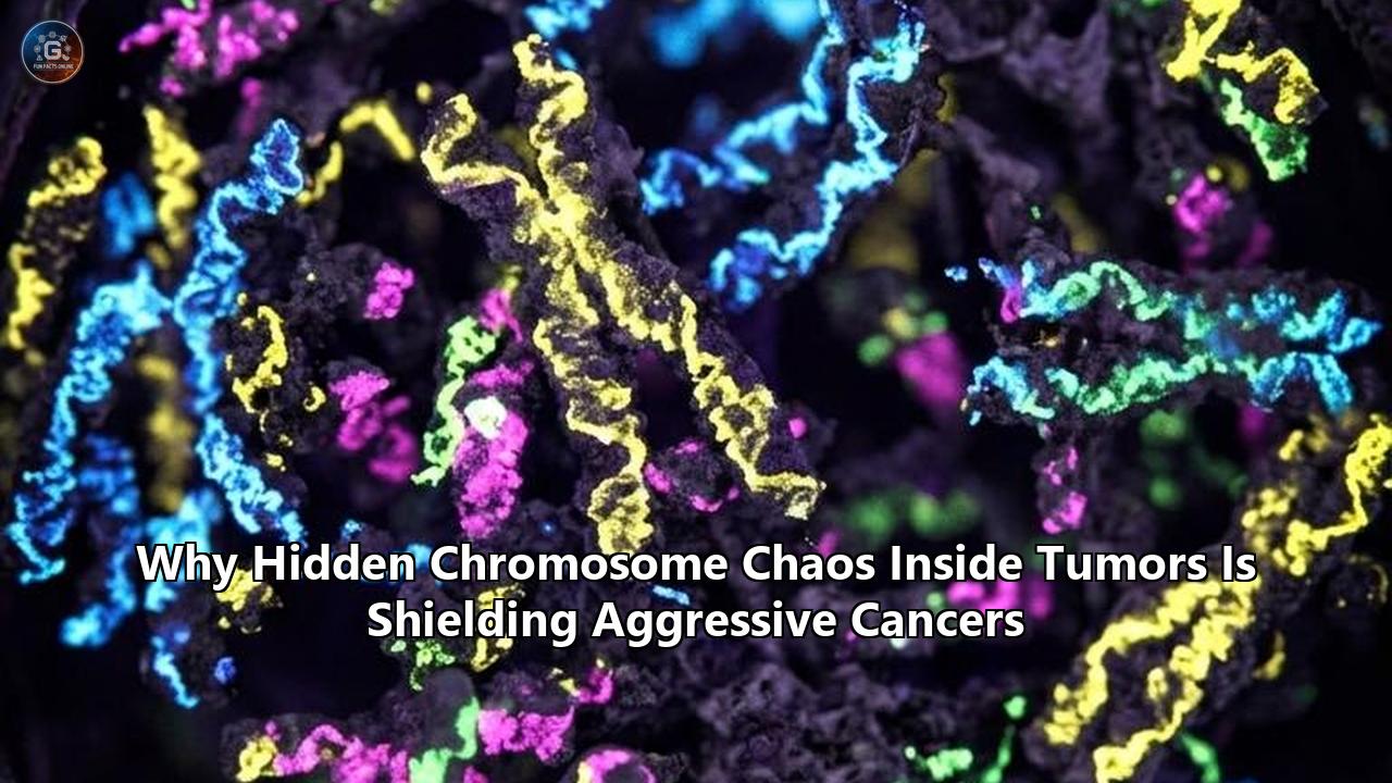
For decades, cancer geneticists tracking aggressive tumors have faced a frustrating paradox: while common cancers like luminal breast or localized prostate disease often yield to targeted drugs against single mutated genes, the deadliest cancers do not. In diseases like basal-like breast cancer (BLBC)—an aggressive, hard-to-treat subtype of triple-negative breast cancer—tumors routinely lack targetable receptors and exhibit massive genomic disarray. Inside these cells, entire chromosomes shatter, duplicate, or vanish, carrying hundreds of genes away with them.
For years, clinical genomics largely treated these sweeping structural alterations as secondary "background noise"—chaotic downstream scars of a cell cycle gone wrong, rather than actionable targets. That paradigm has now been directly challenged.
In a landmark study published in Nature, a research team led by Dr. Daniel Schramek and Dr. Jeff Wrana at Sinai Health’s Lunenfeld-Tanenbaum Research Institute (LTRI), alongside Dr. Khalid Al-Zahrani at the University of Toronto, untangled this chromosomal chaos in living breast tumors. Utilizing a new dual-action gene-editing technology known as CRISPR-KOALA (Knockout and Activation Linked Assay), the team conducted a systematic in vivo functional screen across more than 3,700 genes situated on routinely altered chromosomal regions.
The results exposed a hidden layer of cancer biology: the researchers uncovered 81 previously unrecognized driver genes embedded within these reshuffled chromosomal regions. Crucially, nearly 90 percent of these drivers were completely invisible in conventional Petri dish cell cultures, manifesting their tumor-promoting power only within the complex, living tissue microenvironment.
This discovery provides a clear lens through which to re-examine how chromosome instability in cancer serves not merely as a biomarker of advanced disease, but as an active, self-shielding evolutionary engine. By examining the mechanics behind the Schramek lab’s discovery alongside parallel clinical advances in kinesin inhibition, extrachromosomal DNA profiling, and chemotherapy response signatures, we can extract key principles governing how structural genomic chaos protects aggressive tumors—and how precision oncology can finally dismantle it.
The Architecture of Chaos: Structural Alterations vs. Point Mutations
To understand why aggressive tumors resist targeted therapies, oncology must move beyond the classical "Somatic Gene Mutation Theory"—the view that cancer is primarily driven by a linear sequence of individual point mutations (such as BRAF V600E or EGFR T790M). While single-gene drivers explain early tumor initiation in select tissues, advanced refractory malignancies are dominated by karyotypic structural chaos.
CLASSICAL POINT MUTATION MODEL vs. CHROMOSOMAL INSTABILITY MODEL

[ Normal Genome ] ---> Single Base Mutation ---> Targeted Drug Sensitivity
                      (e.g., BRAF V600E)        (High initial response)

[ Normal Genome ] ---> Chromosome Missegregation ---> Aneuploidy & ecDNA Hubs
                       & Mitotic Catastrophe        (Massive dosage shifts &
                                                     nongenetic plasticity)
Chromosomal instability (CIN) is defined as an elevated rate of chromosome missegregation during cell division. Rather than reproducing two identical daughter cells with 23 pairs of chromosomes, a cell experiencing high CIN routinely misallocates entire chromosomes or major chromosomal arms. This missegregation leads to several distinct structural configurations:
- Aneuploidy: An abnormal whole-number count or partial-arm imbalance of chromosomes, altering the dosage of hundreds of genes simultaneously.
- Whole-Genome Doubling (WGD): A sudden endoreduplication event where a cell duplicates its entire complement of chromosomes, creating tetraploid or polyploid states that cushion the cell against lethal mutations while amplifying oncogene copy numbers.
- Chromothripsis: A localized genomic catastrophe where one or several chromosomes shatter into dozens or hundreds of fragments during mitosis and are randomly stitched back together by error-prone DNA repair machinery.
- Extrachromosomal DNA (ecDNA): Circular, megabase-sized loops of DNA that break free from chromosomes, lacking centromeres and segregating stochastically into daughter cells.
When a tumor exhibits ongoing chromosome instability in cancer, it operates under a model of punctuated macroevolution. Single-cell sequencing studies from UT MD Anderson Cancer Center, led by Dr. Nicholas Navin, demonstrated that tumors do not merely accumulate genetic changes through slow, steady Darwinian drift. Instead, they undergo sudden, early bursts of copy-number alterations (CNAs) that instantly spawn distinct subclonal lineages.
These distinct subpopulations take up residence in specific spatial niches throughout the tumor microenvironment. When a patient receives targeted monotherapy or chemotherapy, the drug destroys the dominant sensitive clones, but the high structural heterogeneity guarantees that a pre-existing subclone with a protective chromosomal configuration will survive, proliferate, and drive relapse.
Deep-Dive Case Study: Decoding PLGRKT and Metabolic Survival Hubs
The power of looking at structural genomic chaos through functional models is vividly illustrated by the Schramek team’s discovery of PLGRKT.
Basal-like breast cancer is notorious for carrying recurrent gains of chromosome arm 9q. Because chromosome 9q contains hundreds of genes, traditional bulk sequencing could not determine which gene was driving tumor growth, or whether the entire arm gain was merely an collateral event.
The CRISPR-KOALA Innovation
To solve this, Dr. Khalid Al-Zahrani developed CRISPR-KOALA. Conventional CRISPR tools could either knock out genes (CRISPRko) or activate them (CRISPRa), but could not perform both actions simultaneously in a single, living animal model. CRISPR-KOALA combined both modalities into a single vectors system, enabling researchers to systematically simulate chromosomal loss (by knocking out genes on deleted regions) while simultaneously simulating chromosomal gain (by activating genes on amplified regions) directly inside mouse mammary glands.
CRISPR-KOALA WORKFLOW IN LIVING TISSUE

 [ Gene Loss Region ]   ---> CRISPRko (Silences hundreds of genes)  \
                                                                     ===> In Vivo Tumor Screen
 [ Gene Gain Region ]   ---> CRISPRa  (Activates targeted genes)    /     (Identified 81 Drivers)
Out of 3,700 genes tested on altered chromosomal regions, 81 emerged as strong functional drivers of tumor growth. The single most prominent oncogenic driver identified was PLGRKT (Plasminogen Receptor C-Terminal Lysine Transmembrane Protein), located on chromosome 9.
The Survival Mechanism
When PLGRKT was amplified via chromosomal gain, it did not accelerate cell proliferation in a standard culture dish. Instead, its true function emerged deep within the physical architecture of a solid tumor.
As solid tumors expand, they outgrow their local blood supply, creating core regions of severe hypoxia (oxygen deprivation) and nutrient deprivation. Healthy cells—and ordinary cultured cancer cells—undergo apoptotic death or metabolic arrest when deprived of oxygen. However, high expression of PLGRKT re-engineered the cancer cell’s mitochondria.
PLGRKT acts as a metabolic switch, allowing cancer cells to shift energy production mechanisms under hypoxic stress, generating cellular ATP and maintaining mitochondrial membrane potential without requiring high oxygen tension.This single finding exposes why cell culture screens failed for decades: 90% of the 81 driver genes identified by CRISPR-KOALA required functional interactions with the surrounding extracellular matrix, stromal cells, or hypoxic physical gradients to reveal their survival benefits. The chromosomal gain of 9q was not random; it was selected because it provided the tumor with a localized metabolic shield against severe microenvironmental stress.
METABOLIC PROTECTION DRIVEN BY CHROMOSOMAL GAIN

  Solid Tumor Core (Severe Hypoxia)
                 │
                 ▼
  Chromosomal Gain of 9q (PLGRKT Up-regulation)
                 │
                 ▼
  Mitochondrial Metabolic Rewiring
                 │
                 ▼
  Resistance to Hypoxic Apoptosis & Continued Growth
The Immunological Paradox: Cytosolic DNA and Immune Evasion
One of the most complex aspects of chromosomal chaos is how it manipulates the immune system. Intuitively, a cell that missegregates its chromosomes should be easily recognized and destroyed by immune surveillance. The biological reality inside solid tumors is far more insidious.
When a cell with continuous chromosome instability in cancer undergoes aberrant mitosis, lagging chromosomes frequently get isolated outside the primary daughter nuclei. These isolated chromosomes form tiny structures called micronuclei. The nuclear envelope surrounding a micronucleus is structurally fragile and regularly ruptures, exposing double-stranded DNA (dsDNA) directly to the cytoplasm.
THE MICRONUCLEUS / cGAS-STING PARADOX

  Mitotic Missegregation ---> Fragile Micronucleus Formation ---> Rupture into Cytoplasm
                                                                       │
                                                                       ▼
                                                          Cytosolic dsDNA Exposed
                                                                       │
                                                                       ▼
                                                            cGAS-STING Pathway
                                                                       │
                   ┌───────────────────────────────────────────────────┴───────────────────────────────────────────────────┐
                   ▼                                                                                                       ▼
         Acute Phase Activation                                                                                  Chronic Phase Adaptation
 (Type I Interferons, CXCL10, NK/T-cell recruitment)                                                    (STING Desensitization, ENPP1 Cleavage,
                                                                                                        Treg Infiltration, Metastatic Spreading)
In non-transformed cells, cytosolic DNA binds to the enzyme cyclic GMP-AMP synthase (cGAS), which synthesizes 2'3'-cGAMP. This second messenger activates the Stimulator of Interferon Genes (STING) protein on the endoplasmic reticulum, initiating a robust inflammatory cascade:
$$\text{Cytosolic dsDNA} + \text{cGAS} \longrightarrow \text{2'3'-cGAMP} \longrightarrow \text{STING Activation} \longrightarrow \text{Type I Interferons (IFN-}\beta\text{)} + \text{Pro-inflammatory Cytokines}$$
In acute contexts, this pathway triggers NK cell and cytotoxic T-lymphocyte cytotoxicity, clearing damaged cells. However, in advanced tumors with continuous, chronic CIN, the cGAS-STING pathway undergoes a fundamental functional shift:
- STING Desensitization: Continuous, low-grade cGAMP stimulation leads to downstream pathway desensitization or loss of canonical Type I interferon signaling.
- ENPP1 Upregulation: High-CIN tumor cells frequently upregulate the cell-surface ectonucleotidase ENPP1, which rapidly hydrolyzes extracellular cGAMP into adenosine. Adenosine acts as a potent immunosuppressive signal, paralyzing surrounding effector T-cells.
- Immunosuppressive Cell Recruitment: Non-canonical STING signaling pathways downstream of sustained CIN selectively drive the secretion of chemokines that attract regulatory T cells (Tregs) and myeloid-derived suppressor cells (MDSCs) into the tumor bed.
- Metastatic Priming: Chronic low-level inflammation driven by cytosolic micronuclei fragments activates non-canonical NF-$\kappa$B transcription factors, promoting epithelial-to-mesenchymal transition (EMT) and enabling cancer cells to migrate into the bloodstream.
As a result, high-CIN tumors construct an immunological "shield". Although they possess high levels of structural mutations and neoantigens, the persistent cytosolic DNA signaling actively reorganizes the microenvironment into an immune-cold or immunosuppressive sanctuary, rendering standard anti-PD-1 and anti-PD-L1 checkpoint inhibitors ineffective.
Extrachromosomal DNA (ecDNA) and Circular Super-Enhancer Hubs
While structural changes on linear chromosomes provide dosage adjustments, extrachromosomal DNA (ecDNA) represents an even more volatile form of genomic chaos.
According to cross-cancer genomic analyses published by the international eDyNAmiC consortium, ecDNA is detected in over 17% of all solid tumors, with its frequency spiking significantly following cytotoxic chemotherapy or targeted treatments.
LINEAR INHERITANCE vs. ecDNA STOCHASTIC INHERITANCE

  Linear Chromosomes (Mendelian)          ecDNA Circles (Non-Mendelian)
       [ Centromere ]                          [ No Centromere ]
             │                                       │
      Equal Division                           Random Segregation
    (1 Copy to each cell)                 (Cell A: 18 copies; Cell B: 2 copies)
The unique structural characteristics of ecDNA explain why it acts as a primary engine of rapid treatment resistance:
1. Non-Mendelian Stochastic Inheritance
Unlike linear chromosomes, ecDNA circular structures lack centromeres. During mitosis, they cannot attach to the mitotic spindle via kinetochores. Instead, ecDNAs passively "hitchhike" near condensed chromosomes and are randomly divided between daughter cells.
One daughter cell may receive 2 copies of an ecDNA ring carrying an oncogene (such as MYC or EGFR), while the sister cell receives 25 copies. Under drug selection pressure, the cell with 25 copies survives effortlessly by overproducing the target protein, driving rapid adaptation within a single cell division cycle.
2. Chromatin Unwrapping and High Transcriptional Volume
Linear chromosomal DNA is tightly wound around histone octamers and packed into dense heterochromatin. Circular ecDNA, by contrast, displays ultra-accessible chromatin architecture. The circular topology reduces physical torsional strain, allowing RNA Polymerase II and transcription factors open access across the entire circle.
3. Spatial Condensates and Enhancer Hijacking
Work published in Cancer Cell showed that ecDNA circles do not operate in isolation. Dozens of individual ecDNA molecules physically cluster together inside the cell nucleus, forming specialized liquid-liquid phase-separated transcription condensates.
Within these hubs, enhancer elements located on one ecDNA circle activate high expression of oncogenes located on completely different ecDNA circles—a phenomenon termed trans-acting enhancer hijacking. Furthermore, recent findings in Nature Cell Biology confirmed that transposable elements (transposons) embedded within ecDNAs can mutate into novel synthetic enhancers, creating custom regulatory circuits that fuel therapy resistance.
| Feature | Linear Chromosomal DNA | Extrachromosomal DNA (ecDNA) |
|---|---|---|
| Structure | Linear, highly condensed histones | Circular, open, hyper-accessible chromatin |
| Centromere | Present (ensures equal segregation) | Absent (random stochastic segregation) |
| Inheritance | Mendelian (1:1 daughter cell ratio) | Non-Mendelian (extreme copy-number variations) |
| Regulation | Local promoter-enhancer interactions | Nuclear condensate clustering & trans-enhancer activation |
| Adaptation Speed | Slow (accumulation over generations) | Instantaneous (dosage shifts in 1-2 divisions) |
Targeted Vulnerabilities: Exploiting Structural Dependencies
For years, structural chromosome instability was viewed as an untargetable feature of advanced cancer. However, forcing an abnormal number of chromosomes through mitosis imposes extreme mechanical and proteotoxic stress on the cell. To survive ongoing chaos without undergoing fatal mitotic catastrophe, cancer cells develop selective dependencies on specific molecular machinery—creating druggable synthetic lethal vulnerabilities.
SYNTHETIC LETHALITY IN CHROMOSOMALLY UNSTABLE CELLS

   Diploids Cell (Normal)                    High-CIN / WGD Tumor Cell
  ┌───────────────────────┐                 ┌───────────────────────────┐
  │ 23 Pairs Chromosomes  │                 │ Extra Chromosomes & Poles │
  │ Low Mechanical Stress │                 │ High Spindle Assembly Strain│
  └───────────────────────┘                 └───────────────────────────┘
              │                                           │
   Add KIF18A Inhibitor                        Add KIF18A Inhibitor
              │                                           │
              ▼                                           ▼
   Normal Division Succeeded                  Mitotic Catastrophe & Cell Death
   (Minimal Toxicity)                         (Selective Tumor Clearance)
1. KIF18A Inhibition and Whole-Genome Doubling
A prime translation of this principle is the development of small-molecule inhibitors targeting KIF18A, a kinesin-8 family motor protein. KIF18A localizes to the plus-ends of kinetochore microtubules, where it controls chromosome alignment at the metaphase plate during mitosis.
Healthy diploid cells possess normal spindle machinery and can complete mitosis cleanly even if KIF18A activity is partially reduced. However, cancer cells that have undergone Whole-Genome Doubling (WGD) or possess high levels of chromosome instability in cancer carry excess chromosomes and abnormal multipolar centrosomes. These altered cells depend heavily on KIF18A to manage microtubule dynamics and prevent chromosome alignment errors from triggering cell death.
At the 2026 conference of the Society for Laboratory Automation and Screening (SLAS), updates were presented on ATX-295, a potent, orally bioavailable small-molecule KIF18A inhibitor developed by Accent Therapeutics. Clinical trials (Phase 1/2) are evaluating ATX-295 in patients with advanced platinum-resistant ovarian cancer and high-CIN solid tumors, with the FDA granting Fast Track designation.
By inhibiting KIF18A, ATX-295 causes selective multipolar spindle collapse and prolonged mitotic arrest specifically in high-CIN cancer cells, inducing selective apoptosis while sparing normal diploid host tissues.
2. Targeting ecDNA Nuclear Hubs
Rather than trying to inhibit every individual protein produced by amplified genes on ecDNA (such as MYC, HER2, or EGFR), researchers are developing strategies to disorganize the ecDNA machinery itself.
By targeting phase-separating condensate proteins or topoisomerase complexes that physically maintain ecDNA clusters, novel agents can disrupt ecDNA hub structures. Without functional condensate clustering, ecDNA circles undergo silencing or fail to segregate during mitosis, effectively "unplugging" the tumor's amplified driver signals.
3. Spindle Assembly Checkpoint (SAC) Modulation
High-CIN tumors rely on a delicate balance: they need enough chromosome missegregation to generate adaptive diversity, but not so much that it leads to widespread lethal damage. Therapeutic interventions are testing two opposing strategies:
- CIN Suppression: Inhibiting mitotic kinases like Aurora B or Mps1/TTK to enforce rigid fidelity, eliminating the tumor's ability to evolve drug resistance under treatment.
- CIN Hyper-elevation: Overwhelming the tumor's coping mechanisms using SAC inhibitors or targeted spindle poisons, driving chromosome missegregation rates past the survivable limit into terminal mitotic disaster.
Diagnostic Innovations: Decoding Karyotypic Signatures in the Clinic
Unlocking precision oncology for structural chaos requires clinical diagnostic tools that measure chromosome dynamics in real-time. Standard Next-Generation Sequencing (NGS) panels focus almost exclusively on point mutations and short insertions/deletions across small gene sets. These tools are fundamentally blind to large-scale structural rearrangement dynamics and active CIN rates.
To address this diagnostic gap, a team at the Spanish National Cancer Research Centre (CNIO), led by Dr. Geoff Macintyre in collaboration with the University of Cambridge and spinout Tailor Bio, developed a computational platform that identifies specific signatures of chromosomal instability.
HISTORICAL SCARS vs. ONGOING CHROMOSOMAL INSTABILITY

  Bulk Tumor Sequencing                     Single-Cell WGS (scWGS)
 ┌──────────────────────┐                  ┌───────────────────────────┐
 │ Measures Historical  │                  │ Detects Active, Cell-     │
 │ Static Scars         │                  │ Unique Copy Number Changes│
 └──────────────────────┘                  └───────────────────────────┘
            │                                            │
            ▼                                            ▼
 Cannot predict active response             Accurately predicts active resistance
 to ongoing drug challenges                to taxanes, platinums, and PARPi
Published in Nature Genetics, the CNIO team’s diagnostic framework moves beyond counting chromosome copies. It categorizes complex chromosomal structural features into distinct operational signatures:
- Breakpoint distribution and spatial clustering
- Copy-number segment size variability
- Whole-genome doubling history
- Active single-cell copy-number heterogeneity
By evaluating these structural signatures across patient biopsy samples, the CNIO/Tailor Bio diagnostic algorithm accurately predicts which patients will fail to respond to standard chemotherapies—specifically platinum agents, taxanes, and anthracyclines. Patients identified as having specific non-responsive CIN signatures can be spared unnecessary cytotoxic side effects and immediately routed to targeted clinical trials, such as KIF18A inhibitors or DNA damage response (DDR) combinations.
Simultaneously, single-cell whole-genome sequencing (scWGS) platforms are resolving a critical distinction: historical CIN versus ongoing active CIN. Historical CIN reflects past catastrophic events that are now fixed across all tumor cells, whereas ongoing CIN represents active, uncoordinated chromosome missegregation occurring cell-by-cell in real-time.
Recent scWGS clinical studies in triple-negative breast cancer demonstrated that measuring active, cell-unique copy-number events predicts rapid relapse and PARP inhibitor resistance far more accurately than measuring static historical scars alone.
Comparative Analysis: How Structural Dynamics Drive Resistance
To understand why traditional targeted therapies continuously fail in high-CIN cancers, we must contrast how point mutations and structural alterations manage drug selection pressure:
THERAPY SELECTION PRESSURE & ADAPTATION PATHWAYS

  POINT MUTATION ADAPTATION (Slow, Target-Specific)
  [ Tumor ] ──(Drug Exposure)──> Kills 99% ──(Months/Years)──> Rare Point Mutation ──> Relapse
                                                               (e.g., T790M)

  CHROMOSOMAL INSTABILITY ADAPTATION (Rapid, Multi-Pathway)
  [ Tumor ] ──(Drug Exposure)──> Kills 80% ──(Days/Weeks)───> ecDNA / Aneuploid Hub ──> Relapse
                                                               (Dosage Shift + Hypoxia
Shield + ENPP1 Evasion)- Point Mutation Resistance: Operates through target site modification. For example, an EGFR-mutated lung tumor treated with an EGFR inhibitor eventually develops a second site mutation (T790M or C797S) that prevents drug binding. This process requires a specific nucleotide substitution to occur randomly within a surviving cell.
- Chromosomal Instability Resistance: Operates through multi-pathway network remodeling. When an aggressive tumor faces drug pressure, pre-existing subclonal populations with altered chromosome counts or ecDNA copy numbers adjust gene dosage across multiple pathways at once. A single chromosome arm gain can simultaneously upregulate drug efflux pumps, activate alternative growth factor receptors, alter mitochondrial survival mechanisms (as with PLGRKT), and release ENPP1 to suppress local immune cells.
This biological reality explains why combination therapies targeting two point mutations often fail in high-CIN cancers: the tumor does not need to mutate individual genes sequentially. Through continuous karyotypic shuffling, it presents a dynamic array of adaptive solutions that bypass drug blockades.
Lessons for Next-Generation Precision Oncology
The insights gained from the July 2026 Nature study by Schramek et al., alongside concurrent advances in CIN biology, establish key principles that redefine how researchers and oncologists approach refractory cancers:
1. In Vitro Cell Culture Screening Is Fundamentally Incomplete
For decades, high-throughput drug screening relies on immortalized cancer cell lines grown on flat plastic dishes. The discovery that nearly 90% of the 81 drivers of basal-like breast cancer were completely undetectable in dish cultures explains why so many candidate drugs fail in human clinical trials.
Key structural drivers depend on physical matrix stiffness, hypoxic oxygen gradients, and complex stromal interactions to exert their oncogenic function. Future discovery screens must utilize in vivo CRISPR assays (like CRISPR-KOALA) or patient-derived organoid-stromal co-cultures.
2. Chromosome Instability Is an Active Target, Not an Untreatable Biomarker
Viewing CIN merely as an indicator of poor prognosis is an obsolete strategy. Chromosomal chaos generates mandatory structural dependencies—such as extreme reliance on spindle alignment motors (KIF18A), centrosome clustering proteins, topoisomerases, and metabolic stress adaptors. Target discovery must focus on identifying the synthetic lethal vulnerabilities created by chromosome chaos.
3. Precision Medicine Must Integrate Karyotypic Profiling
Genomic profiling panels must evolve beyond reporting simple gene mutation lists. Clinical diagnostics should routinely incorporate single-cell CIN signatures, ecDNA quantification, and whole-genome doubling assays. Identifying a tumor’s structural signature provides immediate actionable insight regarding whether conventional chemotherapy will fail and which structural-targeted therapies should be deployed.
What to Watch Next: Milestones and Future Directions
As the oncology field shifts from cataloging gene mutations to dismantling structural genome chaos, several upcoming clinical and scientific milestones will define the next decade of cancer treatment:
- Clinical Translation of KIF18A Inhibitors (2026–2028): Initial safety, biomarker selection, and efficacy data from Phase 1/2 trials of ATX-295 and competing mitotic kinesin candidates will determine whether targeting whole-genome doubling can deliver durable responses in platinum-resistant cancers.
- Clinical Trial Validation of CIN Signature Diagnostic Tests: The rollout of CNIO/Tailor Bio’s CIN-signature diagnostic test in prospective clinical trial cohorts will demonstrate whether computational karyotype profiling can reliably guide treatment decisions and eliminate non-beneficial chemotherapy regimens.
- Expansion of In Vivo Functional Genomics Platforms: The application of dual CRISPR-KOALA functional screens to other notoriously high-CIN malignancies—including glioblastoma, pancreatic ductal adenocarcinoma, and small cell lung cancer—will likely expose dozens of additional tissue-specific structural driver genes.
- Therapies Target ecDNA Hub Disruption: Preclinical validation of small molecules that disrupt ecDNA phase-separated condensates or block non-Mendelian ecDNA segregation during mitosis will pave the way for a novel class of "ecDNA-unplugging" therapeutics.
Deciphering the hidden mechanisms of chromosome instability in cancer has reshaped our understanding of the tumor genome. By mapping the structural drivers buried inside scrambled chromosomes and exploiting the cellular dependencies created by genomic chaos, precision oncology is moving closer to turning cancer's greatest evolutionary shield into its ultimate vulnerability.
Reference:
- https://www.cnio.es/en/news/cnio-scientists-develop-test-that-predicts-which-patients-will-not-respond-to-cancer-chemotherapy/
- https://temertymedicine.utoronto.ca/news/scientists-untangle-chromosome-chaos-and-uncover-81-hidden-genes-driving-hardest-treat-breast
- https://moleculargenetics.utoronto.ca/news/mogen-faculty-identify-new-genetic-drivers-aggressive-breast-cancer
- https://eacr.org/conference/chromosomesincrisis2026/index
- https://www.earth.com/news/scientists-identify-dozens-of-potential-treatment-targets-for-aggressive-breast-cancer/
- https://www.vjoncology.com/video/2b6tvxjv6pw-chromosomal-instability-as-a-driver-of-cancer-progression-therapy-resistance/
- https://www.cd-genomics.com/biomedical-ngs/resources/ecDNA-eccDNA-2025-breakthroughs.html
- https://www.drugdiscoverynews.com/targeting-chromosomal-chaos-17128
- https://www.mdpi.com/journal/cancers/special_issues/821405F066
- https://www.annualreviews.org/content/journals/10.1146/annurev-cancerbio-071124-101613
- https://pubmed.ncbi.nlm.nih.gov/41997126/
- https://www.researchgate.net/publication/403888996_A_recipe_for_chaos_Extrachromosomal_DNA_and_the_hallmarks_of_cancer
- https://www.mdanderson.org/newsroom/research-newsroom/many-cancers-evolve-through-early-bursts-of-chromosome-changes-and-originate-from-a-single-cancer-cell.h00-159856134.html
- https://www.sinaihealth.ca/news/scientists-untangle-chromosome-chaos-and-uncover-81-hidden-genes-driving-the-hardest-to-treat-breast-cancer
- https://www.frontiersin.org/journals/immunology/articles/10.3389/fimmu.2024.1462496/full
- https://www.cancerresearchhorizons.com/news-and-events/our-articles/on-the-horizon-extrachromosomal-dna
- https://www.cancergrandchallenges.org/challenges/active-challenges/extrachromosomal-dna
- https://www.researchgate.net/publication/353713577_Chromosomal_instability_accelerates_the_evolution_of_resistance_to_anti-cancer_therapies
- https://oamonitor.ireland.openaire.eu/national/search/publication?pid=10.1101%2Fcshperspect.a029611
- https://www.biorxiv.org/content/10.1101/2025.11.26.690781v1