A medical milestone in functional neurosurgery has quietly bypassed the traditional operating room, replacing the steel of the scalpel with the imperceptible vibration of high-frequency sound waves. In clinical suites across the United States, neurosurgeons are eliminating debilitating hand tremors in awake, talking patients within a matter of minutes. This shift has progressed from an experimental alternative into a highly sought-after clinical reality.
The momentum behind this technology reached a major turning point following a landmark decision by the U.S. Food and Drug Administration (FDA). The agency approved the use of the Exablate Neuro platform—the primary therapeutic system developed by medical technology pioneer Insightec—for staged bilateral pallidothalamic tractotomy (PTT) in patients with advanced Parkinson’s disease. This regulatory expansion allows clinicians to perform the procedure on both sides of the brain, addressing motor symptoms that affect both sides of the body.
Previously, patients with severe, drug-resistant Parkinson’s disease or essential tremor could only receive treatment on one side of the brain, leaving the other half of the body to suffer from progressive shaking, stiffness, and motor control loss. The new approval unlocks comprehensive, head-to-toe relief without requiring a single incision, borehole, or permanent implant.
Early medical center rollouts of this expanded approach—including procedures performed at Columbia University Irving Medical Center, NewYork-Presbyterian/Weill Cornell Medicine, and the University of Miami Health System—confirm that functional neurosurgery is undergoing a quiet, acoustic-driven reorganization. Insightec’s global database shows that more than 30,000 patients have undergone focused ultrasound treatments for movement disorders.
To understand why surgeons are increasingly turning to sound waves to treat tremors, it is necessary to examine the complex biophysics, neuroanatomy, and clinical economics driving this medical transition.
What is Focused Ultrasound Tremor Treatment?
To the uninitiated, the phrase "ultrasound" evokes the gentle, low-intensity diagnostic imaging used to monitor fetal development or visualize internal organs. That diagnostic modality relies on wide, scattered sound waves that bounce off internal structures to generate an image without altering the tissue.
By contrast, a focused ultrasound tremor treatment is an entirely different physical intervention. It utilizes high-intensity, therapeutic acoustic energy. Rather than dispersing waves across a broad area, the therapeutic system concentrates multiple intersecting beams of ultrasound energy onto a microscopic point deep within the brain’s motor circuitry.
The basic physics is comparable to using a magnifying glass to focus diffuse sunlight onto a dry leaf. While individual rays of sunlight pass harmlessly through the air, their convergence at a single focal point generates intense, localized heat.
Similarly, when more than 1,000 individual ultrasound beams pass through a patient's scalp, skull, and healthy brain tissue, they do so without causing damage. However, at the exact geometric point where those beams intersect, their combined kinetic energy converts into thermal energy, raising the local temperature to between 55°C and 60°C. This localized heat creates a tiny, highly precise lesion, destroying the hyperactive, misfiring brain cells responsible for generating the tremor.
               [ 1,024-Element Acoustic Helmet ]
                         \   |   /
                          \  |  /  <-- Diffuse, low-energy sound waves
                           \ | /       pass harmlessly through skull
                            \|/
                      ==============   <-- Skull
                            |
                            |          <-- Millimeter-scale convergence
                            *          <-- Focal point (55°C - 60°C)
                                           Ablates target tissue (Vim or PTT)
Surgeons call this "incisionless surgery" or "an electronic scalpel". Because the skull does not need to be opened, the procedure does not require general anesthesia, a sterile operating theater, or an extended hospital stay. The patient remains fully awake throughout, lying inside a specialized magnetic resonance imaging (MRI) scanner.
The integration of MRI guidance is what elevates the technology from a blind acoustic projection into a highly controlled neurosurgical tool. Known clinically as Magnetic Resonance-guided Focused Ultrasound (MRgFUS), this platform allows neurosurgeons to see the exact structural anatomy of the patient’s brain in real time while simultaneously measuring local temperatures at the target site with sub-millimeter precision.
The Deep Science: How Physics Pierces the Skull
The greatest obstacle to performing any noninvasive brain intervention is the human skull. Evolution designed the cranium as a highly dense, variable, and protective helmet of bone specifically engineered to absorb and deflect external kinetic energy. For decades, this natural armor made transcranial ultrasound delivery an engineering impossibility.
When sound waves encounter bone, they do not travel in straight lines. The acoustic impedance mismatch between soft tissue and bone causes the waves to refract (bend), scatter, and absorb. This absorption can dangerously overheat the skull bone while dissipating the energy of the sound waves, leaving them too weak and disorganized to form a clean, therapeutic focal point inside the brain.
Overcoming this barrier required advancements in phased-array transducer technology, computational power, and personalized skull modeling.
Phased-Array Transducers and Phase-Shift Correction
The Insightec Exablate Neuro system solves the skull barrier by using a hemispherical helmet embedded with 1,024 individual ultrasound transducer elements. Instead of emitting a single, massive acoustic wave, the helmet projects 1,024 discrete, low-power sound waves from different angles around the head.
Because every human skull is unique in its geometry, thickness, and density, a sound wave traveling through a thick section of the parietal bone will slow down and lag behind a wave passing through a thinner section of the temporal bone. If all 1,024 elements were fired simultaneously, they would arrive at the target out of phase, resulting in destructive interference where the waves cancel each other out.
To prevent this, every patient undergoes a high-resolution computed tomography (CT) scan of their skull prior to the procedure. This scan measures the exact thickness, density, and local mineral composition of the bone along the projected pathway of every single one of the 1,024 acoustic beams.
Using this diagnostic data, the system’s software calculates the precise time delay (known as a phase shift) required for each individual transducer. The computer instructs the elements aligned with thicker bone to fire a fraction of a microsecond earlier, and those aligned with thinner bone to fire slightly later.
This micro-calibration ensures that despite traveling through highly irregular skull structures, all 1,024 acoustic waves arrive at the target site at the exact same instant, perfectly in phase. This synchronization produces constructive interference, magnifying the acoustic pressure at the focal spot while keeping the energy passing through the skull bone safe and diffuse.
Skull Density Ratio (SDR)
Even with phase-shift correction, the structural composition of a patient's skull determines whether they are a candidate for focused ultrasound. Clinicians evaluate this using the Skull Density Ratio (SDR), which is calculated by comparing the density of the trabecular (spongy, middle layer) bone to the cortical (compact, outer/inner layers) bone of the skull on the pre-operative CT scan.
- High SDR (>0.45): Indicates a highly uniform, dense skull that permits excellent transmission of acoustic energy. These patients require less acoustic power to achieve therapeutic temperatures at the target site.
- Low SDR (<0.40): Indicates a skull with a highly porous, uneven middle layer. This structure scatters and absorbs sound waves like a sponge, converting too much acoustic energy into heat within the bone itself. If a surgeon attempts to treat a patient with a very low SDR, they risk overheating the skull bone—potentially causing thermal pain or bone damage—before reaching the 55°C required to create a therapeutic lesion in the brain.
Currently, approximately 10% of patients evaluated for focused ultrasound are excluded from treatment due to an unfavorable SDR, highlighting the physical boundaries that still govern this noninvasive approach.
Real-Time MR Thermometry
Once the acoustic beams safely cross the skull barrier, the surgeon must have a reliable way to monitor the temperature of the target tissue. This is accomplished through MR thermometry, a technique that exploits the temperature-dependent properties of water molecules in the brain.
As tissue heats up, the hydrogen bonds between water molecules stretch and weaken, causing a subtle, predictable shift in the resonance frequency of the proton nuclei. The MRI scanner detects this shift and translates it into a real-time, color-coded thermal map overlaid on the patient’s structural brain scans.
This thermal feedback loop allows the surgical team to view the exact boundaries of the heat signature. If the heat begins to drift toward critical adjacent structures, the surgeon can instantly halt the acoustic energy, providing a level of safety and precision that traditional open-brain surgery cannot replicate.
The Patient Experience: A Walkthrough of Incisionless Surgery
For a patient accustomed to the sterile, high-stress environment of traditional surgery, undergoing a focused ultrasound tremor treatment is an unusual experience. There are no IV lines for general anesthesia, no intubation tubes, and no post-operative recovery rooms filled with monitors. Instead, the entire process takes place in an outpatient MRI suite over the course of two to three hours.
Step 1: Head Preparation and Frame Placement
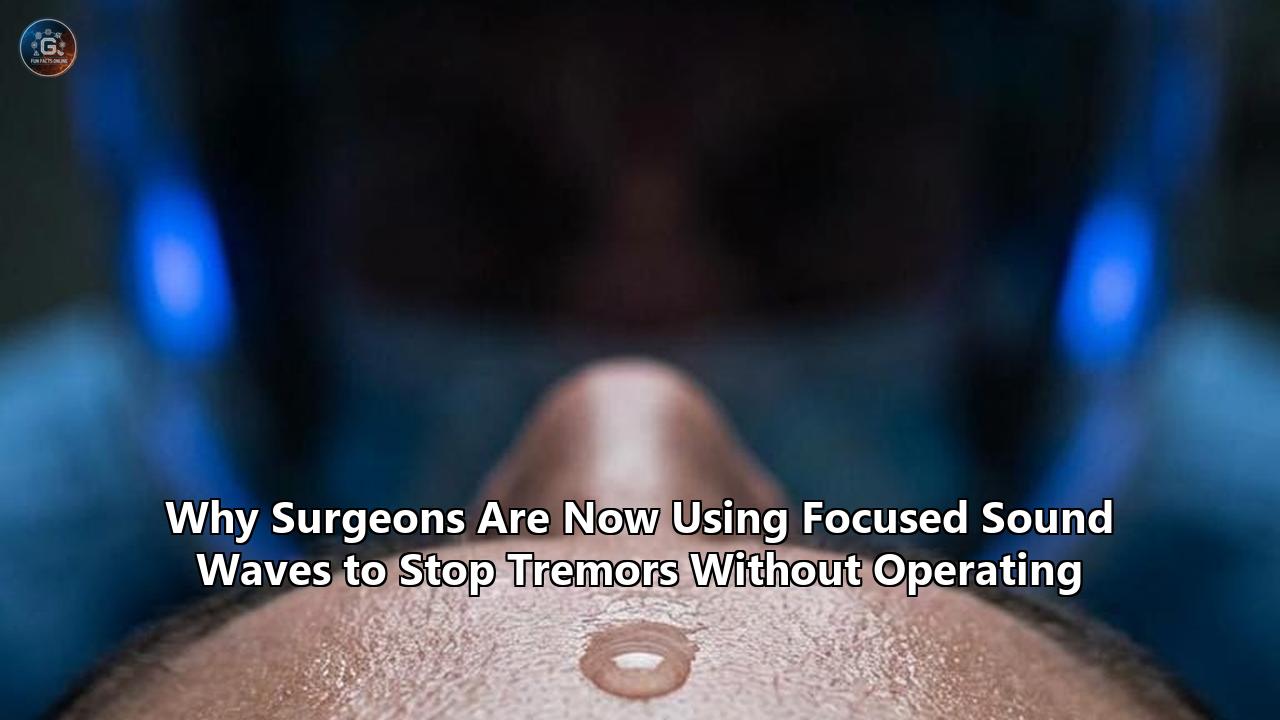
The procedure begins with a complete shaving of the patient’s head. While this can be emotionally difficult for some, it is a critical safety requirement. Even a single pocket of air trapped in a hair follicle can act as a barrier to ultrasound, absorbing the acoustic energy and causing a localized skin burn.
Once the scalp is clean, the neurosurgeon applies a local anesthetic to four points on the skull and secures a stereotactic head frame. This rigid metal frame is bolted directly to the outer table of the skull. While uncomfortable, the frame is essential to prevent even millimeter-scale movement of the patient's head during the procedure, ensuring the acoustic beams remain locked onto the target coordinate.
Step 2: The Water-Cooling Helmet
With the stereotactic frame secured, the patient lies down on the MRI table. A flexible, dome-shaped silicone membrane is sealed around the patient’s head, and the 1,024-element transducer helmet is positioned over it.
The space between the scalp and the transducer helmet is filled with chilled, degassed water. The water serves two vital purposes:
- Acoustic Coupling: Sound waves travel poorly through air but propagate exceptionally well through water, which has an acoustic impedance closely matching human tissue.
- Thermal Protection: The water is continuously circulated and cooled to approximately 15°C to dissipate any heat that builds up on the surface of the scalp as the sound waves pass through, protecting the skin from thermal injury.
+-------------------------------------------------------------+
|                     MRI SCANNER BORE                        |
|                                                             |
|     +-------------------------------------------------+     |
|     |            Focused Ultrasound Helmet            |     |
|     |  +-------------------------------------------+  |     |
|     |  |          Circulating Chilled Water        |  |     |
|     |  |  +-------------------------------------+  |  |     |
|     |  |  |           Silicone Membrane         |  |  |     |
|     |  |  |  +-------------------------------+  |  |  |     |
|     |  |  |  |          Patient Scalp        |  |  |     |  |
|     |  |  |  |                               |  |  |     |  |
Step 3: Test Shots (Sublethal Sonications)
The patient is rolled into the bore of the MRI scanner. The neurosurgeon sits in an adjacent control room, separated by a glass window, looking at a bank of monitors displaying high-resolution structural MRI scans of the patient’s brain.
The surgeon first delivers a series of low-energy "test shots," or sublethal sonications. These initial bursts raise the temperature at the target site to approximately 40°C to 45°C. This temperature is not high enough to destroy brain tissue, but it is warm enough to temporarily stun the neurons at the focal point.
After each test shot, the patient is rolled out of the MRI bore. The clinical team performs a rapid neurological assessment, asking the patient to draw a spiral on a tablet, hold a cup of water, or touch their finger to the doctor's hand.
This interactive step is crucial. If the target is correct, the patient’s tremor will temporarily vanish or improve significantly. If the patient reports transient side effects, such as a tingling sensation in their fingertips or lips (paresthesia) or minor slurring of speech, the surgeon knows the focal spot is slightly off-target and can adjust the electronic coordinates by a fraction of a millimeter before delivering any permanent energy.
Step 4: Therapeutic Ablation (Lethal Sonication)
Once the precise target is confirmed and any side effects are ruled out, the patient is rolled back into the scanner. The surgeon titrates the energy of the transducers upward, delivering a therapeutic sonication that lasts between 10 and 20 seconds.
The temperature at the target site rises rapidly to 55°C–60°C, causing focal thermal coagulation necrosis—permanently disabling the targeted pathway. During these few seconds, the patient may experience a brief sensation of warmth, mild headache, or transient dizziness, but the ablation itself is largely painless.
Step 5: The Immediate Transformation
Immediately after the therapeutic sonication, the patient is rolled out of the scanner. The transformation is visible. A hand that was violently shaking just minutes prior is now steady.
This dramatic, on-the-table clinical resolution is a hallmark of the procedure. Patients who have spent decades unable to write their names, button their shirts, or feed themselves are handed a pen and paper.
This immediate restoration of function was documented by Oscar-winning screenwriter and Downton Abbey creator Lord Julian Fellowes, who underwent focused ultrasound therapy to treat his severe essential tremor. Fellowes, whose writing career was threatened by his inability to write legibly, described the immediate regain of physical control as a profound return to normalcy.
Once the procedure is complete, the stereotactic frame is removed from the patient’s skull, the pin sites are dressed, and the patient is monitored for a few hours. Most are discharged and return home the same afternoon.
Anatomical Targets: Redesigning the Brain's Circuitry
To understand why ablating a tiny spot of brain tissue can stop a tremor, it is necessary to examine the underlying neural pathways that dictate human movement. Tremor disorders are not diseases of the muscles; they are diseases of the brain's internal communication networks.
The brain coordinates movement through a series of feedback loops connecting the cerebral cortex, the basal ganglia, the cerebellum, and the thalamus. In conditions like essential tremor and Parkinson's disease, these feedback loops become hyper-synchronized, generating rhythmic, oscillatory electrical patterns that travel down the spinal cord and manifest as involuntary shaking.
Focused ultrasound works by strategically interrupting these hyperactive neural pathways. By destroying a tiny "relay station" along the loop, the abnormal feedback loop is broken, allowing the rest of the motor network to function normally.
Depending on the patient's specific diagnosis, surgeons target different regions of the brain.
                +----------------------------+
                |       Cerebral Cortex      |
                +----------------------------+
                 ^                          |
                 | Motor Signals            | Descending Motor
                 |                          v Commands
         +---------------+          +---------------+
         |   Thalamus    | <======  | Basal Ganglia |
         | (Vim Nucleus) |  Abnormal| (Globus       |
         +---------------+  Feedback| Pallidus)     |
                 ^          Loop    +---------------+
                 |                          |
                 +==========================+
The Ventral Intermediate Nucleus (Vim) of the Thalamus
For patients with essential tremor (ET), the primary surgical target is the Ventral Intermediate Nucleus (Vim) of the thalamus. The Vim serves as the principal relay station for the cerebellothalamocortical tract, a pathway that transmits sensory and motor coordination signals from the cerebellum to the primary motor cortex.
In essential tremor, this tract becomes hyperactive. When a patient attempts to perform a voluntary action, such as reaching for a glass, the faulty loop floods the motor cortex with rhythmic, alternating signals, causing an "action tremor".
An acoustic ablation of the Vim (known as a thalamotomy) cuts this hyperactive connection. This stops the oscillatory signals from reaching the motor cortex, providing highly effective, long-lasting tremor control.
The Pallidothalamic Tract (PTT)
While essential tremor is defined primarily by involuntary shaking, Parkinson's disease is a much more complex, systemic neurodegenerative disorder characterized by a constellation of motor symptoms, including:
- Tremor: Shaking that typically occurs at rest.
- Rigidity: Severe, painful stiffness in the muscles and joints.
- Bradykinesia: Extreme slowness of physical movement.
- Dyskinesia: Involuntary, erratic, writhing movements often triggered by long-term use of levodopa medications.
Because Parkinson's disease involves a different pathway than essential tremor, targeting the thalamus's Vim nucleus is often insufficient for managing its broader motor complications. Instead, surgeons target the basal ganglia loop, specifically the Globus Pallidus Interna (GPi) or the Pallidothalamic Tract (PTT).
The late 2025 FDA approval of staged bilateral pallidothalamic tractotomy (PTT) represents a major advancement in this targeting strategy. The PTT consists of a bundle of nerve fibers (including the ansa lenticularis and the lenticular fasciculus) that project from the hyperactive GPi to the thalamus.
Under normal physiological conditions, the GPi acts as an inhibitory brake on the motor signals passing through the thalamus. In Parkinson's disease, the loss of dopamine-producing neurons in the substantia nigra causes the GPi to become hyperactive, applying an excessive "brake" on the motor system. This excessive inhibition results in the stiffness, slowness, and freezing characteristic of the disease.
By placing an acoustic lesion precisely within the PTT, neurosurgeons disrupt this overactive inhibitory pathway. This eases the pathological "braking" action, restoring motor coordination, reducing muscle rigidity, relieving dyskinesia, and calming tremors.
The clinical value of targeting the PTT lies in its efficiency: because it is a highly concentrated bottleneck of nerve fibers, a very small lesion can yield broad improvements across multiple symptoms.
| Target Structure | Primary Indication | Main Motor Symptoms Addressed | Typical Clinical Approach |
|---|---|---|---|
| Vim (Ventral Intermediate Thalamus) | Essential Tremor (ET), Tremor-Dominant Parkinson's | Hand & arm tremors during action | Unilateral or staged bilateral |
| PTT (Pallidothalamic Tract) | Advanced Parkinson’s Disease | Rigidity, bradykinesia, tremor, dyskinesia, muscle pain | Staged bilateral |
| GPi (Globus Pallidus Interna) | Advanced Parkinson’s Disease | Levodopa-induced dyskinesia, rigidity | Unilateral |
Focused Ultrasound vs. Deep Brain Stimulation (DBS)
For decades, Deep Brain Stimulation (DBS) has been the gold-standard surgical therapy for patients with severe, medication-refractory movement disorders. DBS operates on a different clinical model than focused ultrasound. Rather than destroying overactive brain tissue to disrupt faulty circuits, DBS modulates those circuits using electrical impulses.
This functional difference introduces a complex set of clinical trade-offs that patients and their multidisciplinary medical teams must navigate.
The Mechanics of Deep Brain Stimulation
DBS is a major, invasive neurosurgical procedure. It requires a surgeon to drill a hole in the patient’s skull (craniotomy) and manually guide a thin metal wire (lead) embedded with electrical contacts deep into the brain.
This lead is connected via an insulated wire tunneled beneath the skin of the neck to an Implanted Pulse Generator (IPG)—essentially a pacemaker for the brain—implanted under the collarbone.
[ Deep Brain Stimulation (Invasive) ]     [ Focused Ultrasound (Incisionless) ]

        Drill hole in skull                       No incisions or implants
                |                                            |
         Lead wire inserted                                  |
         deep into brain                                     |
                |                                            |
         Battery implanted                                   |
         under collarbone                                    |
                |                                            |
        Physician tunes voltage                    Permanent acoustic lesion
        over multiple sessions                     created in 10-20 seconds
The primary clinical advantage of DBS is its adjustability and reversibility. The electrical parameters—including voltage, frequency, and pulse width—can be noninvasively adjusted by a neurologist during post-operative clinical visits.
If the patient's disease progresses, or if they experience side effects, the stimulation can be tuned up, down, or turned off entirely. Furthermore, because no brain tissue is destroyed, the procedure is theoretically reversible; if a cure for Parkinson's is developed, the hardware can be removed.
The Advantages of Focused Ultrasound
While DBS is highly effective, its invasive nature carries inherent risks and long-term burdens that many patients are unable or unwilling to accept. This is where focused ultrasound offers a compelling alternative.
- No Hardware or Implants: Because focused ultrasound is incisionless, patients carry no foreign hardware inside their bodies. This eliminates the risk of hardware-related infections, lead migration, or wire breakage. It also spares the patient from undergoing future surgeries to replace depleted batteries.
- No Risk of Surgical Infection or Bleeding: Opening the skull and inserting a physical probe carries a small but serious risk of intracranial hemorrhage, stroke, or infection. Focused ultrasound bypasses these surgical complications entirely, as no physical instrument penetrates the brain tissue.
- No General Anesthesia: DBS often requires the patient to undergo parts of the surgery under general anesthesia, which carries cognitive and cardiovascular risks—particularly for elderly or frail patients. Focused ultrasound is performed entirely on awake patients, making it accessible to individuals who are medically ineligible for open-brain surgery.
- Immediate Outpatient Recovery: Following focused ultrasound, patients typically walk out of the hospital the same day and return to normal daily activities within 24 to 48 hours. DBS patients face a multi-day hospital stay and a recovery period of several weeks to allow the surgical incisions to heal.
The Disadvantages of Focused Ultrasound
The primary trade-off of focused ultrasound is its irreversibility. When the acoustic waves heat the target tissue to 55°C–60°C, they create a permanent lesion. Once that tissue is ablated, it cannot be recovered.
If a patient experiences a persistent side effect, such as balance issues or sensory numbness, that side effect may be permanent if the lesion was slightly misplaced or too large. Additionally, because focused ultrasound is not adjustable, physicians cannot tune the treatment over time as the patient's disease progresses.
Defining the Ideal Candidate Profiles
The choice between these two advanced interventions is not a matter of one being universally superior to the other; rather, it is a clinical decision tailored to the patient’s medical profile.
- The Ideal Focused Ultrasound Candidate:
Typically older (70+ years of age) with significant medical comorbidities.
Taking blood thinners (anticoagulants) that make open neurosurgery highly dangerous.
Has a high Skull Density Ratio (SDR > 0.45).
Suffers from a severe needle or surgical phobia.
Seeks immediate symptom relief without the burden of ongoing hardware management or frequent clinical adjustment visits.
- The Ideal Deep Brain Stimulation Candidate:
Typically younger (under 65 years of age) with many years of life ahead, where preserving brain tissue for future disease-modifying therapies is a priority.
Has rapidly progressive Parkinson’s disease where highly adjustable, bilateral, and dynamic symptom management is required.
Has an unfavorable Skull Density Ratio (SDR < 0.40), making focused ultrasound physically impossible.
Requires treatment for complex motor patterns that cannot be addressed by a static lesion alone.
The Economics of Incisionless Neurosurgery
Behind the medical and physical science of focused ultrasound lies an equally complex economic reality. The adoption of any advanced medical technology is ultimately governed by its capital cost, clinical efficiency, and insurance reimbursement frameworks.
High Capital Expenditure for Hospitals
For a healthcare system, establishing a focused ultrasound program requires a substantial upfront financial investment. The specialized Insightec Exablate Neuro platform itself carries a price tag exceeding $2 million.
Furthermore, because the system must operate inside an MRI scanner, a hospital must possess a compatible, high-field MRI system (typically a 1.5-Tesla or 3-Tesla scanner). A dedicated clinical MRI scanner costs between $1.5 million and $3 million, bringing the total initial capital hardware cost to upwards of $4 million to $5 million.
This high capital barrier explains why focused ultrasound tremor treatment is initially concentrated within major academic medical centers and large, well-funded regional hospital networks.
Cost-Effectiveness and Dominance over DBS
Despite the steep initial hardware cost, health economics researchers have found that focused ultrasound is highly cost-effective, and in some clinical pathways, economically "dominant" over deep brain stimulation. In health economics, an intervention is termed "dominant" when it is both more effective at improving quality-adjusted life years (QALYs) and less expensive over a given time horizon.
A study published in the British Journal of Radiology modeled the cost-effectiveness of unilateral focused ultrasound compared to unilateral DBS for medically refractory essential tremor within the UK National Health Service (NCS). The researchers found that over a 5-year time horizon, focused ultrasound was significantly less expensive than DBS (£19,779 versus £62,348, respectively) while generating slightly better clinical utility outcomes (+0.03 QALYs).
This economic benefit is driven by several key clinical factors:
- Elimination of Operating Room Costs: Traditional DBS surgery requires multiple hours of dedicated operating room time, a full surgical staff, an anesthesiology team, and highly expensive sterile surgical consumables. Focused ultrasound bypasses the operating theater entirely, utilizing a standard outpatient MRI suite.
- No Post-Operative ICU or Inpatient Stay: DBS patients typically require at least one night in an intensive care unit (ICU) or a step-down ward to monitor for intracranial bleeding or infection. Focused ultrasound patients are monitored in an outpatient recovery bay for a few hours before going home, saving thousands of dollars per procedure in hospital bed utilization.
- No Long-Term Hardware Maintenance: DBS requires ongoing, lifetime clinical follow-up. Neurologists must spend hours programming and fine-tuning the device’s electrical parameters. Over time, the implanted batteries must be surgically replaced, or the hardware must be repaired if wires break or migrate. Focused ultrasound has zero ongoing maintenance costs once the single-day treatment is complete.
The Insurance and Reimbursement Landscape
Historically, patients seeking focused ultrasound had to pay out of pocket, with self-pay prices ranging from $30,000 to $45,000 per procedure. However, the economic and clinical evidence has driven a major shift in insurance coverage.
In the United States, Medicare now provides universal coverage for unilateral focused ultrasound tremor treatment (specifically targeting the Vim nucleus for essential tremor and tremor-predominant Parkinson’s) across all 50 states.
For the newer, staged bilateral procedures approved in late 2025 and early 2026, the reimbursement landscape is currently in transition. Following the FDA’s greenlight, major commercial insurers and regional Medicare Administrative Contractors (MACs) are establishing dedicated local coverage determinations.
Medical device manufacturers and clinical advocacy groups, such as the Focused Ultrasound Foundation, employ reimbursement support managers to help patients navigate the complex prior-authorization process, demonstrating that financial navigation remains a key part of the patient journey.
Limitations, Risk Factors, and Side Effects
No medical procedure—no matter how advanced or noninvasive—is entirely free of risk. While focused ultrasound eliminates the immediate physical dangers of open-brain surgery, it still creates a permanent, physical lesion inside a patient’s central nervous system.
Understanding the adverse event profile of focused ultrasound is essential for establishing realistic clinical expectations.
Adverse Events from the Pivotal Clinical Trials
The safety and efficacy profile of focused ultrasound was established in a landmark, multi-center, randomized, double-blind clinical trial led by Dr. W. Jeffrey Elias, Director of Stereotactic and Functional Neurosurgery at the University of Virginia. The study, published in The New England Journal of Medicine*, evaluated 76 patients with moderate-to-severe essential tremor who were randomized in a 3:1 ratio to undergo active focused ultrasound thalamotomy or a "sham" procedure (where patients underwent the exact same setup but without the delivery of acoustic energy).
The trial demonstrated immediate, dramatic tremor control, with hand-tremor scores improving by 47% at three months and maintaining a 40% improvement at the 12-month mark. However, the study also documented several persistent side effects:
+---------------------------------------------------------+
|     ADVERSE EVENT SPREAD (12 Months Post-Procedure)     |
|                                                         |
|  Sensory Disturbances / Numbness (Paresthesia)  [14%]   |
|  ====================                                   |
|                                                         |
|  Gait Disturbance / Unsteadiness (Ataxia)       [ 9%]   |
|  =============                                          |
|                                                         |
|  Persistent Moderate-to-Severe Deficits        [ 1-2%]  |
|  ==                                                     |
+---------------------------------------------------------+- Paresthesia or Numbness: Immediately after the procedure, 38% of patients reported some degree of sensory change, such as tingling or numbness in the treated hand or face. By 12 months, this resolved in the majority of patients, but persisted in 14% of the cohort.
- Gait Disturbance or Unsteadiness (Ataxia): At the 3-month mark, 36% of patients experienced issues with balance, walking, or unsteadiness. This represents a temporary "stunning" of the nearby cerebellar pathways. At 12 months, the rate of persistent gait disturbance had dropped to 9%.
- Speech and Swallowing Difficulties: A small percentage of patients experienced transient dysarthria (slurred speech) or dysphagia (difficulty swallowing) due to the local swelling (edema) surrounding the ablation site. In rare cases, minor slurring can persist long-term.
These side effects occur because the target structures in the brain—the Vim nucleus and the PTT—are positioned adjacent to major sensory and motor tracts. If the acoustic lesion is slightly too large, or if the heat diffuses beyond the intended boundaries, it can encroach upon the internal capsule (causing weakness) or the sensory thalamus (causing numbness).
The Progressive Nature of Parkinson's Disease
It is critical to distinguish between treating a symptom and curing a disease. Focused ultrasound is highly effective at stopping tremors, stiffness, and slowness, but it does not stop the underlying neurodegenerative process.
In Parkinson’s disease, the progressive loss of dopamine-producing neurons in the substantia nigra continues unabated. Over time, patients may develop new symptoms—such as cognitive decline, depression, autonomic dysfunction, or swallowing difficulties—that cannot be treated by acoustic ablation. Patients must understand that while the procedure can restore motor function, it is a symptomatic management tool rather than a disease-modifying cure.
The Future Horizon: Beyond Ablation to Neuromodulation and BBB Opening
The success of focused ultrasound in treating movement disorders has proven that acoustic energy can safely and precisely cross the human skull to alter deep brain function. However, creating permanent tissue lesions is only the first step.
Biomedical engineers and neuroscientists are developing new applications for focused ultrasound that do not require destroying brain cells. These emerging modalities are transforming functional neurosurgery from a field of destructive lesioning into one of dynamic, noninvasive cellular access.
Low-Intensity Focused Ultrasound (LIFU) and Neuromodulation
While high-intensity focused ultrasound (HIFU) uses thermal energy to ablate tissue, Low-Intensity Focused Ultrasound (LIFU) utilizes much lower acoustic power levels. Rather than generating heat, LIFU relies on mechanical shear stress to temporarily and reversibly alter the electrical excitability of neurons.
This is known as acoustic neuromodulation. By directing low-intensity acoustic waves to specific brain circuits, scientists can temporarily "turn down" hyperactive pathways or "excite" underactive ones.
This approach acts like a temporary, noninvasive deep brain stimulator. Clinicians are researching LIFU for the treatment of:
- Depression and Anxiety: Modulating the amygdala or anterior cingulate cortex to restore normal emotional regulation.
- Obsessive-Compulsive Disorder (OCD): Quieting hyperactive corticostriatal loops.
- Chronic Pain: Temporarily blocking pain transmission pathways in the sensory thalamus.
- Epilepsy: Suppressing hyper-excitable seizure focus zones in real-time.
Because LIFU is completely reversible, it eliminates the primary risk of focused ultrasound—the permanent loss of brain tissue. If a patient experiences a side effect, the acoustic energy is turned off, and the brain returns to its baseline state immediately.
                     +---------------------------------------+
                     |        FOCUSED ULTRASOUND PATHS       |
                     +---------------------------------------+
                                         |
                +------------------------+------------------------+
                |                                                 |
                v                                                 v
  [ High-Intensity (HIFU) ]                         [ Low-Intensity (LIFU) ]
  - Thermal Ablation (55°C - 60°C)                  - Non-thermal / Mechanical
  - Permanent Tissue Lesion                         - Reversible Neuromodulation
  - Treats Tremors & Parkinson's                    - Explored for Depression, OCD, Pain
Blood-Brain Barrier (BBB) Opening
Perhaps the most promising frontier in neuro-oncology and neurodegenerative medicine is the use of focused ultrasound to open the blood-brain barrier (BBB).
The BBB is a highly selective semipermeable border of endothelial cells that prevents harmful toxins and pathogens from entering the brain from the bloodstream. However, this protective barrier also blocks more than 98% of therapeutic molecules—including chemotherapy drugs, monoclonal antibodies, and gene therapies—making it exceptionally difficult to treat brain tumors, Alzheimer's disease, or Amyotrophic Lateral Sclerosis (ALS).
Focused ultrasound can safely, precisely, and temporarily open this barrier. The procedure involves injecting microscopic, gas-filled lipid bubbles (microbubbles) into the patient’s bloodstream.
When low-intensity ultrasound waves are focused on a specific target area of the brain, the acoustic pressure causes the microbubbles to rapidly expand and contract (cavitate) within the local capillaries. This mechanical oscillation gently stretches the tight junctions between the endothelial cells of the blood-brain barrier.
                             [ Focused Ultrasound Wave ]
                                         |
                                         v
   +-------------------------------------v-------------------------------------+
   |  Capillary Wall (Endothelial Cells)                                       |
   |                                                                           |
   |   [Cell]====[Cell]====[Cell]  -Tight Junctions-  [Cell]====[Cell]====[Cell] |
   |                                      |                                    |
   |                                      v  Microbubble Cavitation            |
   |                                                                           |
   |   [Cell] -- -- -- [Cell]   <-- Barrier Opened Temporarily -->   [Cell]     |
   |                                                                           |
   |                  [ Chemotherapy / Antibodies / Genes Enter ]               |
   +---------------------------------------------------------------------------+
For a window of approximately 4 to 6 hours, the barrier in that specific millimeter-sized region remains open, allowing therapeutic drugs in the bloodstream to cross directly into the brain tissue. The barrier then naturally closes, protecting the brain once again.
Clinical trials are underway to evaluate this technique:
- Alzheimer's Disease: Opening the BBB in the hippocampus to allow the patient's immune system or monoclonal antibodies to more effectively clear amyloid-beta plaques.
- Glioblastoma (Brain Cancer): Temporarily opening the BBB to deliver therapeutic doses of chemotherapy drugs (like temozolomide or carboplatin) directly into the tumor, sparing the rest of the body from systemic toxicity.
- ALS and Parkinson's: Delivering neurotrophic growth factors or gene-editing vectors directly to degenerating motor neurons.
What to Watch Next
As focused ultrasound enters its second decade of clinical availability, several key milestones will shape its trajectory:
- Long-Term Efficacy Data: Because the staged bilateral approvals for Parkinson's and essential tremor are relatively recent, researchers are monitoring patients to evaluate the multi-year durability of bilateral lesions. Understanding whether tremors recur after several years will define how early in the disease process this treatment should be offered.
- Expansion of Bilateral Treatment Sites: Following the initial wave of adoptions at academic medical centers, watch for how quickly smaller regional hospitals establish staged bilateral focused ultrasound programs. This expansion will depend heavily on the pace of local insurance approvals and MAC coverage decisions.
- Developments in Frameless Technology: Researchers are designing systems that eliminate the need for the rigid stereotactic head frame. Replacing the skull bolts with advanced optical tracking systems and real-time motion-correction software would improve patient comfort during the procedure.
- Clinical Trial Readouts for Psychiatric Indications: Look for key trial results evaluating focused ultrasound for severe OCD, treatment-resistant depression, and opioid addiction. Positive data could prompt the next wave of FDA approvals, moving focused ultrasound from movement disorders into the mainstream of interventional psychiatry.
Surgeons are choosing focused sound waves to stop tremors because the physical reality of the technology matches its clinical promise: it delivers rapid, deep-brain therapeutic relief while sparing patients the physical trauma, long recovery times, and potential complications of open-brain surgery. The transition from physical metal scalpels to precise acoustic energy is no longer a future concept—it is a routine clinical treatment that continues to expand our understanding of how we can interact with the human brain.
Reference:
- https://southfloridahospitalnews.com/focused-ultrasound-available-at-uhealth-for-patients-with-tremor-disorders/
- https://www.neurosurgery.columbia.edu/news/columbia-neurosurgery-expands-parkinsons-treatment-options-new-bilateral-ptt-focused-ultrasound-procedure
- https://www.sciencenews.org/article/parkinsons-ultrasound-brain-shake-pain
- https://www.itnonline.com/content/mri-guided-focused-ultrasound-effective-treat-essential-tremor
- https://www.tandfonline.com/doi/abs/10.1080/17434440.2025.2489493
- https://www.pennmedicine.org/physicians-hub/clinical-briefing/focused-ultrasound-therapy-for-the-treatment-of-medication-refractory-essential-tremor
- https://www.prnewswire.com/news-releases/insightec-announces-fda-approval-of-staged-bilateral-focused-ultrasound-treatment-for-parkinsons-disease-302499479.html
- https://refreshmiami.com/news/insightec-receives-new-fda-approval-of-parkinsons-treatment/
- https://www.nyp.org/advances/article/neurology-neurosurgery/fda-approves-bilateral-focused-ultrasound-to-treat-symptoms-of-advanced-parkinsons-disease
- https://www.youtube.com/watch?v=HGL90zVkskk
- https://columbiamedrev.wixsite.com/cu-medical-review/post/focused-ultrasound-engineering-sound-into-a-therapeutic-tool
- https://www.fusfoundation.org/diseases-and-conditions/essential-tremor/
- https://www.fusfoundation.org/the-technology/mechanisms-of-focused-ultrasound/
- https://www.fusfoundation.org/for-patients/how-it-works/
- https://pmc.ncbi.nlm.nih.gov/articles/PMC6835146/
- https://practicalneurology.com/news/the-fda-has-approved-a-focused-ultrasound-device-for-essential-tremor/2475661/
- https://neurologytoday.aan.com/doi/10.1097/01.NT.0000502826.66257.ac
- https://www.uvahealth.com/news/focused-ultrasound-trial-finds-promise-for-parkinsons-patients
- https://www.researchgate.net/figure/Focused-ultrasound-FUS-for-noninvasively-delivering-acoustic-energy-to-the-brain-A_fig1_342934866
- https://pubmed.ncbi.nlm.nih.gov/23944301/
- https://www.fusfoundation.org/posts/the-top-10-focused-ultrasound-newsletter-stories-of-2025/
- https://www.precisionhealth.com.au/healthcare-services/ultrasound-brain/mr-guided-focused-ultrasound-mrgfus-for-parkinsons-disease/
- https://academic.oup.com/bjr/article/7448870
- https://cdn.fusfoundation.org/2024/02/22100533/2024-FUSF-BrainTargeting-Whitepaper-5-pages-web.pdf
- https://www.neurology.org/doi/10.1212/WNL.0000000000211364
- https://clinicaltrials.gov/study/NCT04996992
- https://aquariusph.com/portfolio/fus-et-pub/
- https://www.mdedge.com/familymedicine/article/111928/movement-disorders/mri-guided-thalamotomy-significantly-reduces-hand
- https://pmc.ncbi.nlm.nih.gov/articles/PMC9733625/
- https://aquariusph.com/news/fus-et-news/
- https://insightec.com/news/nejm-publishes-results/
- https://pubmed.ncbi.nlm.nih.gov/27557301/
- https://pubmed.ncbi.nlm.nih.gov/40184524/