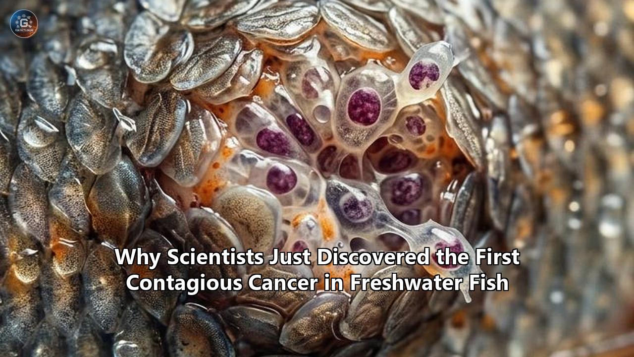
In late July 2026, a team of geneticists and aquatic biologists at the University of Vermont published findings in Nature that upended a foundational axiom of veterinary oncology. For over a decade, black, tar-like skin tumors afflicting brown bullhead catfish (Ameiurus nebulosus) in Lake Memphremagog—a 30-mile glacial lake straddling the border between Vermont and Quebec—were assumed to be the byproduct of toxic pollution or viral infection. Instead, deep whole-genome sequencing revealed something far more extraordinary: the raised melanoma lesions are not the fish's own mutated cells. They belong to an immortal, parasitic cell lineage jumping from living host to living host across the watershed.
This discovery marks the identification of a contagious cancer in fish—the first transmissible malignancy ever documented in a freshwater ecosystem and only the fourth known transmissible cancer lineage in the animal kingdom.
The finding shatters the long-held assumption that cancer is purely an internal evolutionary dead end that dies alongside its host. It also resolves a baffling ecological cold case that began in 2012, when local anglers first pulled bottom-dwelling catfish out of the lake covered in disfiguring jet-black patches. What was long viewed as a localized environmental toxicology crisis has turned out to be an unprecedented event in aquatic evolutionary biology.
[ Solitary Mutation in Original Host ]
                 │
                 ▼
 [ Cell Lineage Acquires Immune Evasion ]
                 │
                 ▼
 [ Mechanical / Mucosal Transfer During Spawning ]
                 │
                 ▼
 [ Clonal Tumor Engraftment in New Host ]
The 2012 Cold Case: Why Toxicologists Looked in All the Wrong Places
To understand why this discovery took fourteen years to reach print, one must understand how wildlife disease investigations normally unfold. When high rates of skin cancer appear in bottom-feeding fish, environmental toxicologists follow a standard playbook.
Brown bullheads are benthic organisms; they spend their lives plowing through soft mud and organic sediment, vacuuming up invertebrates, detritus, and small fish. Because heavy metals, polycyclic aromatic hydrocarbons (PAHs), polychlorinated biphenyls (PCBs), and agricultural runoff settle into lakebed sediments, benthic fish serve as ecological indicators. When bullheads in polluted industrial waterways like Ohio's Cuyahoga River developed liver and skin carcinomas in the late 20th century, chemical mutagens were the proven culprit.
So when reports of black skin lesions surged in Lake Memphremagog—reaching a staggering 30 percent prevalence among adult bullheads by 2015—regulatory agencies in both the U.S. and Canada launched standard ecotoxicological surveys. Water quality testing searched for carcinogenic compounds. State and provincial biologists evaluated industrial discharge, agricultural pesticides, naturally occurring geological arsenic, and leachate from the nearby regional landfill in Coventry, Vermont.
Simultaneously, virologists ran assays for oncogenic pathogens. Retroviruses and herpesviruses are well-known triggers for skin papillomas and lymphocystis in freshwater species like walleye. Yet every viral screen came back clean. Electron microscopy revealed no viral particles within the tumor matrix, and high-throughput RNA sequencing failed to isolate viral transcripts capable of driving cellular transformation.
The ultraviolet radiation hypothesis was equally unviable. Melanoma in humans and terrestrial mammals is primarily driven by UV radiation damage to melanocytes. But brown bullheads inhabit dark, turbid waters, often buried in mud at depths where UV light cannot penetrate. Toxicologists were left with an infuriating paradox: a high-prevalence melanoma epidemic in an aquatic species without a clear mutagenic driver, a viral vector, or solar exposure.
Genomic Forensics: Unmasking the Clonal Lineage
The turning point came when Julie Dragon, a genomic scientist at the University of Vermont Cancer Center, and Mark Henderson, a fisheries biologist at UVM's Rubenstein School of Environment and Natural Resources, shifted their analytical framework. Rather than asking what external factor was triggering cancer in individual catfish cells, they analyzed the genomic architecture of the tumors themselves.
In a conventional cancer, every tumor arises independently from the host's normal somatic tissues. If you sequence the DNA of a normal skin cell from Host A and compare it to Host A's tumor, the two genomes will be virtually identical, differing only by specific driver and passenger somatic mutations acquired during oncogenesis. Furthermore, Host A's tumor DNA will look far more similar to Host A's healthy tissue than it does to a tumor taken from Host B.
When Dragon’s team performed whole-genome sequencing (WGS) comparing tumor tissue and healthy muscle tissue across dozens of affected brown bullheads from Lake Memphremagog and adjacent waters, the canonical pattern collapsed.
The tumor DNA did not match the host fish carrying it.
Instead, tumors collected from disparate fish possessed a unique, highly conserved genetic signature. The analysis uncovered hundreds of thousands of single nucleotide polymorphisms (SNPs), copy number variations, and structural genetic variants that were shared across every single tumor sample—yet completely absent from the healthy host fish tissue.
The statistical probability of independent tumors acquiring hundreds of thousands of identical non-coding and coding genetic variants across dozens of individual fish is zero. The conclusion was inescapable: every tumor analyzed was composed of cells derived from a single ancestral progenitor cell that had escaped its original host years ago.
This single cell line has transformed into a unicellular, transmissible entity—a biological parasite operating under the guise of cancer. The identification of this contagious cancer in fish confirmed that the black lesions sweeping through Lake Memphremagog were not an outbreak of environmental mutations, but an infectious epidemic of autonomous cells.
The Exclusive Transmissible Cancer Club
Transmissible cancers represent one of the rarest evolutionary phenomena on Earth. Prior to the UVM discovery, scientists had identified only three distinct types of naturally occurring transmissible malignancies across all animal life:
- Canine Transmissible Venereal Tumor (CTVT): Discovered in dogs, CTVT is an ancient continuous cell line that originated in a wild canid approximately 11,000 to 12,000 years ago. It spreads directly through physical contact during coitus.
- Devil Facial Tumor Disease (DFTD): First observed in Tasmania in 1996, DFTD consists of two distinct clonal lineages (DFTD1 and DFTD2) that spread between Tasmanian devils via aggressive biting during territorial disputes. DFTD has reduced wild devil populations by up to 90% in impacted zones.
- Bivalve Transmissible Neoplasia (BTN): Found in marine mollusks, including soft-shell clams (Mya arenaria), mussels, and cockles. BTN manifests as a leukemia-like blood cancer where malignant hemocytes proliferate in the animal's circulatory fluid and shed into seawater, drifting on currents to infect neighboring filter feeders.
The brown bullhead melanoma (BBM) lineage represents the fourth major class of transmissible cancer, but it occupies a unique ecological and physiological niche.
Unlike CTVT and DFTD, which exist in warm-blooded mammalian hosts with complex adaptive immune systems, BBM operates in a cold-blooded ectothermic vertebrate. Unlike BTN, which exists in marine invertebrates lacking an adaptive immune system based on major histocompatibility complexes (MHC), brown bullheads are jawed vertebrates with fully developed T-cells, B-cells, antibodies, and cellular rejection machinery.
| Biological Feature | Canine CTVT | Tasmanian DFTD | Marine BTN | Brown Bullhead Melanoma (BBM) |
|---|---|---|---|---|
| Host Taxonomy | Mammalian (Canid) | Mammalian (Marsupial) | Marine Invertebrate (Bivalves) | Freshwater Vertebrate (Teleost Fish) |
| Cellular Origin | Histiocytic | Schwann Cell | Hemocyte (Circulatory) | Melanocyte (Pigment Cell) |
| Primary Vector | Sexual Contact | Territorial Biting | Passive Seawater Filtration | Spawning Aggregations / Benthic Contact |
| Medium | Direct Tissue | Direct Tissue | Isotonic Seawater | Hypotonic Fresh Water |
| Host Immunity | Adaptive (MHC-based) | Adaptive (MHC-based) | Innate Only | Adaptive (MHC-based) |
The Biophysical Wall: Surviving the Freshwater Environment
One of the most complex scientific puzzles surrounding the discovery is how living cancer cells survive the leap between hosts in a freshwater environment.
In marine shellfish, cellular transfer is facilitated by the chemistry of ocean water. Seawater is isotonic relative to the internal fluid of marine invertebrates; its salt concentration (roughly 3.5%) prevents cells from bursting or collapsing due to osmotic pressure. Cancerous clam cells can float in open ocean water for days, remaining viable until pulled into the siphon of a neighboring clam.
Fresh water presents a severe biophysical barrier. It is hypotonic relative to vertebrate intracellular fluid. If an isolated mammalian or fish skin cell is dropped into pure fresh water, water rapidly rushes across the cell membrane via osmosis, causing the cell to swell, lyse (burst), and rupture within minutes.
How, then, does a brown bullhead melanoma cell move from one fish to another through Lake Memphremagog's waters?
The UVM research team points to two primary mechanisms: direct physical contact during spawning and micro-environment localized transmission within benthic sediment.
               [ Spawning Aggregation ]
                          │
          ┌───────────────┴───────────────┐
          ▼                               ▼
[ Direct Physical Contact ]    [ Mucus-Protected Shedding ]
  - Courting abrasions           - Sloughed skin clusters
  - Biting & skin contact        - Settles in soft sediment
          │                               │
          └───────────────┬───────────────┘
                          ▼
           [ Cellular Engraftment ]
Spawning Aggregations and Courtship Abrasion
Brown bullheads spawn during late spring and early summer. During courtship, adults congregate in shallow, muddy bays, creating dense aggregations. Courtship behavior in Ameiurus nebulosus is notably physical: males and females entwine their bodies, bite each other’s flanks, and vigorously rub their skin together in shallow nests cleared in the sediment.
Unlike scaled fish (such as perch or bass), bullheads are smooth-skinned catfish lacking hard dermal scales. Their outer epidermis is delicate and rich in mucus-secreting goblet cells. Intimate physical contact, paired with biting and skin abrasions during spawning, creates a direct portal of entry. Tumor tissue protruding from an infected fish can be mechanically sheared off and rubbed directly into fresh wounds on an uninfected partner, allowing intact cancer cells to bypass the osmotic stress of open water entirely.
Sediment Micro-Reservoirs and Mucus Protection
For short-distance waterborne transmission, the tumor cells rely on protective mucus matrices. Brown bullhead skin secretes a viscous, proteinaceous mucus layer containing antimicrobial peptides and structural lipids. When melanoma cells shed from raised, ulcerated lesions, they are often cast off in sloughed clumps of epidermal mucus rather than as bare individual cells.
This mucus layer acts as an osmotic buffer, shielding the underlying cancer cells from freshwater dilution long enough for them to settle into the benthic sediment layer. Because bullheads spend hours resting quietly on the lake floor with their bellies resting on muddy substrates, these localized cellular reservoirs in the sediment provide another point of contact for scaleless skin.
Immunological Cloaking: Bypassing Graft Rejection
The existence of any transmissible cancer requires an evolutionary breakthrough in immune evasion. Under normal physiological conditions, an animal’s immune system recognizes foreign tissue from another individual of the same species and destroys it through allograft rejection. This process is driven by the Major Histocompatibility Complex (MHC)—a set of cell-surface proteins that showcase internal cellular peptides to circulating T-lymphocytes.
When organ transplants occur in humans without immunosuppressive drugs, T-cells identify foreign MHC molecules and initiate an aggressive cellular attack. For a contagious cancer in fish to succeed, the rogue melanoma clone must possess a mechanism to render itself invisible to the bullhead immune system.
Genomic sequencing of the brown bullhead melanoma clone revealed how the lineage achieves this cellular camouflage:
- Downregulation of MHC Class I Machinery: Much like Tasmanian devil facial tumor cells, the fish melanoma clone has silenced key genes responsible for displaying MHC Class I molecules on its cell surface. Without surface MHC Class I expression, host CD8+ cytotoxic T-lymphocytes cannot recognize the invading tumor cells as foreign tissue.
- Immunosuppressive Microenvironment: RNA sequencing of tumor biopsy samples showed elevated expression of immunosuppressive signaling proteins, including transforming growth factor-beta (TGF-β) homologues. These proteins inhibit localized inflammatory responses, dampening the activity of host macrophages and natural killer (NK) cells attempting to infiltrate the tumor margin.
- Genetic Bottlenecks in Host Populations: Lake Memphremagog was carved out during the last glacial retreat roughly 10,000 to 12,000 years ago. Post-glacial freshwater fish populations in landlocked northern basins often exhibit reduced genetic diversity across their MHC loci. When host animals share highly similar MHC profiles across a watershed, the barrier to cell engraftment drops significantly, facilitating the initial establishment of a clonal lineage.
Pollution as a Catalyst: The Ecotoxicological Paradox
The discovery that the black patches are caused by a contagious cell line resolves one enigma while introducing another: what role, if any, did environmental pollution play in spawning this disease?
For decades, environmental advocates and local citizens pointed to industrial activities around Lake Memphremagog as the driver behind the fish tumors. The lake serves as a binational reservoir, providing drinking water to over 175,000 residents in Quebec and Vermont. Upstream municipal wastewater, historical industrial operations along tributary rivers, agricultural runoff, and the operation of the regional landfill in Coventry, Vermont, have fueled long-standing political debates between U.S. and Canadian stakeholders.
When toxicological studies initially failed to find a direct chemical trigger for the skin tumors, local environmental groups voiced skepticism. The discovery of a clonal, transmissible cancer provides a nuanced middle ground that reshapes both environmental policy and disease ecology.
Pollution was not the direct mutagen that caused thousands of individual fish cells to mutate into independent skin tumors. However, chemical stressors likely acted as an environmental catalyst.
Sublethal exposure to heavy metals, naturally occurring geological arsenic, and synthetic industrial chemicals is known to induce chronic immunotoxicity in freshwater teleosts. In an unstressed aquatic ecosystem, a bullhead's mucosal immune system might successfully destroy an incoming foreign tumor cell before it can engraft. But in a watershed stressed by chemical runoff, suppressed cell-mediated immunity dramatically lowers the physiological threshold required for transmissible cancer cells to take root.
Environmental degradation likely created the physiological window of opportunity that allowed a single, chance oncogenic mutation in an individual catfish to cross the threshold from a solitary, fatal tumor into an infectious, transmissible clone.
Ecosystem Impact vs. Human Health Concerns
Whenever a novel infectious agent is uncovered in a public water supply, public concern escalates rapidly. The lead researchers at the University of Vermont and state environmental health officials have been explicit regarding human safety risks.
There is zero risk of transmission to humans.Transmissible cancers are extraordinarily host-specific. The physiological, thermal, and immunological barriers preventing a cold-blooded catfish melanoma cell from surviving in a human body are absolute:
- Body Temperature Mismatch: Brown bullheads thrive in water temperatures ranging from 4°C in winter to 24°C in summer. The cellular machinery of the melanoma clone is adapted to these lower temperatures; human core body temperature (37°C) induces rapid thermal shock and protein denaturation in teleost cellular tissue.
- Xenograft Rejection: The human immune system possesses a multi-layered xenograft rejection response. Human T-cells, NK cells, and complement pathways recognize non-human animal cell surface markers instantaneously, destroying foreign cells upon contact.
- Water Treatment Efficacy: Standard municipal water filtration and chlorination protocols utilized for the 175,000 residents relying on Lake Memphremagog destroy intact biological cells effortlessly. The disease is not a waterborne virus or chemical solute; it requires the physical transfer of intact, viable eukaryotic cells.
While human health is not threatened, the long-term ecological trajectory of the brown bullhead population remains an active area of study.
When Devil Facial Tumor Disease swept through Tasmania, it decimated devil populations by more than 80 to 90 percent, pushing the species toward endangered status. DFTD is nearly 100% fatal because facial tumors rapidly grow so large that infected animals can no longer feed.
In contrast, brown bullheads in Lake Memphremagog appear to live with their melanoma tumors for extended periods. Field surveys conducted by Mark Henderson’s team showed that while infected catfish display severe, visually dramatic black skin lesions, many infected individuals maintain normal body condition scores and continue to reproduce.
This suggests that the brown bullhead melanoma clone may be evolving toward an ecological equilibrium with its host—behaving less like an explosive mortality event and more like an endemic parasite that balances high transmissibility with moderate host impact.
What Comes Next: Unanswered Questions in Aquatic Oncology
The publication of the UVM team's findings in Nature marks the beginning of a broader effort in aquatic wild oncology. Researchers across North America and Europe are now re-evaluating historical records of unexplained wildlife tumor outbreaks.
Several critical technical questions dominate current investigations:
1. Determining the Founder Event Date
Using molecular clock analysis—tracking the slow accumulation of neutral somatic mutations across tumor samples collected over different years—genomicists are working to pinpoint the exact decade when the original founder cancer arose. Did this clone emerge recently in response to 20th-century industrial development, or has it been circulating silently in northern watersheds for decades?
2. Watershed Mapping and Geographic Boundaries
Is this contagious cancer in fish confined strictly to Lake Memphremagog, or has it spread down the Saint-François River system into the Saint Lawrence River watershed? Biologists are expanding genetic screening to brown bullhead populations across New England, New York, Ontario, and Quebec to map the geographical boundaries of the clonal lineage.
3. Molecular Targets for Comparative Cancer Research
From a biomedical perspective, studying how the bullhead melanoma clone evades immune destruction and maintains genomic stability across hundreds of host generations offers valuable insights. Transmissible cancers are biological extremes: they are cancer lineages that have achieved cellular immortality, surviving long past the death of the organism that generated them.
Unraveling the genetic pathways that allow these fish tumor cells to downregulate MHC molecules, resist environmental osmotic stress, and maintain cellular integrity could uncover novel targets for human cancer immunotherapies.
What began as a localized fish disease puzzle on the Vermont-Quebec border has expanded our understanding of how cancer cells adapt, survive, and cross the boundary between an internal disease and an infectious parasite. As surveillance efforts expand to other freshwater species, scientists are confronting a striking reality: in freshwater ecosystems, cancer cells can occasionally achieve a life of their own.
Reference:
- https://www.reddit.com/r/science/comments/1v4eczw/scientists_have_found_the_first_known/
- https://www.uvm.edu/uvmnews/news/more-parasites-tumors-vermont-scientists-discover-transmissible-cancer-catfish
- https://www.forbes.com/sites/johndrake/2026/07/27/scientists-just-discovered-a-contagious-cancer-in-a-catfish-the-fourth-known-to-science-meet-the-other-three/
- https://www.fox10phoenix.com/news/freshwater-contagious-cancer-spreading-catfish-vermont-lake
- https://timesofindia.indiatimes.com/health/worlds-first-transmissible-cancer-in-a-freshwater-fish-discovered-what-scientists-know-about-this-rare-melanoma/articleshow/132642243.cms
- https://sciencemediacentre.es/en/transmissible-melanoma-type-cancer-has-been-discovered-fish-fourth-known-contagious-tumor-among
- https://www.theweathernetwork.com/en/news/nature/animals/first-transmissible-cancer-found-in-freshwater-fish-in-canada-u-s-lake