The Unconscious Listeners: A Scientific Breakthrough Under the Scalpels
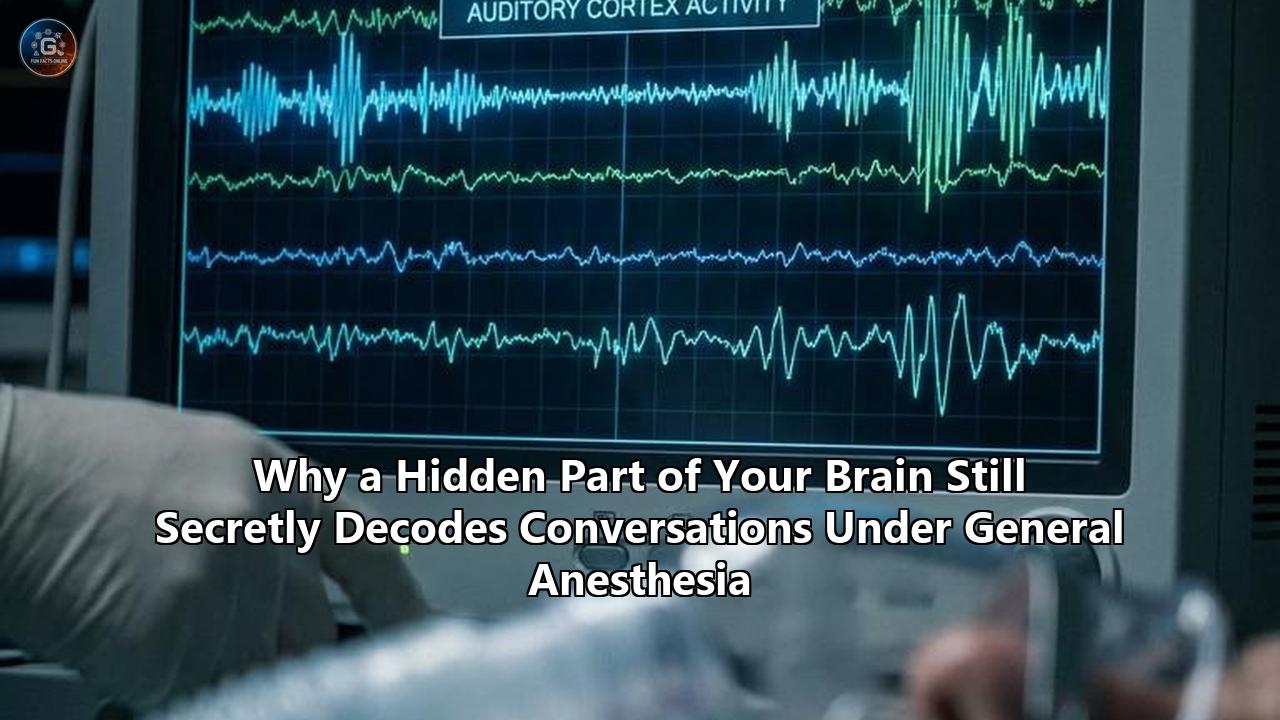
While a patient lies completely motionless on an operating table, their breathing controlled by a ventilator, their eyes taped shut, and their mind plunged into the deep, drug-induced void of general anesthesia, a hidden part of their brain is still listening. Not only is it listening, but it is also actively sorting spoken words into grammatical categories, identifying the emotional tone of the speaker, and predicting the final words of sentences before they are even uttered.
This unexpected reality is the core finding of an unprecedented neuroscientific study published in Nature. Led by neurosurgeon Kalman Katlowitz, alongside senior authors Sameer Sheth and Benjamin Hayden at the Baylor College of Medicine, a team of researchers successfully recorded the activity of individual human neurons deep inside the brain during general anesthesia. By utilizing ultra-advanced, high-density neural probes in patients undergoing surgery, they discovered that the hippocampus—the brain's primary engine for memory and learning—continues to process complex language and adapt to sensory patterns while its host is completely unconscious.
Historically, general anesthesia has been viewed as a biological "off switch." It was believed that the cocktail of sedatives administered during surgery shut down higher-order cognitive processing entirely, isolating the sensory organs so that signals from the outside world died on the vine before reaching the brain’s processing hubs. The Baylor study directly challenges this long-held clinical dogma. It demonstrates that the unconscious brain is not a dark, silent house; rather, it is a highly active computational machine that continues to model the external environment, perform predictive linguistics, and undergo neural changes associated with learning—all without generating a shred of conscious awareness or lasting memory.
This discovery holds profound implications for how we understand human consciousness, memory formation, and the physiological nature of anesthesia itself. It forces us to confront a fundamental question in cognitive science: if a deeply anesthetized, unconscious brain can decode complex language and anticipate speech patterns, what is the evolutionary purpose of conscious awareness?
The Surgical Mystery: What Happens When the Lights Go Out
To appreciate the scale of this discovery, it is necessary to examine how general anesthesia affects the human nervous system. Every year, millions of people undergo surgery under the influence of general anesthetics like propofol, sevoflurane, or desflurane. To the casual observer, an anesthetized patient looks as though they are merely asleep. However, anesthesiologists are quick to point out that general anesthesia is not sleep; it is a drug-induced, reversible coma.
During natural sleep, the brain remains highly dynamic. It cycles through rapid eye movement (REM) and non-REM stages, with brainwaves fluctuating in predictable patterns. A sleeping person can be easily roused by a loud noise, a physical touch, or a sudden change in environment.
In contrast, general anesthesia creates a state of profound sensory decoupling and behavioral unresponsiveness. When a drug like propofol enters the bloodstream, it binds to $\text{GABA}_\text{A}$ receptors throughout the central nervous system. GABA ($\gamma$-aminobutyric acid) is the brain's primary inhibitory neurotransmitter. By enhancing GABAergic activity, anesthetics effectively quiet neuronal firing, shifting the brain's electrical activity from high-frequency, complex communication into slow, highly synchronized, high-amplitude waves.
This shift alters how different regions of the brain communicate with one another. Under normal, awake conditions, your brain operates like a bustling global communications network, with sensory cortices, the thalamus, and the prefrontal cortex constantly exchanging high-speed information. When anesthetic drugs saturate the system, this network breaks down. The thalamus—the brain's central relay station—becomes heavily suppressed, and long-range connectivity across the cerebral cortex is disrupted.
[Awake Brain]
   Sensory Cortex <====== High-Speed Information ======> Prefrontal Cortex (Conscious Integration)
         ^                                                     ^
         \==================== Thalamus =======================/

[Anesthetized Brain (Propofol)]
   Sensory Cortex   - - - - X (Local Processing Only) X - - - -  Prefrontal Cortex (Disconnected)
         ^
         X (Thalamocortical Relay Blocked)
         ^
      Thalamus (Suppressed)
Until now, the prevailing theory was that this breakdown in cortical connectivity prevented any meaningful processing of complex external stimuli. It was assumed that while the primary auditory cortex might register a sound, the signal could not travel any further. Without the coordinated participation of higher-order association cortices and frontal networks, the brain was thought to be blind and deaf to the semantic meaning of the world.
The Tool of Discovery: How Neuropixels Probes Pierced the Silence
The reason this silent, deep-brain linguistic processing went unnoticed for so long lies in the limitations of our standard neuroimaging tools.
Traditionally, scientists monitoring brain activity under anesthesia have relied on tools like electroencephalography (EEG) or functional magnetic resonance imaging (fMRI). While useful, these methods have significant blind spots:
- Scalp EEG measures the electrical activity of millions of neurons averaged together through the thick barrier of the skull. It is like standing outside a massive football stadium and trying to understand individual conversations based on the collective roar of the crowd.
- fMRI measures changes in blood flow as a proxy for neural activity. While it offers excellent spatial resolution, it operates on a delay of several seconds, making it far too slow to track the millisecond-by-millisecond processing of natural human speech.
To bypass these limitations, Katlowitz and his team seized a rare clinical opportunity. They recruited seven adult patients who were undergoing temporal lobe resection surgery at Baylor St. Luke's Medical Center to treat drug-resistant temporal lobe epilepsy. Because these patients were already having portions of their brain surgically removed to control their seizures, surgeons were able to temporarily insert ultra-precise, high-density recording devices directly into the tissue slated for extraction.
The technology that made this possible is called a Neuropixels probe. Developed by a global consortium of research institutes, a Neuropixels probe is a silicon microelectrode array thinner than a human hair, yet packed with roughly 1,000 individual recording sensors. When slid gently into the brain, it can record the electrical action potentials of hundreds of individual, isolated neurons simultaneously.
Traditional Electrodes vs. Neuropixels Probes

Traditional Microelectrodes:
[====] ---------> Records 1-2 neurons at a time (low resolution)

Neuropixels Probe (Hair-thin silicon shank):
[•|•|•|•|•|•|•|•|•|•|•|•|•|•|•|•|•|•|•|•|•|•|•] ---> Over 1,000 sensors recording hundreds of individual
                                                   neurons across different cortical layers simultaneously
Using these probes, the researchers targeted the human hippocampus. Located deep within the medial temporal lobe, the hippocampus has long been known as the gatekeeper of memory. It was chosen not only because of its accessibility during this specific epilepsy surgery, but because it sits at the very apex of the brain's sensory processing hierarchy, far downstream from the primary sensory cortices. If the hippocampus was actively responding to external sounds, it would mean sensory information was successfully traveling through the deeply anesthetized brain, bypassing the blockades erected by propofol.
Deciphering the "Oddball" Tones: How the Sleeping Brain Learns
The researchers began their investigation with a classic neurophysiological test known as the Auditory Oddball Paradigm.
While the seven patients were maintained in a stable, deep state of general anesthesia using propofol, researchers placed headphones on them. They played a continuous stream of repetitive, high-frequency "beep" sounds (the standard tones). Occasionally, and at random intervals, they interspersed a different, low-frequency "boop" sound (the deviant, or "oddball," tone).
In an awake human, the brain easily notices this difference. This detection produces a characteristic electrical signature on an EEG called the mismatch negativity (MMN), which reflects the brain's automatic attention-grabbing mechanism when a pattern is broken.
When Katlowitz and his colleagues analyzed the real-time electrical chatter of individual neurons in the anesthetized hippocampi, they observed something remarkable. More than 70 percent of the monitored neurons registered the auditory tones, and they fired in distinctly different patterns when an oddball tone was played compared to the standard tones.
Even more surprising was how this neural response changed over time. Over a ten-minute session, the hippocampal neurons actually grew better at telling the standard tones and the oddball tones apart. As the experiment progressed, the neurons' firing rate in response to the standard tones steadily decreased (a process called habituation), while their response to the rare, deviant tones became sharper and more pronounced.
Hippocampal Neuron Firing Rates Over Time (Oddball Paradigm)

First 2 Minutes:
Standard Tone: [||||||||||] (10 spikes)
Oddball Tone:  [||||||||||] (10 spikes)  --> Low initial distinction

After 10 Minutes:
Standard Tone: [|||]        (3 spikes)   --> Neuron habituates (ignores repetitive sound)
Oddball Tone:  [||||||||||||] (12 spikes) --> Neuron sensitizes (focuses on the novelty)
In cognitive science, this type of shift is called representational plasticity. It is the fundamental neural mechanism underlying learning and adaptation. To confirm that this was a genuine, systematic neural process and not a statistical fluke, the researchers built a biologically plausible recurrent neural network (RNN) model designed to simulate these biological networks. The model confirmed that this learning behavior emerged naturally from the flexible tuning of individual auditory-responsive connections.
The fact that this plastic rewiring was happening deep within the brain under propofol anesthesia was a revelation. It proved that brain activity under anesthesia is not static. Even when a person is entirely unconscious, their brain is still tracking statistical patterns in their environment, updating its internal predictions, and physically altering its neural responses based on experience.
Decoding a Podcast: The Deep-Brain Grammar Nerd
With proof that the anesthetized brain could learn to distinguish simple tones, the researchers pushed the experiment further. They decided to test whether the unconscious mind could handle the complex, fast-moving structural demands of natural human language.
While the patients remained deeply anesthetized, the team played 10- to 20-minute audio clips of natural speech through their headphones. The clips were taken from educational videos and storytelling podcasts, specifically episodes of The Moth Radio Hour.
Using the Neuropixels probes, the team recorded the firing of hundreds of hippocampal neurons while the spoken stories unfolded. When they later analyzed the neural data alongside the transcript of the audio, the results were stunning:
1. Parts of Speech Discrimination
The individual neurons in the hippocampus did not treat the podcast as an undifferentiated stream of sound. Instead, they responded selectively to the grammatical structure of the language. Certain groups of neurons spiked vigorously when the speaker uttered a noun, while an entirely different set of neurons fired in response to verbs, pronouns, prepositions, or adjectives. The brain was silently parsing the syntax of the sentences in real time.
2. Semantic Mapping
Beyond grammar, the unconscious brain was categorizing the meaning of the words. By training an algorithm to analyze the patterns of neural firing, the researchers found they could partially decode what kind of word had just been spoken. The neurons categorized words into twelve distinct semantic themes, showing clear, unique electrical signatures for words relating to places, names, physical actions, numbers, or emotional expressions.
3. Predictive Coding
Most shockingly, the researchers discovered that the hippocampal neurons were performing predictive coding. In a conscious human, the brain is a prediction engine; when someone says "I take my coffee with milk and...", your brain automatically anticipates the word "sugar" before it is even spoken.
The Neuropixels data revealed that this exact anticipatory firing was occurring under general anesthesia. The firing patterns of the hippocampal neurons shifted and aligned themselves to predict the semantic properties of the upcoming word in a sentence fractions of a second before the speaker actually said it.
Predictive Firing Under Anesthesia:

Audio Timeline:        "...I took a walk in the..." --------> (Pause) -------> "...forest."
                                                                  |
Hippocampal Firing:    [=======================] ------------> [|||||||] ------> [||]
                                                                  |
                                              Neurons pre-emptively fire in patterns
                                              for "place/nature" semantic category
                                              BEFORE the word "forest" is spoken.
To establish a baseline, the researchers compared these unconscious recordings with data from awake, conscious controls listening to the exact same audio. The linguistic processing, semantic mapping, and predictive coding in the deeply anesthetized, unresponsive brain were nearly identical in accuracy to those of an awake, fully attentive listener.
The brain was reading ahead, analyzing the narrative, and preparing for the next line of the story—all while there was absolutely "no one home" to consciously experience it.
The Hippocampal Enigma: Why the Memory Hub is Processing Speech
The discovery that the hippocampus is highly active and processing complex language under anesthesia presents a profound neuroanatomical puzzle.
In standard medical textbooks, sensory processing is depicted as a strict linear pathway:
[Ear] ---> [Brainstem] ---> [Thalamus] ---> [Primary Auditory Cortex] ---> [Association Cortices] ---> [Hippocampus]Under this classic model, the hippocampus sits at the very end of this pipeline, receiving highly processed, refined sensory data and packaging it into long-term memories. It was assumed that if you administered a powerful anesthetic like propofol, the flow of information would be cut off at the level of the thalamus or primary sensory cortices, leaving deep structures like the hippocampus completely isolated from external inputs.
The Baylor study reveals that this is not what happens. Instead, the primary auditory pathways and their connections to deep temporal structures like the hippocampus remain remarkably functional. Even when the thalamus is suppressed and the global cortical networks required for conscious integration are fractured, a dedicated, low-level sensory pipeline remains wide open, feeding complex auditory data directly into the medial temporal lobe.
Why would the brain keep this deep pathway open during a state of total unconsciousness?
The answer likely lies in the evolutionary origin of the hippocampus. Long before it evolved into the primary organ of human autobiographical memory, the hippocampus served as a spatial mapping and navigation tool in primitive animals. To navigate an environment safely, an organism must constantly build internal models of its surroundings, track changes in sensory inputs, and predict what lies around the next corner.
This suggests that the predictive mechanisms we use to understand language—such as anticipating the next word in a sentence—are not unique to human consciousness. Rather, language processing co-opted the ancient, unconscious, predictive mapping systems of the hippocampus. The hippocampus acts as an unprompted, automated modeling engine. It is hardwired to organize incoming data, track statistical patterns, and predict what comes next, regardless of whether we are awake, asleep, or chemically knocked out.
Consciousness vs. Computation: What is the Mind Actually For?
For decades, cognitive scientists and philosophers have debated the boundaries of the unconscious mind. Many prominent theories of consciousness argued that while simple sensory processing (like hearing a sound or feeling a touch) could occur unconsciously, higher-order cognitive tasks (such as syntax parsing, semantic classification, learning, and predictive modeling) strictly required a conscious mind.
The results of the Baylor study fundamentally challenge these frameworks. As Athena Akrami, a neuroscientist at University College London who was not involved in the research, noted: "The computations look nearly identical to those in awake brains, yet they produce no awareness, no memory, no ability to act."
This disconnect forces a conceptual re-evaluation of major neurobiological models of consciousness:
| Theory of Consciousness | Core Premise | How the New Discovery Challenges It |
|---|---|---|
| Global Neuronal Workspace Theory (GNWT) | Consciousness occurs when sensory information is "broadcast" across a wide network of frontal and parietal brain regions, making it globally available. | Confirming that highly complex processing, predictive coding, and semantic decoding can occur locally within the hippocampus without being globally broadcast or generating awareness. |
| Predictive Processing Framework | The primary purpose of conscious thought is to minimize prediction errors by continuously modeling and anticipating our sensory environments. | Demonstrates that highly accurate predictive coding of language occurs entirely within the unconscious state. Predictive modeling does not require consciousness; it is an automated, subconscious default. |
| Integrated Information Theory (IIT) | Consciousness is a product of the structural integration of information across the entire brain (${\Phi}$). | Suggests that complex information can be parsed, categorized, and modeled in localized, modular, and compartmentalized networks (like the hippocampal-auditory loop) without requiring global integration. |
If the unconscious brain is capable of such high-level computations on its own, it raises a challenging question: What is the evolutionary function of consciousness?
If we do not need consciousness to parse grammar, decode meaning, learn environmental patterns, or predict the future, what does conscious awareness actually add to the equation?
The study’s findings suggest that consciousness may not be defined by what information the brain processes, but by how that information is integrated, generalized, and made available for voluntary action. While the unconscious hippocampus can run highly sophisticated linguistic algorithms in isolation, it remains a closed loop during anesthesia. It cannot share that information with the motor cortex to allow the patient to speak, nor can it coordinate with the prefrontal cortex to allow the patient to make voluntary, deliberate decisions based on what they hear.
Consciousness, then, may not be the engine of computation, but the global conductor—the system that allows isolated computational modules to talk to one another, create a unified experience of reality, and translate raw processing power into deliberate behavior.
The Paradox of Memory: Why We Process but Do Not Remember
One of the most mind-bending aspects of this research is the complete dissociation between processing and memory.
If a patient's hippocampus is actively listening to a podcast, analyzing its structure, and altering its neural pathways to "learn" patterns under anesthesia, why do they wake up with absolutely no recollection of it? Upon entering the recovery room, every single patient in the study had zero explicit, reportable memory of the stories played through their headphones.
To resolve this paradox, we must distinguish between two different types of memory and the distinct biological steps required to form them:
- Implicit Memory: This is an unconscious form of memory where past experiences influence our behavior or performance without our conscious awareness.
- Explicit (Declarative) Memory: This is the conscious, reportable recollection of facts, events, and experiences.
The hippocampal activity observed under anesthesia represents implicit processing and representational plasticity. When the neurons changed their firing patterns over ten minutes to isolate the "oddball" tones, they were exhibiting a form of localized, short-term synaptic adaptation. This type of learning does not require conscious awareness, and it can occur entirely within local circuits.
The Two-Step Memory Process

Step 1: Immediate Processing & Synaptic Plasticity (ACTIVE under anesthesia)
   [Spoken Words] ===> [Auditory Loop] ===> [Hippocampal Neurons Fire & Adjust Connections]

Step 2: System Consolidation & Conscious Integration (BLOCKED under anesthesia)
   [Local Hippocampal Signal] ===x=== (Blocked by Propofol) ===x===> [Widespread Neocortex]
                                                                        |
                                                             Permanent, Conscious Memory
For these localized synaptic changes to be converted into a permanent, conscious, and recallable explicit memory, they must undergo a complex biological process called system consolidation. This process requires a coordinated, reciprocal dialogue between the hippocampus and the widespread neocortex, typically mediated by synchronized brain rhythms (such as theta and gamma oscillations) over several hours.
Anesthetics like propofol directly disrupt this delicate hippocampal-neocortical dialogue. While the drug leaves the immediate, local sensory processing pathways intact within the hippocampus, it completely severs the long-range connections required to broadcast those signals to the rest of the brain. The information is processed in real time, but the "save" button is never pressed. The trace of the podcast is processed locally, but it is never integrated into the wider cortical network, causing it to quickly fade away once the stimulus stops.
The History of "Unconscious Hearing" in Medicine
While the resolution of this study is unprecedented, the concept that patients can process auditory information under anesthesia is not entirely new. For decades, anesthesiologists have documented strange instances of "implicit recall" in post-surgical patients.
In classical psychology experiments, researchers played lists of specific, uncommon words to patients under clinically adequate anesthesia. Upon waking, the patients had no conscious memory of the words. However, when given a word-association or word-stem completion task (for example, being asked to complete the letters "COSM..." with the first word that came to mind), they were statistically far more likely to choose the words they had heard while unconscious (such as "COSMOS") compared to control groups.
Similarly, large-scale clinical meta-analyses have shown that playing soothing music or positive therapeutic suggestions to patients under general anesthesia can significantly reduce their self-reported postoperative pain and lower their requirements for opioid pain medications after surgery.
The Baylor study provides the first definitive, cellular-level explanation for these clinical phenomena. It proves that the human brain possesses a highly functional, unconscious auditory processing network that can decode real-world sounds, form implicit associations, and trigger biological responses, even when the patient is completely unconscious.
Monitoring Brain Activity Under Anesthesia: Next-Generation Clinical Implications
Understanding the precise nature of brain activity under anesthesia is not just a fascinating academic exercise; it has urgent, real-world clinical applications in modern medicine.
1. Preventing Intraoperative Awareness and PTSD
Every year, a small but significant number of patients experience a terrifying phenomenon known as intraoperative awareness with recall. This occurs when a patient is given paralytic drugs (to keep them still during surgery) but does not receive an adequate dose of anesthetic to render them completely unconscious. The patient may find themselves awake, paralyzed, and able to feel pain or hear conversations in the operating room, but completely unable to speak or move to alert the surgical team.
Even when patients do not experience explicit pain, hearing fragmented conversations while paralyzed and vulnerable can lead to severe, long-term psychological trauma, including post-traumatic stress disorder (PTSD).
Currently, anesthesiologists monitor the depth of anesthesia using scalp-based EEG monitors (such as the Bispectral Index, or BIS monitor). However, these devices rely on coarse, macroscopic brainwaves and can sometimes be misled by muscle activity or individual variations in drug sensitivity.
By mapping exactly how brain activity under anesthesia transitions from structured, predictive processing to a state of complete silence, researchers can develop a new generation of highly precise, algorithmic anesthesia monitors. If a monitor could track the micro-level coordination between the auditory cortex and the hippocampus, it could alert anesthesiologists the very instant a patient's brain begins to transition from deep, isolated processing back toward integrated, conscious awareness, allowing for immediate dosage adjustments.
How Next-Generation Anesthesia Monitors Could Work

Current EEG Monitors (Coarse Scalp Signals):
[==============================] (Measures overall voltage changes. Slow to catch light anesthesia states.)

Future Predictive Monitors (Micro-Coherence Tracking):
[Auditory Cortex] <===== Monitor analyzes real-time alignment =====> [Hippocampus]
                                      |
              Alert triggers immediately if predictive alignment
              is detected, indicating imminent return to consciousness.
2. Advanced Brain-Computer Interfaces (BCIs)
The discovery that the hippocampus can decode grammatical structures and predict word meanings without conscious awareness opens up exciting new avenues for Brain-Computer Interfaces.
If the brain can perform complex linguistic computations while unconscious, BCI devices could be designed to read these deep-brain signals in patients who are otherwise entirely unresponsive—such as those in deep comas, vegetative states, or suffering from severe locked-in syndrome. By tapping into the brain's automated, unconscious predictive processing, researchers could potentially decode what a patient is hearing or trying to express, even if their conscious mind is temporarily inaccessible or permanently damaged.
3. Optimizing the Surgical Environment
Because we now know that the hippocampus is actively listening and undergoing physical learning changes in response to the operating room environment, the medical community may need to rethink surgical protocols.
If the anesthetized brain is a "grammar nerd" that continues to decode and catalog speech, the environment in the operating room matters far more than previously assumed. Negative, dismissive, or clinical discussions of a patient's prognosis while they are on the operating table could be processed by the unconscious hippocampus, potentially influencing their emotional recovery, pain perception, and psychological well-being after surgery.
This reinforces the clinical value of playing structured, soothing auditory stimuli—such as calming music or positive therapeutic tracks—during major surgeries, actively using the unconscious mind's capacity for learning to promote physical healing and reduce post-operative distress.
The Uncharted Territories of the Unconscious Mind
The landmark study by Katlowitz, Sheth, and Hayden has permanently altered our understanding of the boundary between the conscious and unconscious mind. By demonstrating that a hidden part of our brain can decode language, parse grammar, learn statistical patterns, and predict the future while under general anesthesia, it has shown that some of our most sophisticated cognitive skills are completely independent of conscious awareness.
However, like all great scientific breakthroughs, this discovery raises as many questions as it answers:
- Does this apply to other anesthetics? The study was conducted primarily on patients anesthetized with propofol. Do different classes of anesthetics—such as inhaled volatile gases (sevoflurane) or dissociative anesthetics (ketamine)—produce the same level of hidden deep-brain language decoding, or do they suppress the hippocampus in different ways?
- What happens during natural sleep or comas? Is the hippocampus equally busy parsing speech while we are in a deep, natural sleep, or when a patient is in a clinically induced coma following a traumatic brain injury?
- Are other sensory systems doing the same thing? If the brain is silently decoding conversations, is it also secretly processing, categorizing, and making predictions about visual patterns, tactile sensations, or changes in ambient temperature while we are under?
As researchers continue to explore these questions, one thing is clear: the human brain is far more active, resilient, and computationally capable than we ever imagined. Even when we are completely gone, plunged into the deepest chemical silence of anesthesia, a quiet sentinel deep inside our minds remains wide awake—listening, learning, and waiting for us to return.
Reference:
- https://time.com/article/2026/05/14/anesthesia-brain-processes-language/
- https://pubmed.ncbi.nlm.nih.gov/42092132/
- https://www.ecoportal.net/en/anesthetized-brains-language-processing/22969/
- https://www.sciencenews.org/article/brain-cells-listen-under-anesthesia
- https://www.bcm.edu/news/researchers-discover-advanced-language-processing-in-the-unconscious-human-brain
- https://www.smithsonianmag.com/smart-news/is-the-unconscious-mind-aware-of-its-surroundings-new-research-suggests-anesthetized-brains-can-process-overheard-words-180988710/
- https://www.youtube.com/shorts/4b1ILIJ9U6E
- https://news.mit.edu/2024/how-sensory-prediction-changes-under-anesthesia-tells-us-how-conscious-cognition-works-1010
- https://www.pnas.org/doi/10.1073/pnas.1917251117
- https://www.medpagetoday.com/neurology/generalneurology/121187
- https://shufflecuriosity.com/scientists-put-a-brain-under-anesthesia-and-played-it-a-podcast-it-understood-every-word/
- https://singularityhub.com/2026/05/18/the-fully-anesthetized-brain-can-still-track-a-podcast/
- https://www.reddit.com/r/consciousness/comments/1t6iqv0/while_patients_lay_unconscious_under_anesthesia/
- https://ki-to-oku.medium.com/the-hippocampus-keeps-learning-under-general-anesthesia-5ecaf659fbeb
- https://www.reddit.com/r/consciousness/comments/1t6iqv0/while_patients_lay_unconscious_under_anesthesia/
- https://www.researchgate.net/scientific-contributions/William-Munoz-2237778300
- https://www.discovermagazine.com/the-unconscious-brain-may-still-process-sound-learn-patterns-and-predict-words-under-anesthesia-49065
- https://www.chosun.com/english/industry-en/2026/05/07/N3ASSRZPWFBAPNL2ESEPKXZGWU/
- https://www.sciencenews.org/article/brain-cells-listen-under-anesthesia
- https://www.chosun.com/english/industry-en/2026/05/07/N3ASSRZPWFBAPNL2ESEPKXZGWU/
- https://pmc.ncbi.nlm.nih.gov/articles/PMC8282577/
- https://www.researchgate.net/figure/Unconsciousness-and-Active-Brain-States-Ketamine-binds-preferentially-to_fig2_49713808