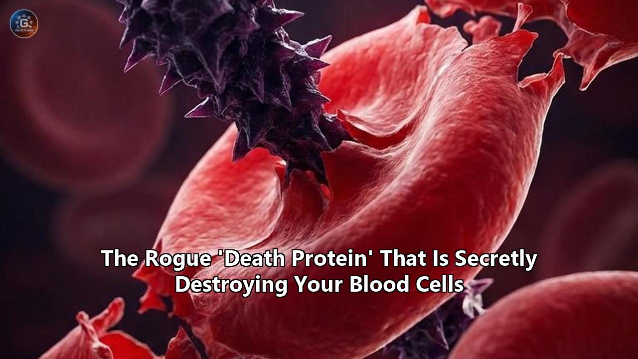
On April 6, 2026, researchers from the University of Tokyo and St. Jude Children’s Research Hospital published findings in Nature Communications that completely dismantled decades of established cellular biology. The research team, led by Dr. Masayuki Yamashita, identified that a well-known molecular executioner—a protein called MLKL (mixed lineage kinase domain-like pseudokinase)—is secretly driving the aging process of the human immune system.
For years, MLKL was understood solely as a biological detonator. When a cell became infected or irreversibly damaged, MLKL would be activated to execute "necroptosis," a highly inflammatory form of programmed cell suicide where the cell membrane is violently ruptured. However, the new findings reveal a stealthy, non-lethal function. In hematopoietic stem cells (HSCs)—the master builders responsible for creating our entire blood and immune system—MLKL activates only transiently. Instead of destroying the cell, it hijacks and damages the mitochondria, sapping the cell's energy and triggering a premature aging cascade.
When researchers deactivated this protein in mice, the results were staggering. The stem cells maintained their youthful regenerative capacity, produced healthier immune cells, and exhibited significantly lower DNA damage, even when subjected to the intense biological stress of advanced age and simulated chemotherapy. As Dr. Yamashita observed, "We are the first to formally demonstrate in primary cells that there is a cell death–independent function of activated MLKL".
This discovery provides a rare, tangible mechanism for why our immune systems degrade as we get older, shifting the focus of anti-aging research toward organelle-level protection. By analyzing this specific news event as a case study, we can extract vital biological principles about how our bodies age, how cellular stress responses are repurposed by evolution, and what the future of regenerative pharmacology will look like.
The Crime Scene: The Slow Collapse of the Hematopoietic System
To understand the magnitude of this discovery, we must first examine the environment where the crime takes place: the hematopoietic system.
Deep within the marrow of our bones lies a finite reservoir of hematopoietic stem cells. These remarkable entities hold a dual mandate. First, they must self-renew, creating exact copies of themselves to ensure the stem cell pool never runs dry. Second, they must differentiate, branching out into the staggering variety of specialized cells that make up our blood—oxygen-carrying red blood cells, clotting platelets, and the diverse armies of white blood cells that form the immune system.
Under optimal conditions in a young, healthy individual, HSCs maintain a pristine balance. They divide quietly and efficiently, churning out a balanced ratio of myeloid cells (such as macrophages and neutrophils, which provide immediate, non-specific immune defense) and lymphoid cells (such as T-cells and B-cells, which provide targeted, long-term adaptive immunity).
As the years pass, this factory begins to fail. The HSCs lose their ability to self-renew efficiently. More disastrously, they undergo a phenomenon known as "myeloid skewing". The stem cells stubbornly refuse to produce adequate numbers of lymphoid cells, leaving the adaptive immune system weakened and highly vulnerable to novel viruses and mutated cancer cells. Instead, they overproduce myeloid cells, which flood the body with low-grade, chronic inflammation. This persistent state of systemic inflammation—often termed "inflammaging"—is heavily implicated in the onset of cardiovascular disease, neurodegeneration, and frailty.
Until April 2026, the exact molecular trigger for this stem cell exhaustion remained obscured. Researchers largely looked to genetic mutations or epigenetic alterations—the gradual degradation of the cell's "software". But the University of Tokyo and St. Jude researchers looked elsewhere. By examining the hardware of the cell—specifically the mitochondria and the proteins that interact with them—they uncovered a highly localized sabotage mission. When analyzing the effects of the death protein, blood cells provide a perfect biological canvas to witness how stress accelerates physical decay.
The Modus Operandi: Sabotage Over Murder
The breakthrough materialized through an unexpected observation during a high-stress biological experiment. Dr. Yamashita and his team were treating genetically modified mice—specifically, mice bred without the MLKL protein (MLKL-knockout mice)—with 5-fluorouracil, a harsh chemotherapy drug known to induce massive cellular stress.
"We discovered an unexpected phenotype in HSCs of MLKL-knockout mice repeatedly treated with 5-fluorouracil, where aging-associated functional changes were markedly attenuated despite no detectable difference in HSC death, prompting us to investigate whether this pathway might induce functional changes beyond cell death," Dr. Yamashita explained.
If MLKL’s only job was to kill cells, then knocking it out should simply result in fewer dead cells. But that is not what happened. The stem cells in both the normal mice and the knockout mice survived the chemotherapy at identical rates. Yet, the cells without MLKL emerged functionally younger, highly energetic, and free from the hallmark traits of myeloid skewing.
This led the researchers to a startling realization: MLKL was not acting as an executioner in these stem cells; it was acting as a vandal.
When a hematopoietic stem cell experiences stress—whether from the toxic assault of chemotherapy, the radiation of a bone marrow transplant, or the slow accumulation of inflammatory signals during natural aging—the MLKL pathway is triggered. In most somatic cells, this would prompt MLKL proteins to oligomerize, form a deadly pore in the outer cell membrane, and cause the cell to burst.
In HSCs, however, the activation of MLKL is fleeting and localized. The protein travels directly to the mitochondria, the energy-producing organelles of the cell. There, it inflicts direct physical damage, reducing the mitochondrial membrane potential, warping their physical structure, and severely impairing their ability to produce energy through glycolysis.
The cell survives the encounter, but it is fundamentally crippled. It is the biological equivalent of a burglar breaking into a factory, ignoring the self-destruct button, and instead smashing the generators. The factory stays standing, but it can no longer produce high-quality goods. To fully understand the impact of this death protein, blood cells must be viewed not just as static entities, but as highly active metabolic engines that lose their efficiency the moment their power source is compromised.
Principle I: The Evolutionary Repurposing of Cellular Pathways
Looking at this specific event as a case study allows us to extract a broader biological principle: the profound pleiotropy of evolutionary design. Pleiotropy occurs when a single gene or protein influences multiple, seemingly unrelated phenotypic traits.
Biology is highly conservative. Evolution rarely invents an entirely new molecular machine when it can simply rewire an existing one. For decades, the scientific community categorized proteins into strict, functional silos. A protein was either a structural building block, a metabolic enzyme, a signaling messenger, or a death effector. MLKL, alongside its upstream regulator RIPK3 (receptor-interacting protein kinase 3), was firmly placed in the "death effector" silo.
This study shatters that rigid categorization. It forces a re-evaluation of how life and death signaling intertwine. The transient, non-lethal activation of a death pathway reveals a highly sophisticated level of cellular triage. When an HSC encounters stress, it must make a calculation. If the damage is catastrophic, the cell must die to protect the organism. But if the damage is moderate, the cell uses the very same death machinery to scale down its own operations.
By damaging the mitochondria via MLKL, the stem cell effectively enforces a metabolic lockdown. It stops heavily dividing. It shifts its output toward myeloid cells, which are cheaper to produce and serve as the body's immediate first responders to the perceived stress. In the short term—say, surviving an acute bacterial infection or a temporary period of starvation—this triage mechanism is highly beneficial. It keeps the organism alive.
The pathology only arises because humans are living long enough for this short-term survival mechanism to become a long-term liability. Chronic, low-grade stress continuously activates MLKL. The temporary metabolic lockdown becomes permanent stem cell exhaustion. This teaches us a critical lesson for future pharmacological research: we cannot assume that a protein has only one function simply because its most dramatic function is the easiest to observe in a petri dish.
Principle II: The Hardware Theory of Cellular Aging
The second major principle extracted from the April 2026 discovery is a shift in how we conceptualize the mechanics of aging.
Much of modern gerontology and anti-aging research has been dominated by genomics and transcriptomics—the study of our DNA and the RNA that translates it. The prevailing theory has been that aging is primarily a software problem. As we age, mutations accumulate in our genetic code, epigenetic markers drift, and the instructions for building a healthy cell become garbled.
While genetic degradation is undeniably a factor in aging, the MLKL case study highlights the equal, if not superior, importance of cellular "hardware"—specifically, the proteome and the organelles.
Dr. Yamashita’s team noted that the damage caused by MLKL was not initially linked to gene expression or epigenetic changes. It was a purely physical, post-transcriptional event. The protein physically translocated to the mitochondria and physically disrupted their membranes. This localized organelle damage subsequently forced the cell to alter its behavior. The software (the DNA) was still intact and capable of giving the correct instructions, but the hardware (the mitochondria) lacked the energy to execute them.
This creates a vicious feedback loop. Mitochondria are not just power plants; they are the central hubs for managing oxidative stress. When MLKL damages the mitochondria, they begin to leak reactive oxygen species (ROS)—highly volatile molecules that act like biological shrapnel. This ROS leakage physically damages the cell's DNA, eventually causing the very genetic mutations that traditional aging theories focus on.
Therefore, by neutralizing the death protein, blood cells can maintain their mitochondrial integrity, thereby preventing the secondary software damage from ever occurring. "Our results show that it can alter biological processes in response to stress, which we directly connected to age-related mitochondrial dysfunction in blood stem cells, suggesting the potential for interventions," Dr. Yamashita stated. This pivots the therapeutic target from trying to rewrite the cell's aging DNA—a monumentally complex task—to simply protecting the cell's power grid from internal sabotage.
Principle III: The Cycle of "Inflammaging"
The third principle this case study illuminates is the self-sustaining nature of age-related inflammation.
One of the great mysteries of aging is why the elderly suffer from chronic, systemic inflammation even in the absence of an active infection. The MLKL discovery provides a missing puzzle piece, demonstrating a closed-loop cycle of degradation.
The cycle begins with external stress—environmental toxins, mild infections, or simply the metabolic byproducts of living. This stress acts upon the hematopoietic stem cells, triggering the transient activation of MLKL. MLKL damages the mitochondria, which impairs the stem cell's ability to produce specialized lymphoid cells.
Because lymphoid cell production drops, the stem cell compensates by mass-producing myeloid cells. Myeloid cells, particularly macrophages, are the body's inflammatory foot soldiers. They patrol the tissues and secrete cytokines, which are chemical alarms that trigger inflammation.
Here is the trap: cytokines are themselves a major source of cellular stress. As the overproduced myeloid cells flood the bloodstream with inflammatory cytokines, those cytokines circulate back to the bone marrow, where they wash over the hematopoietic stem cells. This new wave of inflammatory stress triggers further activation of MLKL, which causes more mitochondrial damage, which causes even more myeloid skewing, which creates even more myeloid cells, which secrete even more cytokines.
The system becomes locked in a death spiral of its own making. The immune system is essentially attacking its own supply lines. By identifying MLKL as the central node where stress translates into mitochondrial damage, researchers have found a place to insert a biological circuit breaker. Interrupting the function of this death protein, blood cells can break the feedback loop, stopping the runaway train of systemic inflammation before it leads to downstream catastrophes like atherosclerosis or Alzheimer's disease.
The Therapeutic Horizon: Necroptosis-Modulating Drugs
The most immediate and profound implications of this case study lie in the realm of clinical medicine. Understanding a biological mechanism is only the first step; the ultimate goal is human intervention.
The University of Tokyo and St. Jude study explicitly opens the door for a new category of pharmaceuticals: necroptosis-modulating drugs. Because the researchers proved that MLKL-driven aging is a responsive, protein-level event rather than a permanent genetic mutation, it is theoretically reversible—or at least preventable—using targeted inhibitors.
Protecting Chemotherapy and Transplant Patients
The first frontier for these therapies will not be generalized anti-aging pills for the public, but acute interventions for highly vulnerable medical patients.
When a patient undergoes aggressive chemotherapy for cancer, the drugs do not just kill the tumor; they devastate the bone marrow. The intense chemical stress triggers massive MLKL activation in the surviving hematopoietic stem cells. Even if the patient survives the cancer, their immune system is frequently aged by decades. They suffer from chronic fatigue, severe immunodeficiency, and a highly skewed myeloid-to-lymphoid ratio that leaves them susceptible to secondary infections and new cancers.
A similar crisis occurs during bone marrow transplants. The transplanted stem cells are subjected to the massive stress of being harvested, processed, and infused into a new, heavily irradiated host body. Many of these stem cells fail to engraft properly or rapidly exhaust themselves due to stress-induced aging.
"In the longer term, this research could lead to therapies that preserve the function of hematopoietic stem cells, ultimately improving recovery and long-term health for patients undergoing chemotherapy, radiation, or transplantation," noted Dr. Yamashita.
If a patient could be administered a temporary MLKL-inhibitor during their chemotherapy regimen or transplant procedure, doctors could chemically shield the stem cells' mitochondria from the stress of the treatment. "Our study suggests that investigating ways to transiently inhibit necroptosis during stressful events may be a strategy to retain those abilities and prevent premature blood stem cell aging and related diseases," the researchers confirmed. The stem cells would weather the chemical storm without undergoing metabolic lockdown, emerging on the other side fully capable of rebuilding a healthy, youthful immune system.
The Nuances of Pharmacological Inhibition
Developing an MLKL inhibitor is not without significant challenges. We must recall the evolutionary origin of the necroptosis pathway: it exists to kill severely damaged or virally infected cells. It is a fail-safe against cancer and uncontrollable pathogens.
If a systemic drug completely and permanently blocks MLKL throughout the entire human body, it might successfully prevent stem cell aging, but it could simultaneously disable the body's ability to destroy pre-cancerous cells. The organism would trade a slowed aging process for a drastically increased risk of malignant tumors.
Therefore, the pharmacology of the future must be heavily nuanced. Drug developers will need to design targeted delivery systems—such as lipid nanoparticles or antibody-drug conjugates—that specifically deliver the MLKL inhibitor only to the hematopoietic stem cells in the bone marrow, leaving the necroptosis pathways in the rest of the body's tissues fully operational.
Alternatively, researchers must focus on the precise mechanism of the transient activation. Why does MLKL form a deadly membrane-bursting pore in normal cells, but only inflict mild mitochondrial damage in stem cells? If scientists can identify the specific co-factors or post-translational modifications that guide MLKL to the mitochondria in HSCs, they can design drugs that block only the non-lethal, aging-inducing function while leaving the lethal, cancer-fighting function intact.
"By revealing how non-lethal activation of cell-death pathways drives stem cell aging, these findings may inspire new classes of mitochondrial-protective or necroptosis-modulating drugs," Dr. Yamashita suggested, pointing directly toward this highly specialized pharmacological future.
Transforming the Case Study into a Blueprint
The April 2026 revelation regarding MLKL is more than just a single scientific achievement; it serves as a blueprint for how we must approach the study of cellular decay moving forward.
For too long, the medical community has viewed aging as an inevitable, generalized wear-and-tear process—a macroscopic degradation that cannot be halted any more than one can stop a car's engine from eventually rusting. This case study forcefully rejects that notion. It proves that aging, at least within the hematopoietic system, is not just passive decay. It is an active, regulated, and highly specific molecular program driven by identifiable proteins acting upon identifiable organelles.
The fact that the very machinery designed to protect us from acute threats (cellular suicide to prevent infection) is the exact machinery driving our chronic decline is a poetic and cruel biological irony. Yet, it is an irony we can now decipher.
As we look toward the immediate future, the race is on. Laboratories worldwide will now pivot to examine other known "death pathways"—such as apoptosis, pyroptosis, and ferroptosis—to see if their executing proteins are moonlighting in similar, non-lethal capacities across different types of stem cells, such as those in the brain, the skin, or the gut.
Furthermore, human clinical trials assessing MLKL biomarkers are the logical next step. If doctors can measure the level of MLKL activation in a patient's blood stem cells, they may possess a highly accurate, predictive "biological clock" capable of determining a patient's true immune age, allowing for early interventions long before the clinical symptoms of frailty appear.
The discovery that a rogue protein is secretly dismantling our blood cells from the inside is a formidable reminder of how much of our own biology remains uncharted. But by dragging this molecular vandal out of the shadows and into the light, science has secured a crucial upper hand in the fight to preserve human health and vitality into advanced age.
Reference:
- https://neurosciencenews.com/mlkl-protein-stem-cell-aging-30539/
- https://www.stjude.org/research/progress/2026/death-defying-mechanism-drives-premature-aging-in-blood-stem-cells-after-transplantation.html
- https://www.sciencedaily.com/releases/2026/04/260416071951.htm
- https://www.wehi.edu.au/news/freeze-executioner-protein-caught-act/
- https://www.miragenews.com/death-protein-could-slow-aging-at-its-source-1657262/
- https://www.sciencedaily.com/releases/2016/07/160714110918.htm