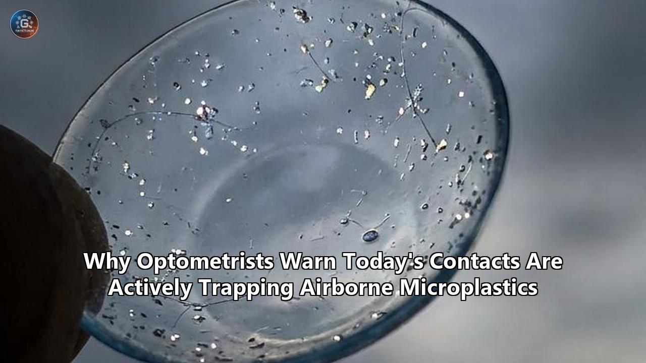
An estimated 90,698 microscopic plastic particles. That is the volume of synthetic debris a single pair of silicone hydrogel contact lenses can shed and trap against the human cornea over a standard year of 10-hour daily wear. While the optical industry has long relied on the safety profile of hydrogel polymers, new clinical data presented throughout 2025 has triggered explicit warnings from optometrists and environmental toxicologists. Modern lenses are not merely degrading under ultraviolet light; they are operating as active, localized electrostatic traps for airborne microplastics, pinning environmental pollutants directly against the vulnerable tissues of the eye.
The intersection of environmental pollutants and contact lenses microplastics retention has forced a rapid reassessment of ocular health protocols. At recent optometry conferences, including the 2025 Optometry's Meeting in Minneapolis, researchers detailed how microplastic toxicity in the eye mimics severe dry eye disease. More critically, a 2025 study published in the Journal of Hazardous Materials definitively confirmed the presence of microplastics—predominantly polyethylene (PE)—in the tear fluid and meibum of human patients. The data demonstrates a statistically significant correlation between the volume of these trapped microparticles and the measurable clinical degradation of the tear film.
With over 45 million Americans and 140 million people globally relying on contact lenses for visual correction, the scale of this synthetic exposure is unprecedented. The localized accumulation of microscopic plastics on the ocular surface is currently driving a measurable spike in conjunctival inflammation, goblet cell depletion, and structural damage to the corneal epithelium.
The Physics of the Trap Mechanism
To understand why optometrists are issuing formal warnings regarding airborne microplastics, one must examine the specific fluid dynamics and mechanical physics of the human eye. Under normal physiological conditions, a healthy tear film surrounds the eye, providing lubrication and flushing out microscopic debris. The human eyelid blinks approximately 15 to 20 times per minute, operating as a biological windshield wiper that sweeps particulate matter into the tear ducts for safe drainage.
The introduction of a silicone hydrogel or polymethyl methacrylate (PMMA) contact lens fundamentally disrupts this baseline hydrodynamic clearance. Modern soft lenses maintain a water content ranging from 33% to 58%, utilizing porous matrices to allow oxygen permeability. However, these same material properties create an ideal localized trap for airborne particulates.
Airborne microplastics, defined as plastic fragments smaller than 5 millimeters, fall at a rate of roughly 3,000 to 10,000 particles per square meter per day in typical urban indoor environments. When these particles—which include transparent fibers, irregular fragments, and microscopic resin pellets—land on the ocular surface, the contact lens blocks the mechanical flushing action of the eyelid. Instead of being swept away by the tear film, the microplastics become lodged at the interface between the lens and the corneal epithelium.
Quantifying the exact volume of contact lenses microplastics exposure requires analyzing both the shedding rate of the polymer itself and the ambient environmental load. The localized retention creates a high-friction environment. Every blink grinds the trapped polyethylene and polypropylene fragments against the cornea. Furthermore, the electrostatic charge inherent in many synthetic lens materials actively attracts smaller nanoplastics (particles smaller than 1 micrometer) directly out of the immediate airspace, compounding the accumulation over a standard 10-to-14-hour wear cycle.
Quantifying Ocular Surface Damage: The Biological Metrics
The physical trapping of microplastics yields immediate, measurable biological consequences. The 2025 Journal of Hazardous Materials study evaluated 45 patients diagnosed with dry eye disease (DED), mapping the precise presence of microplastics in their tear fluid and meibomian glands. The researchers categorized the identified microplastics into five distinct typologies, isolating polyethylene (PE) as the dominant contaminant.
The clinical metrics extracted from this cohort provide a stark quantitative picture of the damage. PE levels in the tear fluid exhibited significant negative correlations with two foundational optometric measurements:
- Schirmer I Test Scores: This test measures basal and reflex tear production using sterile filter paper strips placed in the lower eyelid. Patients with higher concentrations of trapped microplastics demonstrated a marked reduction in millimeter tear production over the standard 5-minute testing window.
- Fluorescein Tear Film Break-Up Time (FBUT): FBUT measures the time it takes for the first dry spot to appear on the cornea after a complete blink. A normal FBUT is over 10 seconds. In the presence of high microplastic loads, the tear film destabilizes rapidly, dropping the FBUT into pathological ranges, leaving the cornea exposed to the atmosphere.
In-vitro screening conducted alongside the human trials isolated human corneal epithelial cells and conjunctival epithelial cells, exposing them to precise, dose-dependent concentrations of polyethylene. The results demonstrated that PE exposure actively reduced cellular viability and induced apoptosis (programmed cell death). The plastic fragments are not merely inert objects causing mechanical friction; they are stressing the cellular architecture to the point of systemic failure.
Further data from murine (mouse) models validated these findings. Researchers applied topical PE drops designed to imitate the exact concentration of airborne PE exposure. The outcome was a statistically significant reduction in the density of conjunctival goblet cells. Goblet cells are highly specialized epithelial cells responsible for secreting mucins—large glycoproteins that form the foundational mucous layer of the tear film. When microplastics trigger the depletion of goblet cells, the eye loses its ability to anchor tears to the corneal surface, leading to chronic, unresolvable inflammation and a clinical presentation that perfectly mimics severe dry eye disease.
The 90-Day Photolytic Degradation Threshold
Airborne exposure is only one half of the equation. The contact lenses themselves actively generate microplastics under specific environmental stressors. A comprehensive study published in the American Chemical Society’s Environmental Science & Technology journal utilized high-content screening to evaluate the structural integrity of contact lenses under varying light conditions. Researchers from Hohai University and Nanjing University collected six different brands of contact lenses with lifespans ranging from one day to one month, subjecting them to rigorous testing.
Using scanning electron microscopy and Fourier transform infrared spectroscopy fingerprint analysis, the research team tracked the exact volume of polymer shedding. In environments devoid of sunlight, no significant microplastic release was detected in the surrounding fluid. However, when the lenses were exposed to a 90-day equivalent of solar irradiation, the structural matrix of the polymers began to fail.
The data revealed a concerning degradation curve. The lenses subjected to 900 hours of irradiation displayed the highest carbonyl indexes, a definitive chemical marker indicating severe polymer aging and oxidative breakdown. Counter to previous hypotheses suggesting that plastic degradation merely creates smaller and smaller fragments, this study found that longer irradiation times resulted in the release of more microplastics with a larger average particle size.
By extrapolating this shedding rate, the study authors modeled that a single pair of contact lenses, worn for 10 hours a day over the course of a year, could release over 90,000 microplastic particles directly onto the ocular surface. For patients utilizing monthly or yearly replacement lenses, the cumulative exposure to ultraviolet light actively weaponizes the lens, transforming it from a protective medical device into a localized source of microplastic pollution.
Secondary Vectors: The Mathematics of Contaminated Eye Drops
As contact lens wearers experience the friction and dryness associated with microplastic accumulation and goblet cell depletion, the standard behavioral response is to apply artificial tears. However, recent toxicological screening indicates that this common remedy serves as a secondary, highly concentrated vector for microplastic exposure.
A 2024 analysis conducted by researchers in South Korea investigated the microplastic contamination levels inside commercially available artificial tear (AT) products. The methodology involved rigorous testing of five different hyaluronic acid-based AT brands, encompassing both multi-use bottles and single-use disposable vials.
The quantitative findings highlight a severe flaw in pharmaceutical packaging:
- Microplastics were detected in 4 out of the 5 artificial tear products.
- The initial drops squeezed from the bottles contained an average of 0.50 ± 0.65 microplastic particles per 30 milliliters.
- The morphological breakdown of these introduced particles showed that 95% were transparent, 55% were irregular fragments, and 35% were sized between 10 and 20 micrometers.
- Chemical analysis confirmed that 95% of the particles were composed of polyethylene (PE).
The researchers modeled the annual exposure rates based on standard patient usage. If a contact lens wearer applies the first drops from a contaminated AT bottle four times a day for a full year, they introduce an additional 730.0 microplastic particles directly onto the surface of their lens. Because the lens blocks the natural hydrodynamic flushing of the tear film, these 730 particles are effectively trapped against the cornea, combining with the 90,000 particles shed by the lens itself and the ambient airborne load.
This secondary contamination vector presents severe implications when viewed alongside historical ocular health crises. In 2023, the Centers for Disease Control and Prevention (CDC) issued severe warnings regarding EzriCare artificial tears, linking the over-the-counter drops to drug-resistant Pseudomonas aeruginosa bacterial infections that resulted in vision loss and death. When microplastics trapped under a contact lens induce micro-abrasions and apoptosis in the corneal epithelium, they compromise the eye’s primary physical barrier. This weakened state drastically lowers the threshold required for opportunistic pathogens, such as Pseudomonas aeruginosa, to establish catastrophic corneal ulcers.
Deep Intraocular Migration: Bypassing the Blood-Eye Barrier
Perhaps the most alarming data emerging from the 2024 and 2025 research cycles is the evidence of microplastic migration deep into the internal architecture of the human eye. The ocular surface was previously assumed to act as an impenetrable biological shield against particulate matter of this size.
The physiological dimensions make this migration mathematically perplexing. The average pore size of the cornea's protective anterior basement membrane is highly restrictive. The conjunctival tissue, while possessing a larger surface area and higher porosity than the cornea, has an average pore size of approximately 3.0 nanometers. By contrast, the smallest microplastic defined in these studies—measuring 1 micrometer—is 3,000 times larger than the conjunctival pore. Based on pure spatial geometry, absorption through ocular tissue should be virtually impossible.
Yet, empirical evidence contradicts this assumption. A 2024 study published in Science of the Total Environment analyzed samples from 49 patients undergoing eye surgery. The researchers discovered more than 1,700 microplastic particles localized within the vitreous humor—the dense, jelly-like fluid that fills the central cavity of the eye between the lens and the retina. Subsequent research published in iScience in 2025 corroborated these findings by detecting microplastics in the aqueous humor, the thinner fluid occupying the front chamber of the eye.
Clinical models attempting to predict the long-term impact of contact lenses microplastics accumulation face a complex variable regarding how these particles bypass the blood-eye barrier. Current hypotheses presented by optometrists at the 2025 Optometry's Meeting suggest two primary pathways. First, the particles may enter systemically through ingestion or inhalation, traveling via the circulatory system before crossing compromised capillary beds in the eye. Second, the chronic inflammation and cellular apoptosis caused by trapped plastics on the corneal surface may physically degrade the epithelial junctions over time, allowing smaller nanoplastics to force their way into the anterior chamber.
The presence of synthetic polymers inside the vitreous and aqueous humors correlates directly with severe internal ocular pathologies. Optometric findings linked to deep intraocular microplastic toxicity include increased intraocular pressure, measurable aqueous humor opacities, and retinopathy. Lab and animal studies have consistently demonstrated that microplastics damage the light-sensing cells of the retina and weaken its protective barrier, directly impairing visual function.
Chemical Hitchhikers and Endocrine Disruption
The mechanical friction and intraocular blockage caused by microplastics represent only the physical dimension of the threat. The biochemical reality is that microplastics function as highly effective transport vectors for a host of toxic chemical compounds.
Plastics are manufactured using complex chemical formulas that include plasticizers, stabilizers, and endocrine-disrupting chemicals (EDCs) such as bisphenol-A (BPA) and various phthalates (commonly used in polyvinyl chloride products). When trapped against the highly vascularized conjunctiva by a contact lens, these particles slowly leach their chemical constituents directly into the ocular environment.
Furthermore, secondary microplastics are notorious in environmental science for their ability to "pick up" or adsorb external pollutants from the atmosphere. Airborne microplastics drifting through urban environments accumulate heavy metal molecules and per- and polyfluoroalkyl substances (PFAS). When a contact lens traps an airborne particle, it is not merely pinning a piece of polyethylene to the eye; it is creating a localized, sustained-release delivery system for PFAS and heavy metals.
The sustained exposure to these chemical hitchhikers induces oxidative stress within the ocular surface cells. The localized toxicity upregulates the expression of inflammatory markers in a time-dependent manner, forcing the biological system into a state of chronic defensive alert. Over months and years of continuous contact lens wear, this chemical onslaught exhausts the regenerative capacity of the corneal epithelium.
Evaluating Demographic Exposure and Industry Standards
Extrapolating these quantitative findings to a global demographic reveals an urgent public health metric. With 45 million active contact lens wearers in the United States alone, the aggregate volume of plastic shedding and airborne particulate trapping is immense.
If half of this demographic relies on monthly or bi-weekly replacement lenses, tens of millions of people are subjecting their eyes to the 90-day photolytic degradation threshold. The high-content screening data proves that as these lenses age under natural ultraviolet exposure, their carbonyl indexes spike, and the structural integrity of the polymer collapses, shedding tens of thousands of microscopic plastic fragments directly onto the human cornea.
The optometric and manufacturing industries are slowly beginning to internalize this data, recognizing that the standards governing medical polymers must evolve. Frameworks such as the Kering Standards, heavily utilized in the luxury eyewear sector to mitigate exposure to airborne microplastics and organic solvents during the manufacturing of acetate optical frames, provide a baseline for industrial responsibility. However, the production of silicone hydrogel contact lenses remains largely insulated from environmental toxicity tracking once the product leaves the sterile manufacturing floor.
Currently, the regulatory apparatus lacks the specific mandates required to address microplastic shedding. While primary microplastics (such as manufactured microbeads in cosmetics) have faced heavy legislative restrictions, secondary microplastics generated by the degradation of larger products remain unregulated. Until pharmaceutical and optometric manufacturing standards adapt to require strict photolytic degradation testing and biodegradable alternatives, the burden of mitigation falls entirely on the prescribing clinician and the individual patient.
Quantitative Mitigation Protocols for Optometrists and Patients
Reducing the threat of contact lenses microplastics toxicity relies on strict adherence to modified clinical guidelines. Optometrists are now deploying data-driven strategies to physically limit the volume of microplastics introduced to the ocular surface, focusing on both the environmental load and the medical devices themselves.
The primary intervention involves altering lens replacement schedules. To avoid the exponential spike in particle shedding observed in lenses exposed to prolonged solar irradiation, optometrists increasingly recommend a strict transition to daily disposable lenses. While daily disposables still act as traps for ambient airborne microplastics during their hours of use, disposing of the lens every 24 hours entirely circumvents the UV-induced polymer degradation documented by the Nanjing University data.
Mitigating the airborne trap effect requires modifying the patient's immediate environment. Since humans spend approximately 90% of their time indoors, reducing the concentration of suspended particulate matter in residential and commercial spaces yields a direct, proportional decrease in the number of particles trapped against the cornea. Implementing high-efficiency particulate air (HEPA) filters, specifically those carrying a Minimum Efficiency Reporting Value (MERV) rating of 13 or higher, captures particles as small as 0.3 micrometers. Running these filtration systems in high-traffic indoor environments effectively removes the transparent fibers and irregular fragments before they can settle onto the ocular surface. In high-dust environments or days with elevated PM2.5 outdoor pollution levels, standard protective eyewear or traditional prescription glasses must replace contact lenses to remove the physical trap entirely.
Finally, the administration of artificial tears requires an immediate protocol update based on the South Korean toxicological screening. Because 80% of tested artificial tear products contained microplastics in their initial drops, the exposure math dictates a change in application technique. The researchers established that simply discarding the first two drops from the vial before applying the solution to the eye reduces the annual microplastic exposure load from 730.0 particles down to 204.4 particles. This single, measurable behavioral adjustment eliminates nearly 72% of the direct microplastic contamination sourced from the eye drops.
Future Milestones and Unresolved Variables
The optometric community is bracing for the long-term clinical manifestations of this continuous synthetic exposure. The next critical milestone in microplastic research will be the development of standardized, automated diagnostic tools capable of measuring intraocular plastic loads directly within the clinic. Current methodologies, such as Raman spectroscopy and scanning electron microscopy, are restricted to high-level research laboratories and post-operative tissue analysis. Developing a non-invasive optical coherence tomography (OCT) setting or tear-film assay that can quantify polyethylene concentrations during an annual eye exam would provide clinicians with the real-time data required to intervene before irreversible goblet cell apoptosis occurs.
Simultaneously, materials science engineers are actively attempting to design biodegradable alternatives to conventional silicone hydrogels. The objective is to formulate a lens matrix that maintains the requisite 50% water content and oxygen permeability without utilizing complex synthetic polymers that shed endocrine-disrupting chemicals upon photolytic breakdown.
The unresolved question hanging over the 2025 and 2026 data sets is the precise transport mechanism utilized by these 10-to-20-micrometer particles to bypass the 3.0-nanometer conjunctival barrier and enter the deep vitreous fluid. Until researchers map the exact biological pathway facilitating this internal migration, the threshold for permanent retinal damage remains dangerously undefined. Optometrists, armed with clear statistical evidence of surface degradation and tear film disruption, must operate under the assumption that every trapped particle represents a compounding physiological risk. The data is definitive: the modern contact lens is capturing the fallout of a synthetic environment, and the human cornea is bearing the structural cost.
Reference:
- https://glance.eyesoneyecare.com/stories/2023-07-17/contact-lenses-may-shed-microplastics-research-finds/
- https://www.optometrytimes.com/view/aoa-2025-microplastic-toxicity-and-its-impact-on-the-eye
- https://www.allaboutvision.com/conditions/related/microplastics-and-your-eyes/
- https://pubmed.ncbi.nlm.nih.gov/40015041/
- https://revisionoptometry.com/blog/are-microplastics-from-contact-lenses-a-health-concern/
- https://pubs.acs.org/doi/10.1021/acs.est.3c01601
- https://www.researchgate.net/publication/385748549_Microplastic_contamination_in_artificial_tears_in_South_Korea_Potential_for_direct_ocular_exposure
- https://iacle.org/wp-content/uploads/2025/04/Microplastic-contamination-in-artificial-tears-in-South-Korea.pdf
- https://scitechdaily.com/cdc-warning-the-eye-drop-danger-lurking-in-your-medicine-cabinet/
- https://pmc.ncbi.nlm.nih.gov/articles/PMC9962686/
- https://www.kering.com/api/download-file/?path=KERING_STANDARDS_V8.pdf