A medical team at University of California, San Francisco (UCSF) published findings in Science on August 6, 2026, revealing why one of the world's most dangerous fungal superbugs can live silently on human skin for months or years without being detected or eradicated.
The pathogen, Candida auris, has infected thousands of hospitalized patients globally, causing severe bloodstream infections with mortality rates approaching 40% to 60%. What has long baffled infectious disease specialists is why current decolonization protocols—such as daily chlorhexidine body washes and aggressive topical treatments—consistently fail to clear the fungus from patient skin.
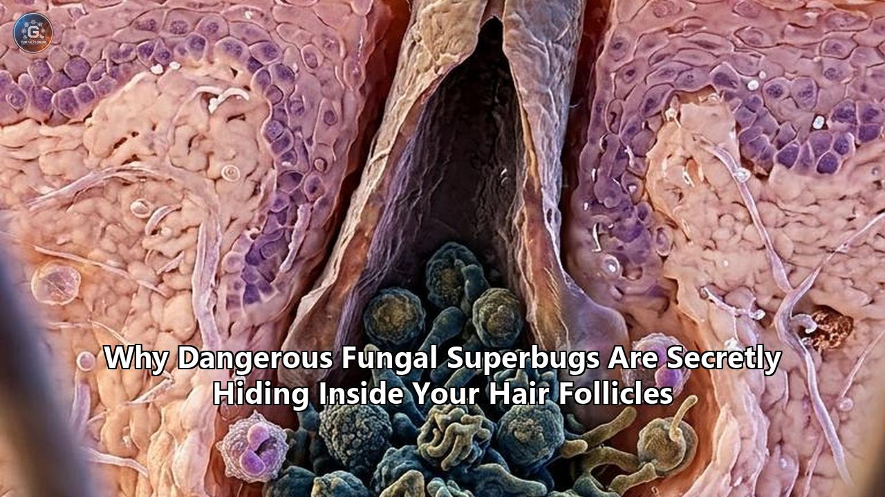
The UCSF study provided the answer: Candida auris does not merely sit on the surface of the skin. It actively burrows into the deep micro-architecture of hair follicles, where it tricks the host’s immune system into turning off its own natural antifungal defenses.
By analyzing how C. auris hijacks hair follicle biology, researchers uncovered a broader principle of fungal pathogenesis. Hair follicles function as anatomical and immunological sanctuary sites for drug-resistant pathogens. From C. auris in hospital intensive care units to the rapid global spread of multidrug-resistant dermatophytes like Trichophyton indotineae, pathogens are hiding inside hair follicles to escape both topical antimicrobials and host immune surveillance.
The UCSF discovery serves as a lens to understand this growing public health crisis, exposing how hair follicle anatomy, fungal cellular manipulation, and modern medical missteps interact to create persistent reservoirs of antifungal-resistant superbugs.
┌─────────────────────────────────────────────────────────────────────────┐
│                     THE HAIR FOLLICLE SANCTUARY                         │
│                                                                         │
│   SKIN SURFACE (Epidermis) ───────────────────────────────────────────  │
│   • Topical washes (chlorhexidine) clear surface cells                 │
│   • Antifungal creams fail to penetrate deeper tissue layers           │
│                                                                         │
│   FOLLICULAR INFUNDIBULUM & ISTHMUS (2-4 mm deep)                       │
│   • Pathogen remodels cell wall, exposing Chitin                        │
│   • Triggers viral-like Immune Response (Interferon-gamma release)      │
│   • Suppresses protective IL-17 antifungal signaling                    │
│   • Stalls epithelial cell turnover; accumulates damaged cells          │
│                                                                         │
│   PROTECTED FUNGAL RESERVOIR                                            │
│   • Shielded from topical baths and sub-inhibitory systemic drugs       │
│   • Re-colonizes skin surface once topical treatment ends              │
└─────────────────────────────────────────────────────────────────────────┘
The UCSF Discovery: How Candida auris Rewires Skin Immunity
The study, led by first author Dr. Dean Merrill, a dermatologist and professor at UCSF, alongside co-senior authors Dr. Ari Molofsky and Dr. Suzanne Noble, set out to solve a persistent clinical dilemma.
When a patient tests positive for Candida auris, hospitals attempt to decontaminate their skin to prevent the yeast from entering open wounds or central venous catheters. Yet, despite repeated antiseptic bathing, patients remain colonized for extended periods.
To understand this persistence, the UCSF team conducted comparative murine and tissue-culture studies contrasting Candida auris with Candida albicans, a far more common yeast species. When C. albicans contacts human or rodent skin, the host immune system recognizes the organism and launches a protective response mediated by interleukin-17 (IL-17).
IL-17 acts as an alarm signal: it triggers rapid turnover and shedding of epidermal cells, recruits neutrophils, and stimulates the production of potent antimicrobial peptides. Within days, the skin effectively scrubs itself clean of C. albicans.
Candida auris operates under a completely different cellular strategy. When it lands on the skin, it retreats down the hair shaft into the follicular infundibulum and isthmus—the upper and middle regions of the hair follicle structure. Once settled inside, C. auris remodels its exterior cell wall, exposing high concentrations of a structural carbohydrate called chitin.Rather than alerting the immune system to deploy IL-17, chitin tricks localized skin-resident immune cells into releasing interferon-gamma (IFN-γ)—an immune signal normally produced to battle intracellular viral infections.
                     Host Immune Response Divergence

   Candida albicans                           Candida auris
          │                                         │
   Triggers IL-17                            Exposes Chitin
          │                                         │
   Activates skin cell turnover               Triggers IFN-γ (Viral signal)
   & antimicrobial peptides                         │
          │                                 Suppresses IL-17 & slows
   Fungus cleared in days                   follicle cell shedding
                                                    │
                                            Persistent Follicular Nest
This biochemical misdirection has two catastrophic consequences for the host:
- Suppression of Antifungal Defense: High levels of localized IFN-γ actively suppress the skin’s IL-17 pathway, shutting down the primary defense mechanism that would otherwise eliminate the yeast.
- Inhibition of Cell Shedding: The elevated IFN-γ signal disrupts normal hair follicle stem cell kinetics, slowing the replacement and shedding of follicular epithelial cells. Worn-out, damaged cells linger in the hair follicle instead of cycling out, creating a stagnant, nutrient-rich environment that functions as a sanctuary for C. auris.
“Candida auris colonizes skin way better than most other fungi, setting it up to invade once the immune system is weakened,” explained Dr. Merrill. “The big clinical problem is that we have no effective way to remove it from the skin.”
Dr. Molofsky emphasized the broader clinical danger: “For most people, Candida auris hangs out on the skin and doesn't cause any problems. It's only when it reaches deeper tissues in medically vulnerable patients that it becomes dangerous. Understanding how it survives on the skin may help explain how it eventually causes serious infections.”
By transforming the hair follicle into a sheltered nest, C. auris secures a permanent foothold. Even if antiseptic washes sterilize the smooth epidermal surface, fungal cells tucked deep inside the hair follicle survive unharmed. As soon as the antiseptic regimen pauses, the surviving yeast cells migrate up the hair follicle lumen and re-colonize the entire body surface.
Anatomy of a Sanctuary: Why Hair Follicles Protect Superbugs
The UCSF discovery highlights a vulnerability in human anatomy: the structure of the pilosebaceous unit. Understanding why hair follicles serve as an ideal microbial hideout requires examining their physical, chemical, and immunological features.
                   CROSS-SECTION OF PILOSEBACEOUS UNIT

     Skin Surface  ──────────────────────────────────────
                   │  │                                 │
                   │  │ Hair Shaft                      │
                   │  │                                 │
     Infundibulum  │ ┌┴┐  ◄── Sebum & Dead Keratin      │
                   │ │ │      (Nutrient-Rich Environment)│
                   │ │ │                                │
                   │ │*│  ◄── * Fungal Superbug Nest    │
     Isthmus       │ │*│      (Shielded from Topicals)  │
                   │ └┬┘                                │
                   │  │                                 │
     Dermis        │  │  Sebaceous                      │
     (2-4 mm deep) │ ┌┴┐ Gland                          │
                   │ └┬┘                                │
                   │  │                                 │
                   └──┴─────────────────────────────────
Depth and Physical Barrier Shielding
The human skin surface (the stratum corneum) is a tight barrier of keratinized, flattened cells designed to keep environmental pathogens out. However, every hair shaft originates from an invagination that extends 2 to 4 millimeters deep into the dermis.
Most topical antiseptic soaps, over-the-counter creams, and body washes operate entirely on the outer 10 to 20 micrometers of the skin surface. They lack the surface tension properties, chemical lipophilicity, or mechanical reach required to penetrate deep into the narrow follicular canal. As a result, standard hygiene measures pass right over these hidden reservoirs.
Lipid-Rich Nutrient Pools
Hair follicles are linked to sebaceous glands, which continuously secrete sebum—a complex mix of triglycerides, wax esters, squalene, and free fatty acids. While sebum contains certain natural antimicrobial lipids, many opportunistic fungi possess lipophilic metabolic enzymes (such as lipases and esterases) that allow them to process these fats for energy.
The follicular canal becomes a warm, humid micro-incubator packed with dead keratin and lipids, providing all the nutrients a fungal colony needs to grow.
Localized Immune Privilege
To prevent destructive autoimmune attacks against regenerating hair shafts, hair follicles maintain a state of "immune privilege." The lower sections of the hair follicle express low levels of Major Histocompatibility Complex Class I (MHC-I) molecules and produce immunosuppressive signaling molecules, including Transforming Growth Factor-beta (TGF-β) and alpha-Melanocyte-Stimulating Hormone (α-MSH).
While this immune privilege protects hair growth, it creates a localized blind spot where host surveillance is inherently suppressed. When a fungus like C. auris actively manipulates host cytokines—turning on IFN-γ to suppress IL-17—it exploits this baseline suppression to establish long-term persistence.
These combined factors make the follicular unit an ideal sanctuary site. Fungi residing in hair follicles are shielded from wash-off mechanisms, protected from immune destruction, and supplied with a steady source of nutrients.
Beyond Yeast: The Trichophyton indotineae Epidemic
While UCSF's research focused on Candida auris, the clinical implications extend across the entire spectrum of medical mycology. The most widespread manifestation of this phenomenon involves dermatophyte fungi—the causative agents behind fungal infections in hair, scalp, skin, and nails.
Over the past decade, a global epidemic of severe, treatment-resistant dermatophytosis has spread across South Asia, Europe, and North America. The primary driver is Trichophyton indotineae (previously classified as Trichophyton mentagrophytes ITS genotype VIII), a dermatophyte species that displays unprecedented resistance to standard antifungal drugs.
┌─────────────────────────────────────────────────────────────────────────┐
│                  THE EMERGING DANGER OF T. INDOTINEAE                   │
│                                                                         │
│   ORIGIN & SPREAD                                                       │
│   • First identified in India; now confirmed in over 40 countries       │
│   • Spreads rapidly via direct contact and shared household items       │
│                                                                         │
│   CLINICAL PROFILE                                                      │
│   • Causes extensive, highly inflamed, itchy skin and hair lesions      │
│   • Invades deep into hair shafts and follicles (Tinea Capitis/Barbae)  │
│   • Can progress to nodular, deep dermal forms (Majocchi's Granuloma)   │
│                                                                         │
│   ANTIFUNGAL RESISTANCE                                                 │
│   • >70-90% resistant to Terbinafine (Squalene Epoxidase mutations)     │
│   • Emerging resistance to oral Triazoles (Itraconazole, Fluconazole)   │
└─────────────────────────────────────────────────────────────────────────┘
Unlike classic ringworm species that stay confined to the dead outer keratin layer of non-hairy skin, T. indotineae exhibits an affinity for hair follicles. When the fungus invades a hair follicle, it causes condition variants such as tinea capitis (scalp infection), tinea barbae (beard area infection), and Majocchi’s granuloma—a deep follicular infection where the fungal hyphae breach the follicular wall, spilling into the surrounding dermis and forming painful, pus-filled nodules.
The main problem with treating these fungal infections in hair is that T. indotineae has acquired mutations in the squalene epoxidase (SQLE) gene, rendering it resistant to oral terbinafine—the global first-line drug for ringworm for more than thirty years.
When patients present with extensive, inflamed rashes, primary care physicians often prescribe standard oral terbinafine or topical antifungal creams. While the drug may clear a fraction of the surface-level fungus, the resistant T. indotineae cells residing inside the hair follicles survive.
Inside the hair shaft lumen, drug concentrations of both topical and systemic antifungals often fall below the Minimum Inhibitory Concentration (MIC) needed to kill the organism. This exposes the deep fungal reservoir to sub-therapeutic drug doses—the exact conditions that drive further genetic mutation and multi-drug resistance.
A May 2024 report in JAMA Dermatology detailing 11 cases of T. indotineae in New York City showed that patients experienced diagnostic delays ranging from 3 to 42 months. During these delays, every single patient was treated with topical antifungals that failed completely as monotherapy.
The fungus survived by retreating into hair follicles and skin folds, spreading within households and resisting standard medical interventions.
The Misuse Engine: Steroids and Misdiagnosis Fuel Follicular Superbugs
The rise of hair follicle fungal superbugs is not purely an evolutionary accident. It is heavily accelerated by human clinical practices—specifically, the widespread misuse of over-the-counter topical corticosteroid-antifungal combination creams and routine diagnostic missteps.
In many parts of the world, combination creams containing strong topical steroids (such as clobetasol propionate) alongside broad-spectrum antifungals and antibacterials are sold over the counter. When patients apply these "triple creams" to an itchy, inflamed fungal rash, the steroid instantly quiets the host immune response, reducing redness and itching within hours.
This temporary relief conceals a dangerous underlying process:
                            THE STEROID MISUSE CASCADE

   Incipient Fungal Infection ──► Applied Topical Steroid ("Triple Cream")
                                                │
                                                ▼
   Impaired Local Immune Defenses ◄── Inflammation Suppressed (Temporary Relief)
                 │
                 ▼
   Fungus Invades Deeper into Hair Follicles & Dermis ("Tinea Incognito")
                 │
                 ▼
Widespread, Recalcitrant Superbug Infection (Majocchi's Granuloma)- Local Immune Paralysis: Corticosteroids shut down local immune cell recruitment and suppress the release of protective cytokines.
- Unchecked Follicular Invasion: Deprived of local immune surveillance, the fungus multiplies unchecked, penetrating past the upper infundibulum down into the deep hair follicle bulb and surrounding tissue.
- Induction of "Tinea Incognito": The steroid strips the rash of its classic ring-like shape, leaving behind a smooth, atypical plaque known as tinea incognito.
When clinicians encounter tinea incognito, they often mistake the lesion for eczema, contact dermatitis, or psoriasis and prescribe even stronger topical or systemic steroids. This further paralyses the skin’s immune system, allowing the superbug to establish a deeply entrenched follicular reservoir.
By the time a correct diagnosis is made via molecular testing or fungal culture, the pathogen is entrenched deep within thousands of pilosebaceous units across the patient's body. The hair follicle has effectively transformed from a simple organ into an encrypted vault, holding drug-resistant superbugs that are shielded from standard medical treatments.
Clinical Lessons from the Follicular Sanctuary
The UCSF Science study on Candida auris, alongside the global surge of T. indotineae, highlights critical lessons for medical mycology, infectious disease control, and dermatological practice.
| Traditional View of Fungal Pathogenesis | Updated Model (Follicular Sanctuary Framework) | Clinical Implication |
|---|---|---|
| Superficial skin fungi reside exclusively in the outer dead keratin layer (stratum corneum). | Pathogens actively target and settle within the deep micro-architecture of hair follicles. | Surface topical therapies (creams, washes) are structurally incapable of eradicating deep reservoirs. |
| Pathogens evade immune recognition passively by hiding from host cells. | Fungi manipulate host immunity (e.g., C. auris chitin exposures trigger IFN-γ to block IL-17). | Therapies must restore host immune pathways, not just target fungal cell walls. |
| Decolonization can be confirmed by surface skin swab cultures. | Negative surface swabs often mask deep follicular colonization, leading to premature treatment stop. | Surveillance protocols require deeper sampling techniques or long-term post-treatment monitoring. |
| Empiric treatment with standard oral antifungals (terbinafine) is sufficient. | High prevalence of SQLE gene mutations makes standard empiric regimens ineffective. | Mandatory molecular diagnostic testing (PCR, sequencing) is needed prior to long-term therapy. |
Lesson 1: Topical Monotherapy Is Ineffective Against Follicular Pathogens
Topical antifungal creams, ointments, and washes cannot reliably penetrate 2 to 4 millimeters into the lipid-filled follicular canal at therapeutic levels. Relying on topical monotherapy to treat fungal infections in hair or colonized skin appendages almost guarantees treatment failure and increases the risk of creating chronic, asymptomatic carriers.
When hair follicles are involved, systemic oral antifungal therapy—often administered for extended durations—is necessary to reach the pathogen via internal blood supply to the follicular papilla.
Lesson 2: Fungi Are Master Immune Manipulators
Fungi are not passive organisms that simply feed on dead keratin; they actively sense their host environment and alter host immune responses.
The finding that C. auris exposes chitin to provoke a viral-like IFN-γ response demonstrates how fungal pathogens exploit host cytokine cross-regulation. By triggering an inappropriate immune signaling cascade, the fungus forces the host skin to build its ideal habitat.
Future anti-infective strategies must account for these host-pathogen interactions rather than relying solely on direct-acting antimicrobial agents.
┌─────────────────────────────────────────────────────────────────────────┐
│                       RE-EVALUATING DECOLONIZATION                      │
│                                                                         │
│   Old Protocol:                                                         │
│   Surface Chlorhexidine Bathing ──► Clears Epidermal Cells              │
│                                 ──► Patient Marked "Cleared"            │
│                                 ──► Fungi Emerge from Hair Follicle     │
│                                 ──► Recurrent Bloodstream Outbreak      │
│                                                                         │
│   New Protocol Needed:                                                  │
│   Follicular Penetrating Delivery + Targeted Antifungal + Immune Modulation│
└─────────────────────────────────────────────────────────────────────────┘
Lesson 3: Surface Surveillance Creates False Security
Standard infection control in hospitals relies on swabbing the skin surface (such as the groin, axilla, or nasal cavity) to verify whether a patient carries Candida auris. However, if the yeast retreats inside hair follicles, surface swabs can easily return false-negative results after bathing.
Once antiseptic bathing stops, the pathogen emerges from its follicular hideout to re-colonize the surface. Infection control protocols must account for deep tissue reservoirs when clearing patients for transfer out of isolation units.
Breakthroughs and Novel Delivery Systems
Recognizing the hair follicle as a primary pathogen sanctuary has shifted research priorities toward drug delivery systems capable of reaching these deep anatomical spaces.
                     NEXT-GENERATION THERAPEUTIC TARGETS

    ┌───────────────────────────┐       ┌───────────────────────────┐
    │  Targeted Nano-Carriers   │       │   Host Immune Modulators  │
    ├───────────────────────────┤       ├───────────────────────────┤
    │ Polymeric Nanoparticles   │       │ Chitin-Blocking Agents    │
    │ Solid Lipid Nanoparticles │       │ Local IFN-γ Antagonists   │
    │ Nanoliposomal Formulations│       │ IL-17 Pathway Restorers   │
    └─────────────┬─────────────┘       └─────────────┬─────────────┘
                  │                                   │
                  └─────────────────┬─────────────────┘
                                    │
                                    ▼
                     Complete Follicular Eradication
Follicle-Targeted Nanoparticles and Liposomes
Pharmacologists are developing nanocarrier systems specifically designed to target the pilosebaceous unit. Polymeric nanoparticles, solid lipid nanoparticles (SLNs), and nanoliposomal gels engineered to sizes between 100 and 300 nanometers naturally accumulate inside hair follicle openings, drawn in by capillary action and movement of the hair shaft.
Research published in International Microbiology demonstrated that nanoliposomal formulations of amphotericin B display improved entry into hair follicles, maintaining drug levels well above the MIC needed to clear resistant T. indotineae and Candida species without causing systemic kidney toxicity.
Immune-Restorative Topicals
The UCSF team’s identification of chitin-induced IFN-γ signaling opens the door for novel immunomodulatory therapies. Researchers are investigating small-molecule inhibitors that block chitin exposure or locally inhibit IFN-γ signaling in the skin.
By preventing C. auris from triggering IFN-γ release, these compounds restore normal IL-17 signaling and allow skin epithelial cells to shed regularly, flushing the fungal yeast out of the hair follicle naturally.
Next-Generation Antifungal Classes
To counter the high rates of resistance to azoles and terbinafine, several novel antifungal classes are entering late-stage clinical development and real-world deployment:
- Fosmanogepix (Gwt1 enzyme inhibitor): Blocks the first step in GPI-anchor biosynthesis, disrupting fungal cell wall integrity and adhesion capabilities across both Candida and dermatophyte species.
- Ibrexafungerp (Triterpenoid glucan synthase inhibitor): Delivers systemic tissue distribution with high concentrations reaching skin appendages and hair follicles while avoiding standard azole resistance pathways.
- Rezafungin (Echinocandin): A long-acting weekly intravenous agent engineered for enhanced stability and deep tissue penetration, providing sustained coverage in hospital settings.
What to Watch Next
As medical science confronts the reality of fungal superbugs hiding inside human hair follicles, several key research benchmarks and policy decisions will shape the public health response over the coming years:
- Infection Control Protocols: Watch for updated Centers for Disease Control and Prevention (CDC) and World Health Organization (WHO) guidance on hospital decolonization. Current surface-based bathing protocols may be phased out in favor of liposomal antiseptic formulations engineered to penetrate follicular canals.
- Mandatory Diagnostic Stewardship: Dermatologists and public health agencies are pushing for rapid PCR diagnostic panels in routine clinical practice. Identifying whether an infection is caused by T. indotineae or standard T. rubrum before initiating treatment is essential to prevent misuse of ineffective oral terbinafine.
- Regulatory Action on Combination Steroid Creams: Global health authorities are re-evaluating the over-the-counter availability of topical steroid-antifungal combination creams. Strict prescription controls could reduce the incidence of steroid-modified tinea incognito and prevent superficial rashes from invading deep hair follicle niches.
- Clinical Trials on Follicle-Targeted Therapeutics: Human clinical trials evaluating nanoparticle-based antifungals and localized chitin-inhibiting agents will offer a test of whether clearing the hair follicle sanctuary can finally eliminate persistent fungal superbugs.
The discovery that dangerous fungi use our own hair follicles as an anatomical hideout and immune shield marks a significant shift in medical mycology. Eradicating these superbugs requires looking deeper—past the surface of the skin, down into the hair follicle, where the battle between host immunity and fungal pathogens takes place.
Reference:
- https://www.miragenews.com/deadly-superbug-lurking-on-skin-invades-1723308/
- https://pmc.ncbi.nlm.nih.gov/articles/PMC10901130/
- https://www.latimes.com/world-nation/story/2025-03-27/deadly-drug-resistant-fungus-cdc-calls-urgent-threat-is-spreading-in-hospitals
- https://www.discovermagazine.com/how-a-deadly-superbug-tricks-the-immune-system-to-survive-in-hair-follicles-49505
- https://www.eurekalert.org/news-releases/1138452
- https://www.researchgate.net/scientific-contributions/Michael-D-Rosenblum-2037344682
- https://pmc.ncbi.nlm.nih.gov/articles/PMC11204959/
- https://www.cdc.gov/mmwr/volumes/72/wr/mm7219a4.htm
- https://kffhealthnews.org/public-health/could-a-rare-deadly-superbug-fungus-be-gaining-a-foothold/
- https://tools.cdc.gov/podcasts/media/pdf/330998_EID_05_22_Trichophyton_in_France.pdf
- https://pmc.ncbi.nlm.nih.gov/articles/PMC12723577/
- https://www.cidrap.umn.edu/antimicrobial-stewardship/drug-resistant-trichophyton-fungus-represents-emerging-threat-us
- https://elischolar.library.yale.edu/cgi/viewcontent.cgi?article=4353&context=ymtdl
- https://www.cidrap.umn.edu/candida-auris/cdc-study-highlights-substantial-antifungal-resistance-candida-auris
- https://www.forbes.com/sites/johndrake/2026/06/30/a-drug-resistant-fungus-is-spreading-through-american-hospitals/
- https://pubs.acs.org/doi/10.1021/acsomega.3c01988
- https://thesunpapers.com/2026/08/05/drug-resistant-fungus-candida-auris-confirmed-in-23-states/