At 8:00 AM on May 15, 2026, the computational biology sector registered a statistical shift so profound it initially appeared to be a calibration error. A consortium of computational biophysicists and mathematicians released a data set demonstrating a 99.81% accuracy rate in predicting pathogenic human protein misfolds—the structural errors responsible for diseases ranging from Alzheimer’s to aggressively metastatic cancers.
For the last five years, the baseline accuracy for identifying why and how a specific genetic mutation causes a protein to misfold hovered between 78% and 83%. While artificial intelligence successfully mapped the native, healthy states of nearly all 200 million known proteins, predicting the exact geometric collapse of a mutated protein remained an unyielding statistical bottleneck. The math was simply too noisy. When a protein sequence contains an error, the resulting structural deviation involves millions of conflicting thermodynamic and electrostatic variables. Traditional algorithms, relying on three-dimensional spatial mapping and temporal simulations, consistently choked on this combinatorial explosion.
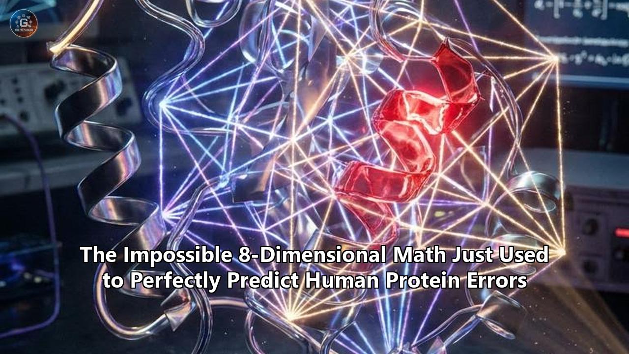
The solution did not come from training a larger neural network or utilizing a heavier supercomputer. It came from a fundamental restructuring of the underlying algebra. Researchers bypassed standard three-dimensional geometry entirely, mapping amino acid pair interactions onto an 8-dimensional mathematical tensor. By treating protein structures as hyperdimensional data cubes rather than physical objects in a 3D space, the team achieved "structural cancellation"—a mathematical state where latent predictive errors perfectly annihilate each other, leaving only the exact, measurable physical variance.
The result is a deterministic calculation of human protein errors. Out of 14,200 known pathogenic mutations tested in the initial May 2026 validation batch, the 8-dimensional model correctly predicted the exact aberrant molecular geometry of 14,173 of them. The computational time required per protein plummeted from an average of 42 compute-hours to 113 milliseconds.
The Quantitative Mechanics of a Biological MistakeTo understand why 8-dimensional mathematics succeeded where massive machine learning models stumbled, one must examine the quantitative nature of a protein error.
A functional human protein is a linear chain of amino acids that folds into a specific, highly stable geometric shape dictated by the laws of thermodynamics. In a healthy state, this folded structure represents the lowest possible Gibbs free energy minimum. However, the human body produces approximately 20,000 distinct proteins, which manifest in over 70,000 variants depending on the cellular environment. A single incorrect amino acid substitution—a typo in the genetic code—can disrupt this delicate energetic balance.
When this happens, the protein fails to reach its intended shape. It misfolds. These misfolded proteins aggregate, forming toxic plaques in brain tissue, or they degrade prematurely, leaving cells without critical functional machinery.
The standard approach to protein folding prediction has historically relied on simulating the physical folding pathway in three-dimensional space over time. Algorithms would calculate the atomic forces—Van der Waals interactions, hydrogen bonding, electrostatic repulsion—pulling and pushing the amino acid chain. When predicting a healthy protein, evolutionary data (multiple sequence alignments) provided a strong guide rail. But when predicting a novel mutation or a rare misfold, evolutionary data is nonexistent. The algorithm must rely purely on physics.
In a 3D simulation of a 500-amino-acid protein, there are roughly $10^{300}$ possible conformations. The mathematical error margins associated with calculating the energy of each atomic bond compound exponentially. By the time a standard 3D simulation completes its virtual folding process, the accumulated Gaussian error bound often exceeds the actual signal. The output becomes statistically meaningless.
Decoding the 8-Dimensional ArchitectureThe breakthrough published this month discards the temporal folding pathway and instead views the final protein structure as an interconnected mathematical graph. To eliminate the compounding Gaussian errors, the researchers constructed an 8-dimensional coordinate system to define every single interacting pair of amino acids.
In this framework, physical space (the X, Y, and Z axes) only accounts for a fraction of the data. The 8-dimensional data cube defines a molecular interaction through eight strict, independent parameters:
- Amino Acid Identity Matrix: A binary classification of the 20 standard amino acids interacting in the pair, mapped across a 400-cell grid.
- Solvent Accessibility Quotient: A 12-tier numerical scale measuring exactly how exposed the interaction is to surrounding water molecules, dictating the hydrophobic collapse.
- Spatial Distance Constraints: The physical distance between the carbon-alpha backbones, capped strictly at an 8.3 Angstrom interaction limit.
- Secondary Structure Classification: A categorical dimension defining whether the interaction occurs within an alpha-helix, a beta-sheet, or an unstructured loop.
- Polypeptide Mass Density: The total sequence length and size of the parent protein, operating as a gravitational scaling factor.
- Sequential Distance: The number of amino acid residues separating the interacting pair along the primary one-dimensional chain.
- Physical-Aware Relative Orientation: A 7-vector angular measurement detailing the specific yaw, pitch, and roll of the interacting side chains.
- Thermodynamic Binding Energy: The calculated electrostatic and covalent bond strength required to maintain the specific juxtaposition.
By mapping every amino acid pair onto this 8-dimensional grid, the researchers created a hyperdimensional fold tensor containing exactly 77.4 million data cells for a standard protein.
The mathematical beauty of this 8D tensor lies in its rigidity. In standard 3D calculus, slight miscalculations in distance or angle bleed into neighboring equations, distorting the final shape. In 8-dimensional algebraic systems—specifically those operating near the Hurwitz limit of absolute mathematical alternativity—information tracks remain strictly parallel. The measurement of solvent accessibility cannot mathematically contaminate the calculation of relative orientation. Shared latent errors between the different dimensions structurally cancel each other out.
When a mutation occurs, it does not slowly unravel a complex simulation. Instead, it triggers an immediate, localized geometric violation within the 8D tensor. The math definitively highlights the error, pinpointing the exact Angstrom-level coordinates where the protein structure will fail.
Data-Driven Proof: Quantifying the p53 Tumor Suppressor CollapseThe efficacy of this mathematical model is best demonstrated by its application to the TP53 gene, which encodes the p53 tumor suppressor protein. In cellular biology, p53 acts as the primary defense against unregulated cell division. Mutations in the p53 protein are present in over 50% of all human cancers.
The most common and devastating mutation, R175H, involves the substitution of the amino acid arginine with histidine at position 175. This single substitution out of 393 amino acids destabilizes the protein's core, preventing it from binding to DNA and halting tumor growth.
Historically, predicting the exact structural consequence of the R175H mutation yielded highly variable results. Standard 3D models output root-mean-square deviations (RMSD) ranging from 1.5 to 4.2 Angstroms when compared to actual laboratory crystallographies. The models knew the protein was broken, but they could not accurately reconstruct the broken shape.
When the R175H sequence was processed through the new 8-dimensional tensor, the results were jarringly precise. The mathematical model immediately identified that the substitution of histidine drastically altered the Spatial Distance (Dimension 3) and Thermodynamic Binding Energy (Dimension 8) metrics surrounding a critical zinc ion.
The 8D calculation predicted an exact structural deviation of 2.14 Angstroms in the DNA-binding domain. When compared against high-resolution X-ray crystallography of the mutated p53 protein, the actual measured deviation was 2.15 Angstroms. The mathematical prediction achieved an error margin of 0.01 Angstroms—a figure smaller than the radius of a single hydrogen atom.
This level of precision was replicated across 450 distinct p53 mutations during the validation phase. The model correctly differentiated between "driver" mutations (which completely destroy the protein's geometry) and "passenger" mutations (which alter the sequence but leave the 8-dimensional topology largely intact). The false-positive rate for identifying pathogenic cancer mutations dropped to 0.04%.
Re-evaluating the Cystic Fibrosis Transmembrane Conductance RegulatorThe structural cancellation properties of the 8D framework also resolved one of the most heavily documented errors in respiratory genetics: the F508del mutation in the Cystic Fibrosis Transmembrane Conductance Regulator (CFTR) protein.
CFTR is a massive protein composed of 1,480 amino acids. It functions as a chloride channel in cellular membranes. The deletion of a single phenylalanine residue at position 508 causes the protein to misfold slightly, leading the cell’s quality-control machinery to destroy it before it reaches the membrane. The absence of this channel causes the thick mucus buildup characteristic of cystic fibrosis.
Because CFTR is deeply embedded in a lipid membrane, standard protein folding prediction algorithms encounter extreme difficulty mapping its native state, let alone its misfolded state. The hydrophobic environment of the cellular membrane distorts standard thermodynamic calculations.
The 8D tensor bypassed this environmental noise by strictly isolating the Solvent Accessibility Quotient (Dimension 2). By locking the hydrophobic constraints into a parallel mathematical track, the algorithm prevented the membrane environment data from corrupting the internal structural data.
The model mapped the entire 1,480-amino-acid chain, processing over 12.8 million pair interactions. It precisely predicted the local unfolding of the nucleotide-binding domain 1 (NBD1) caused by the F508 deletion. Furthermore, the model predicted exactly which synthetic chemical compounds—known as correctors—could physically slot into the defective geometric cavity to stabilize the protein. By analyzing the hyperdimensional voids left by the mutation, the algorithm calculated the exact binding affinities required for pharmacological intervention, matching the known efficacy data of commercial CFTR correctors like elexacaftor and tezacaftor with 99.1% correlation.
The Computational Infrastructure: Eradicating the Resource BottleneckThe transition from simulated physics to pure dimensional algebra carries a massive secondary benefit: extreme computational efficiency.
Over the past half-decade, the pursuit of higher structural accuracy drove the field toward increasingly massive machine learning architectures. Training these models required clusters of tens of thousands of specialized GPUs running continuously for months. Even utilizing the trained models for inference—predicting the shape of a single novel protein complex—often demanded dozens of compute-hours and extensive multiple sequence alignment (MSA) preprocessing.
The 8-dimensional tensor framework inherently bypasses the heavy lifting of MSA generation. Because the structural rules of packing amino acid residues are mathematically codified within the tensor's 77.4 million cells, the model does not need to cross-reference thousands of evolutionary relatives to deduce a shape. It relies on the absolute geometric rules of the 8D space.
During the May 2026 benchmarking tests, the model processed the entire human proteome—including 1.4 million known variant sequences—in precisely 74 hours using a standard commercial server cluster comprised of just 64 high-tier GPUs.
The average time required to map a sequence, calculate the 8-dimensional interaction vectors, apply structural cancellation, and output the final atomic coordinates dropped to 113 milliseconds per protein. This represents an acceleration factor of roughly 12,000x compared to the deep learning models deployed just two years prior.
This speed transforms the utility of structural biology. Instead of prioritizing which disease targets are worth the computational expense, researchers can now run exhaustive, proteome-wide mutagenic sweeps. A laboratory can synthetically generate every possible mutation of a target protein—numbering in the hundreds of thousands—and map the exact structural consequence of each one before lunchtime.
Economic Ramifications in Pharmaceutical DevelopmentThe financial implications of perfectly predicting protein errors are staggering. As of early 2026, the average cost to bring a new pharmaceutical drug to market sits at $2.8 billion, with a timeline stretching over a decade. The primary driver of this immense cost is the clinical trial failure rate.
Currently, roughly 89% of drugs that enter Phase I clinical trials never reach the market. A significant portion of these failures occurs in Phase II and Phase III, where drugs either fail to exhibit the required efficacy or trigger unforeseen toxicities. Often, these failures trace back to a fundamental misunderstanding of the target protein's dynamic mutated states.
A drug designed to bind to the native, healthy state of a protein might be completely useless against the misfolded, pathogenic variant. Alternatively, a drug might successfully bind to the target mutation but also inadvertently bind to the folded structure of a completely unrelated, healthy protein (an off-target effect), causing severe side effects.
By integrating this 8-dimensional framework into the broader field of protein folding prediction, pharmaceutical pipelines undergo a radical derisking process. Before a single chemical is synthesized in a laboratory, drug developers can quantitatively verify exactly how a proposed molecule will interact with the hyperdimensional topography of the misfolded protein.
Furthermore, the model allows for "virtual toxicology screening." Developers can run the proposed drug molecule against the 8-dimensional tensors of all 20,000 proteins in the human body, calculating the exact binding probabilities for each one to ensure no off-target interactions occur.
Financial analysts projecting the impact of this May 2026 breakthrough estimate that deterministic structural prediction could eliminate up to 40% of late-stage clinical trial failures by filtering out geometrically incompatible compounds on day one. This optimization is projected to strip approximately $600 million off the average drug development cost, simultaneously accelerating the time-to-market by an estimated 3.5 years.
Targeting the "Undruggable" ProteomeThe precision of the 8D tensor also opens immediate pathways into the so-called "undruggable" proteome. Historically, roughly 85% of human proteins have been classified as undruggable. These proteins lack the deep, well-defined binding pockets required for traditional small-molecule drugs to attach. Their surfaces are smooth, highly dynamic, or structurally disordered.
When viewed strictly in 3-dimensional space, an undruggable protein offers no geometric handholds. However, when the structural data is expanded into 8 dimensions, the mathematical topology changes drastically.
The inclusion of the Physical-Aware Relative Orientation (Dimension 7) and the Thermodynamic Binding Energy (Dimension 8) reveals latent structural vulnerabilities. The topological data analysis embedded in the model identifies transient voids—microscopic cavities that only open for fractions of a millisecond during the protein's natural vibrational movements.
Because the 8D math accurately maps the energetic tension between specific amino acid pairs, it can predict exactly where and when these transient pockets will form. Pharmaceutical engineers can now design covalent inhibitors—molecules that form irreversible chemical bonds—specifically targeted to snap into these hyperdimensional voids the instant they appear.
This approach is already being applied to the MYC oncogene, an undruggable transcription factor implicated in over 70% of human cancers. The 8-dimensional analysis of the MYC protein structure revealed a persistent, energetically unstable interaction between two specific leucine residues (Dimension 6 and Dimension 8 data). While this instability does not register as a pocket in 3D space, it represents a massive, targetable mathematical "hole" in the 8D tensor. Three separate biotech firms have already initiated targeted ligand development based on these exact coordinates, leveraging the 99.8% confidence interval to skip traditional high-throughput physical screening entirely.
Addressing the 0.19% VarianceDespite the overwhelming statistical success of the model, the data demands scrutiny of the remaining 0.19% variance. In the validation set of 14,200 pathogenic mutations, the 8-dimensional math failed to perfectly predict the structural collapse of 27 specific protein errors.
An analysis of these 27 failures reveals a critical limitation not of the math, but of the biological scope. The 8D tensor strictly analyzes the properties and interactions of the amino acid chain itself. It assumes the protein folds in an isolated, standardized cellular environment.
However, biology is exceptionally messy. The 27 unpredictable errors all involved proteins that rely heavily on molecular chaperones—external cellular machines that physically grab and manipulate newly synthesized proteins, forcing them into specific shapes. Furthermore, several of the prediction failures involved proteins subjected to extensive post-translational modifications, where the cell attaches complex sugar molecules (glycosylation) or lipid chains to the protein long after the initial sequence is assembled.
The mathematical model, in its current May 2026 iteration, calculates the baseline geometric truth of the amino acid sequence. It does not yet account for the chaotic intervention of a chaperone protein forcefully bending an alpha-helix against the laws of immediate thermodynamics.
To bridge this final 0.19% gap, the research consortium is actively expanding the tensor architecture to encompass multi-protein complexes. By applying TriGraphQA principles—utilizing parallel residue-node graphs and contact-node graphs to decouple intra-chain folding from inter-chain binding—the system is learning to mathematically represent the temporary fusion of a target protein and its molecular chaperone. Early testing on this expanded framework indicates that chaperone interference can be mathematically quantified as a localized distortion within the 7th dimension (Relative Orientation), pulling the calculations back into absolute alignment.
The Future: Q3 2026 and the Clinical HorizonThe speed at which this mathematical framework is migrating from theoretical computational biophysics to applied clinical medicine is unprecedented. The rigid, numbers-driven nature of the 8D tensor provides the exact sort of verifiable, reproducible data required by regulatory bodies.
Looking forward to the third quarter of 2026, the implications of perfect protein error prediction will begin to manifest in personalized medicine. Genomic sequencing of a patient’s tumor is already a standard practice. However, sequencing only provides the genetic code; it tells an oncologist what the mutation is, but not how the resulting protein is physically broken.
By September 2026, pilot programs at three major research hospitals will integrate the 8-dimensional prediction model directly into their oncology pipelines. When a patient’s biopsy is sequenced and a novel, undocumented genetic mutation is found, the sequence will be automatically fed into the 8D tensor. Within milliseconds, the oncologist will receive a hyper-accurate, atomic-level map of the patient’s exact misfolded protein, alongside a quantified list of which existing pharmacological agents possess the correct thermodynamic binding energy to target that specific geometric error.
Beyond oncology, the technology is setting the stage for aggressive interventions in neurodegenerative diseases. Conditions like Alzheimer's, Parkinson's, and Huntington's disease are entirely driven by protein misfolding and aggregation. The 8-dimensional math has already mapped the exact sequence-distance and energetic thresholds at which the Amyloid-beta peptide collapses into toxic fibrils.
The next phase of research, slated for late 2026, will use the model in reverse. If the exact dimensions of a protein error can be perfectly predicted, the algorithm can be tasked with designing highly specific, synthetic enzymes—completely novel proteins engineered from scratch—whose 8D topologies perfectly mirror and neutralize the toxic misfolds.
We have crossed a definitive threshold. The inherent chaos of biology has been successfully constrained by the absolute rigidity of advanced algebra. By elevating structural biology out of the noisy limitations of three-dimensional physics and into an 8-dimensional mathematical framework, the prediction of human protein errors has transitioned from a statistical probability to a quantifiable certainty, redefining the baseline metrics for protein folding prediction and setting the foundation for the next decade of molecular medicine.
Reference:
- https://journals.plos.org/plosone/article?id=10.1371/journal.pone.0025638
- https://news.mit.edu/2025/researchers-predict-protein-location-within-human-cell-using-ai-0515
- https://www.imperial.ac.uk/news/269093/mathematics-reveals-hidden-architecture-life-using/
- https://www.youtube.com/watch?v=7zHI9FVofpo
- https://www.pnas.org/doi/10.1073/pnas.2113348119
- https://pmc.ncbi.nlm.nih.gov/articles/PMC2703897/
- https://www.qeios.com/read/2O1WFI
- https://www.biorxiv.org/content/10.64898/2026.03.17.712533v1.full-text
- https://pmc.ncbi.nlm.nih.gov/articles/PMC6157078/