The Unseen World: Unraveling the Physics of Magnetic Resonance Imaging
In the sophisticated realm of medical diagnostics, few technologies capture the imagination quite like Magnetic Resonance Imaging (MRI). It offers a window into the human body, producing exquisitely detailed images of soft tissues, organs, and intricate neural pathways without the use of ionizing radiation. To the patient, the experience might be a noisy sojourn into a narrow tunnel, but behind the hum and clicks of the machine lies a symphony of physics principles, masterfully orchestrated to reveal the body's inner secrets. This article will delve into the profound physics that underpins MRI, from the quantum behavior of atomic nuclei to the complex computational processes that render the final, high-resolution images that have revolutionized medicine.
The Heart of the Matter: The Hydrogen Proton
At the very core of MRI technology is the humble hydrogen atom. Our bodies are composed of approximately 60% water, and each water molecule contains two hydrogen atoms. This makes hydrogen the most abundant element in the body, providing a vast and readily available source for the MRI signal. But it's not the entire hydrogen atom that's of interest; it's the nucleus, which for hydrogen consists of a single, positively charged proton.
This proton possesses an intrinsic quantum property known as "spin." This spin gives the proton a tiny magnetic moment, effectively turning it into a microscopic magnet with a north and south pole. Under normal circumstances, these countless proton magnets within our bodies are randomly oriented, their magnetic fields pointing in all directions, resulting in no net magnetization.
The Power of the Magnet: Alignment and Precession
This is where the "Magnetic" in MRI comes into play. The patient is placed within the bore of the MRI scanner, which houses an incredibly powerful primary magnet. These magnets are typically superconducting magnets, cooled to temperatures near absolute zero with liquid helium, which allows them to conduct enormous electrical currents with no resistance, generating a very strong and stable magnetic field, denoted as B0. The strength of this field is measured in Tesla (T), with clinical scanners typically operating at 1.5T or 3T, thousands of times stronger than the Earth's magnetic field.
When the patient's body is immersed in this powerful B0 field, the randomly oriented proton spins are forced to align with it. They can align in one of two ways: parallel to the magnetic field (a low-energy state) or anti-parallel to it (a high-energy state). A slightly greater number of protons will occupy the lower-energy parallel state. This small excess of protons aligned with the main magnetic field creates a net magnetic vector (NMV) for each small volume of tissue, a tiny but crucial magnetic signal that is the foundation of the MRI image.
In addition to aligning with the B0 field, the protons also exhibit another behavior: they precess, or wobble, around the axis of the main magnetic field. This is analogous to a spinning top wobbling as it slows down. The rate of this precession is not random; it is determined by the strength of the magnetic field and is known as the Larmor frequency. This precise relationship between magnetic field strength and precession frequency is fundamental to MRI.
The "Resonance" in MRI: Exciting the Protons
With the protons aligned and precessing in the main magnetic field, the next step is to manipulate them in a way that generates a detectable signal. This is achieved through the principle of "resonance." The MRI machine uses a radiofrequency (RF) coil to transmit a second, much weaker magnetic field, known as the B1 field, into the patient's body. This B1 field is an RF pulse that oscillates at the Larmor frequency of the hydrogen protons.
When the frequency of the RF pulse matches the precessional frequency of the protons, resonance occurs. The protons absorb the energy from the RF pulse, causing two key things to happen. First, some of the low-energy protons are "excited" and jump to the higher-energy anti-parallel state, which reduces the longitudinal magnetization along the main B0 field. Second, and crucially, the RF pulse forces the precessing protons to move into phase with each other. This coherence of the precessing spins causes the net magnetic vector to tilt away from the main magnetic field and into the transverse plane, perpendicular to B0. The amount the NMV is tilted is called the flip angle, which can be controlled by adjusting the duration and strength of the RF pulse.
The Echo of a Signal: Relaxation and Detection
Once the RF pulse is turned off, the excited protons begin to "relax" back to their lower-energy state, and the net magnetic vector realigns with the main magnetic field. As they do so, they release the energy they absorbed from the RF pulse in the form of a faint radio signal. This signal is detected by the same RF coil that transmitted the pulse (or a separate receiver coil). This decaying signal is known as the Free Induction Decay (FID).
The process of relaxation is not instantaneous and occurs through two distinct and simultaneous mechanisms:
- T1 Relaxation (Spin-Lattice Relaxation): This refers to the recovery of the longitudinal magnetization as the protons release their energy to the surrounding molecular lattice and realign with the main magnetic field. The time it takes for about 63% of the longitudinal magnetization to recover is called the T1 time. T1 times vary for different tissues. For example, fat has a short T1 time, meaning its protons realign quickly, while the cerebrospinal fluid (CSF) has a long T1 time.
- T2 Relaxation (Spin-Spin Relaxation): This describes the decay of the transverse magnetization. After the RF pulse, the protons are precessing in phase. However, they soon begin to dephase due to interactions with the local magnetic fields of neighboring protons. This loss of phase coherence causes the transverse magnetization to decay. The time it takes for the transverse magnetization to fall to about 37% of its initial value is the T2 time. T2 times also differ between tissues. Water and CSF have long T2 times, while more solid tissues have shorter T2 times.
These relaxation times are the key to the remarkable contrast seen in MRI images. By manipulating the timing of the RF pulses and when the signal is collected, we can create images that highlight the T1 or T2 properties of tissues.
Crafting the Image: Pulse Sequences and Image Contrast
An MRI scan doesn't just consist of a single RF pulse. Instead, a series of precisely timed RF pulses and gradients are applied in what is known as a pulse sequence. The parameters of these sequences, such as the Repetition Time (TR) and the Echo Time (TE), can be adjusted to "weigh" the image towards a particular type of contrast.
- T1-Weighted Images: To create a T1-weighted image, a short TR and a short TE are used. The short TR doesn't allow tissues with long T1 times (like CSF) to fully recover their longitudinal magnetization before the next RF pulse, so they produce a weak signal and appear dark. Tissues with short T1 times (like fat) recover quickly, produce a strong signal, and appear bright. T1-weighted images are excellent for visualizing anatomy.
- T2-Weighted Images: For T2-weighted images, a long TR and a long TE are chosen. The long TR allows all tissues to fully recover their longitudinal magnetization, so T1 differences don't dominate the contrast. The long TE allows time for the transverse magnetization to decay. Tissues with long T2 times (like water and CSF) will still have a significant transverse signal at the time of measurement and will appear bright. Tissues with short T2 times will have their signal decay quickly and will appear dark. T2-weighted images are particularly good at highlighting pathology, as many diseases are associated with an increase in water content (edema), which appears bright on a T2-weighted scan.
- Proton Density (PD) Weighted Images: These images are created using a long TR and a short TE. The long TR minimizes T1 contrast, and the short TE minimizes T2 contrast. The resulting image contrast is primarily based on the concentration of protons in the tissue. Tissues with a high density of protons, such as the brain parenchyma, will appear bright.
Finding Your Way: Spatial Encoding and k-Space
So far, we have discussed how a signal is generated, but not how the scanner knows where in the body the signal is coming from. This is the task of the gradient coils. These are additional electromagnets within the MRI scanner that can be turned on and off rapidly to create a slight, predictable variation in the main magnetic field. By altering the magnetic field strength, the Larmor frequency of the protons is also altered in a predictable way.
Spatial localization is achieved in three dimensions:
- Slice Selection: To image a specific "slice" of the body, a gradient is applied along the long axis of the patient (the z-axis). This creates a gradient of Larmor frequencies. An RF pulse is then transmitted with a narrow bandwidth of frequencies that corresponds to the Larmor frequencies of the protons within the desired slice. Only the protons in that slice will be excited.
- Frequency Encoding: After slice selection, a second gradient, the frequency-encoding gradient, is applied in one of the other two dimensions (e.g., the x-axis) while the signal is being read out. This gradient causes the protons along this axis to precess at different frequencies. The frequency of the emitted signal is now directly related to the proton's position along this axis.
- Phase Encoding: To localize the signal in the final dimension (the y-axis), a third gradient, the phase-encoding gradient, is briefly turned on and off before the signal is read out. This gradient imparts a temporary change in the precessional frequency, causing the protons along this axis to accumulate a different amount of phase shift in their precession. This process is repeated multiple times, each time with a slightly different strength of the phase-encoding gradient, to encode the entire slice.
The raw data collected by the receiver coils is not the final image. This data is stored in a temporary mathematical space called k-space. Each point in k-space contains spatial frequency information from the entire slice. The center of k-space contains the low-frequency data, which determines the overall contrast of the image, while the periphery of k-space holds the high-frequency data, which provides the fine details and edges. Once the k-space is filled with data, a mathematical process called a Fourier Transform is applied to convert this frequency and phase information into the final, recognizable anatomical image.
The MRI Machine: A Look Inside the Hardware
An MRI scanner is a highly complex piece of machinery with several key components working in unison:
- The Main Magnet: This is the largest and most expensive component, responsible for creating the strong, uniform B0 magnetic field. Most modern scanners use superconducting magnets.
- Gradient Coils: These are three sets of electromagnets that produce the controlled variations in the magnetic field necessary for spatial encoding. Their rapid switching on and off is what causes the loud knocking and banging sounds heard during a scan.
- Radiofrequency (RF) Coils: These coils transmit the RF pulses to excite the protons and also act as antennas to detect the returning signals. There are large body coils built into the scanner, as well as smaller, specialized coils that can be placed on or around the specific body part being imaged (e.g., a head coil or a knee coil) to improve image quality.
- Computer System: A powerful computer system controls all the components of the scanner, from the timing of the pulse sequences to the reconstruction of the final image from the k-space data.
- Patient Table: This is the movable bed that the patient lies on, which is precisely controlled to position the area of interest in the center of the magnetic field.
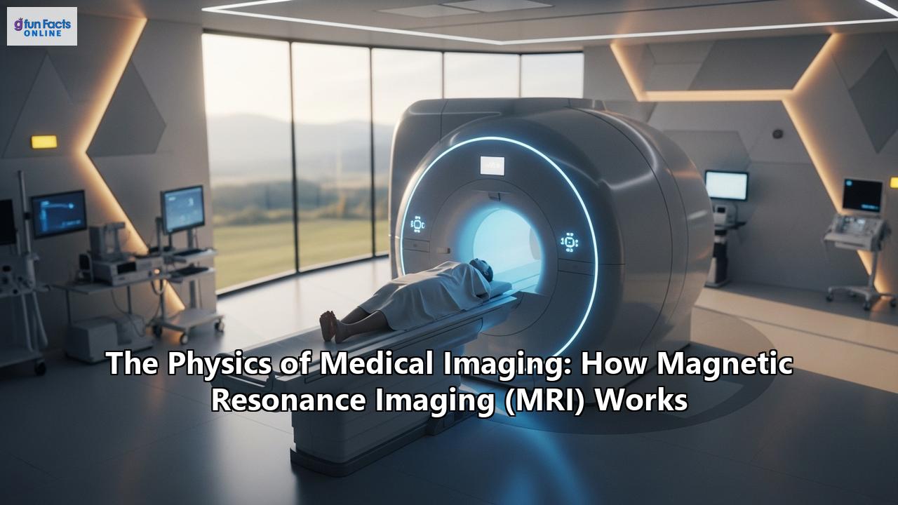
The Patient Experience: What to Expect During an MRI
For the patient, the MRI process begins with thorough screening. Because of the powerful magnetic field, it is crucial to identify any metallic objects or implants in the body. This includes pacemakers, aneurysm clips, cochlear implants, and even some tattoos with metallic ink. Patients will be asked to remove all metal objects, such as jewelry, and will typically change into a hospital gown.
The patient then lies on the scanner table, and the technologist will position them correctly and may place a specialized RF coil over the area being imaged. The table then slides into the bore of the magnet. During the scan, it is vital for the patient to remain as still as possible to avoid blurring the images. The scanner will produce loud noises, and patients are usually given earplugs or headphones. The technologist will be in an adjacent control room but can communicate with the patient via an intercom. In some cases, a contrast agent, typically containing gadolinium, may be injected intravenously to enhance the visibility of certain tissues or blood vessels. The entire scan can take anywhere from 15 minutes to over an hour, depending on the complexity of the examination.
Beyond Anatomy: Advanced MRI Techniques
The versatility of MRI extends far beyond basic anatomical imaging. Advanced techniques allow for the assessment of function, metabolism, and blood flow:
- Functional MRI (fMRI): This technique measures brain activity by detecting changes in blood flow. When a particular area of the brain is active, it requires more oxygen, and the blood flow to that area increases. fMRI can detect the difference in the magnetic properties of oxygenated and deoxygenated blood (the BOLD effect), allowing researchers and clinicians to map brain function.
- Magnetic Resonance Angiography (MRA): MRA is used to visualize blood vessels. It can be performed with or without a contrast agent and is valuable for detecting aneurysms, blockages, or other vascular abnormalities.
- Magnetic Resonance Spectroscopy (MRS): Instead of imaging water, MRS can be tuned to detect the signals from other molecules in the body, such as metabolites. This provides biochemical information about tissues and can be used to help characterize tumors and other metabolic disorders.
- Diffusion-Weighted Imaging (DWI) and Diffusion Tensor Imaging (DTI): These techniques measure the random motion of water molecules in tissue. This is particularly useful in the brain, where the diffusion of water is restricted by the myelin sheaths of nerve fibers. DTI can be used to map out the white matter tracts of the brain, a technique known as tractography. DWI is also extremely sensitive for detecting acute stroke.
A Powerful Diagnostic Tool: Clinical Applications of MRI
MRI's ability to provide excellent soft tissue contrast without using ionizing radiation makes it an invaluable tool in many areas of medicine:
- Neurology and Neurosurgery: MRI is the gold standard for imaging the brain and spinal cord, used to diagnose tumors, stroke, multiple sclerosis, and injuries.
- Musculoskeletal Imaging: It is exceptional for visualizing ligaments, tendons, cartilage, and muscles, making it essential for diagnosing joint injuries, such as torn ligaments in the knee, and for assessing soft tissue tumors.
- Oncology: MRI plays a crucial role in the detection, staging, and monitoring of many types of cancer, particularly in the brain, prostate, and pelvis.
- Cardiovascular Imaging: Cardiac MRI can assess the structure and function of the heart, as well as blood flow through major vessels.
- Abdominal and Pelvic Imaging: It is used to evaluate organs like the liver, kidneys, and pancreas, and is particularly useful for assessing diseases of the biliary tract and pelvic organs.
Safety First: Understanding the Risks of MRI
While MRI is a very safe procedure, there are some potential risks that must be managed:
- The Magnetic Field: The most significant risk comes from the attraction of ferromagnetic objects to the magnet, which can turn them into dangerous projectiles. Thorough screening is the best prevention. The magnetic field can also cause certain medical implants to malfunction or shift.
- Radiofrequency Energy: The RF pulses can cause some heating of the body. In rare cases, this can lead to burns, especially if there are metallic objects on the patient's clothing or if conductive loops are formed by wires or cables.
- Noise: The loud noise from the gradient coils can potentially damage hearing if ear protection is not used.
- Contrast Agents: While generally safe, gadolinium-based contrast agents can cause allergic reactions in some individuals. In patients with severe kidney disease, there is a rare risk of a condition called nephrogenic systemic fibrosis.
- Claustrophobia: The enclosed space of the scanner can induce anxiety and claustrophobia in some patients. Open MRI scanners are an option for some, though they may offer slightly lower image quality.
The Journey of Discovery and the Future of MRI
The history of MRI is a testament to scientific curiosity and innovation. The underlying principles of nuclear magnetic resonance (NMR) were discovered in the 1940s. However, it wasn't until the 1970s that scientists like Paul Lauterbur and Sir Peter Mansfield demonstrated that these principles could be used to create images, an achievement for which they were awarded the Nobel Prize in 2003. The first human MRI scan was performed in 1977, and the technology became commercially available in the 1980s.
The evolution of MRI is far from over. Researchers are continuously developing new techniques to make scans faster, quieter, and more informative. The development of ultra-high-field magnets (7T and beyond) promises even greater image resolution. The integration of artificial intelligence and machine learning is poised to revolutionize image analysis, helping radiologists to detect diseases earlier and more accurately. Hybrid technologies, such as PET-MRI scanners that combine the anatomical detail of MRI with the metabolic information of PET, are opening up new frontiers in medical imaging.
From the subtle dance of a single proton to the intricate tapestry of the human anatomy it reveals, the physics of MRI is a captivating story of scientific ingenuity. It is a technology that has not only transformed medicine but also continues to push the boundaries of what we can see and understand about the remarkable biological machine that is the human body.
Reference:
- https://en.wikipedia.org/wiki/Magnetic_resonance_imaging
- https://www.nibib.nih.gov/science-education/science-topics/magnetic-resonance-imaging-mri
- https://en.wikipedia.org/wiki/K-space_in_magnetic_resonance_imaging
- http://www.sprawls.org/mripmt/MRI02/index.html
- https://www.radiologycafe.com/frcr-physics-notes/mr-imaging/t1-t2-and-pd-weighted-imaging/
- https://www.slideshare.net/slideshow/magnetic-resonance-imaging-contrast-and-weighting-pptx/273950114
- https://www.numberanalytics.com/blog/proton-density-mri-comprehensive-guide
- https://pmc.ncbi.nlm.nih.gov/articles/PMC3097694/
- https://www.radiologycafe.com/frcr-physics-notes/mr-imaging/k-space/
- https://www.umit-tirol.at/data.cfm?vpath=vorlesungen/mri_spatialencoding
- https://www.slideshare.net/slideshow/7-components-of-mri/44312614
- https://umehealth.co.uk/mri-scans-how-they-work/
- https://www.princeedwardisland.ca/en/information/health-pei/magnetic-resonance-imaging-mri-patient-preparation
- https://pmc.ncbi.nlm.nih.gov/articles/PMC10657250/
- https://www.jointcommission.org/resources/news-and-multimedia/newsletters/newsletters/quick-safety/quick-safety-issue-31-strong-mri-safety-programs-prevent-safety-events/strong-mri-safety-programs-prevent-safety-events/
- https://www.ncbi.nlm.nih.gov/books/NBK551669/
- https://www.hopkinsmedicine.org/health/treatment-tests-and-therapies/magnetic-resonance-imaging-mri
- https://www.asnr.org/patientinfo/procedures/mri.shtml
- https://openmedscience.com/understanding-the-different-types-of-mri/
- https://www.mayoclinic.org/tests-procedures/mri/about/pac-20384768
- https://epos.myesr.org/poster/esr/ecr2025/C-23164
- https://www.mdpi.com/2075-4418/14/11/1120
- https://pmc.ncbi.nlm.nih.gov/articles/PMC1306489/
- https://my.clevelandclinic.org/health/diagnostics/4876-magnetic-resonance-imaging-mri
- https://www.fda.gov/apology_objects/abuse-detection-apology.html
- https://sisimaging.com/mri-safety-are-there-any-risks-involved/
- https://mccollege.edu/aas-in-magnetic-resonance-imaging-mri-technology/about-the-mri-technology-career/the-history-of-the-mri-development-of-medical-resonance-imaging/
- https://openmedscience.com/a-short-history-of-magnetic-resonance-imaging-mri/
- https://pmc.ncbi.nlm.nih.gov/articles/PMC9199974/