Cryo-electron microscopy (cryo-EM) has transformed structural biology, enabling scientists to visualize the intricate details of biological molecules like proteins, nucleic acids, and viruses at near-atomic resolutions. This powerful technique offers an unprecedented window into the fundamental mechanisms of life.
At its core, cryo-EM involves rapidly freezing biological samples in a thin layer of vitreous (non-crystalline) ice, typically below -150°C. This flash-freezing process, often using cryogenic liquids like ethane, preserves the molecules in their near-native state, avoiding the potential structural disruptions that can occur with other methods like X-ray crystallography which requires crystallization.
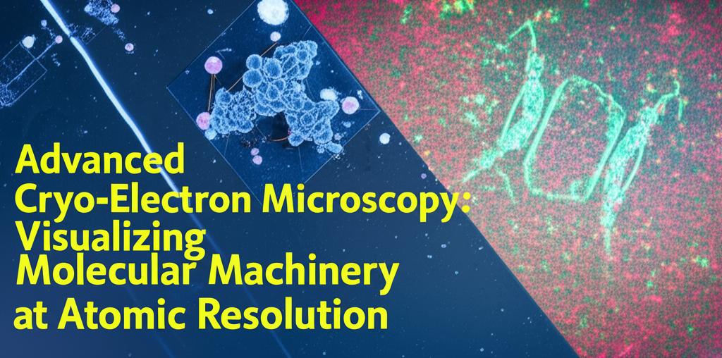
Once frozen, the samples are imaged using a transmission electron microscope. The microscope fires a beam of electrons through the sample, capturing numerous 2D projection images from different angles. These 2D images are then computationally combined and processed using sophisticated algorithms to reconstruct a 3D model of the molecule or complex. This 3D model can achieve near-atomic resolution, revealing the precise arrangement of atoms within the structure.
Recent Breakthroughs and Technological Advancements:The field of cryo-EM is experiencing rapid advancements in hardware, software, and methodology, pushing the boundaries of what can be visualized.
- Enhanced Detectors and Optics: The development of direct electron detectors (DEDs) has been a game-changer. These detectors are significantly more sensitive and faster than previous technologies, leading to higher quality images with improved signal-to-noise ratios. Innovations in electron sources, such as brighter and more coherent beams, and the introduction of phase plates, have further enhanced image contrast and resolution, particularly beneficial for smaller or more challenging samples.
- Sophisticated Image Processing: Advanced image processing algorithms, incorporating Bayesian statistics, maximum-likelihood approaches, and deep learning techniques, are crucial for handling the vast amounts of data generated and overcoming challenges like conformational heterogeneity (where molecules exist in multiple shapes) and low signal-to-noise ratios. Software packages like RELION and cryoSPARC have become indispensable tools, providing user-friendly interfaces and powerful algorithms for particle picking, classification, and high-resolution 3D reconstruction. Artificial intelligence (AI) is playing an increasingly significant role in automating and improving various stages of the cryo-EM workflow, especially in particle picking and structure modeling.
- Automation and Throughput: Automation in sample preparation, data collection, and image processing has dramatically increased the speed and efficiency of cryo-EM. Structures that once took years to solve can now often be determined in weeks or even days. This acceleration is crucial for high-throughput structural studies, such as in structural genomics, where the goal is to determine the structures of many proteins.
- Expanding Applications: Cryo-EM is no longer limited to very large, stable complexes. Technological improvements are making it possible to study smaller proteins, membrane proteins (which are notoriously difficult to crystallize), and dynamic, flexible molecular machines. Cryo-electron tomography (cryo-ET), a related technique, allows for the 3D visualization of molecules within their native cellular environment, providing crucial contextual information.
- Achieving True Atomic Resolution: Recent breakthroughs have pushed the resolution limits of cryo-EM to true atomic levels (around 1.2 Ångströms for some well-behaved samples). This level of detail allows researchers to visualize individual hydrogen atoms and understand subtle structural features critical for molecular function and drug interactions.
The impact of advanced cryo-EM is far-reaching:
- Drug Discovery and Development: Cryo-EM is revolutionizing structure-based drug design. By providing high-resolution structures of disease-relevant proteins and how they interact with potential drug candidates, it enables the design of more potent and selective therapeutics. It is also proving invaluable for understanding drug resistance mechanisms.
- Understanding Fundamental Biological Processes: Cryo-EM is providing unprecedented insights into the workings of complex molecular machines involved in essential cellular processes like DNA replication, transcription, translation, and cellular signaling. The ability to capture molecules in different functional states helps to elucidate their mechanisms of action.
- Integrative Structural Biology: Cryo-EM is increasingly being used in conjunction with other structural biology techniques like X-ray crystallography, Nuclear Magnetic Resonance (NMR) spectroscopy, and computational modeling to provide a more complete understanding of molecular structure and dynamics.
- Expanding to New Frontiers: Efforts are underway to further improve resolution, tackle even more challenging and dynamic systems, and develop methods for time-resolved cryo-EM to capture molecular processes as they happen. The development of more compact and cost-effective cryo-EM instruments aims to make this powerful technology more accessible to a wider range of researchers. The application of cryo-EM is also expanding beyond biology into materials science for imaging beam-sensitive materials.
In conclusion, advanced cryo-electron microscopy continues to evolve at a rapid pace, offering ever-clearer views of the molecular machinery of life. Its ability to visualize complex biomolecules at atomic or near-atomic resolution in their native states is providing profound insights into biology and medicine, paving the way for new discoveries and therapeutic interventions.