The dawn of a new era in medical imaging is not arriving with a blinding flash of light, but with a whisper—a subtle, rhythmic sound generated by light itself. This is the world of Optoacoustic Angiography, a revolutionary modality that invites us to "listen to light" to map the finest rivers of life within our bodies: the microvasculature. By hybridizing the high contrast of optical imaging with the deep penetration of ultrasound, optoacoustic (or photoacoustic) imaging has shattered the long-standing trade-off between resolution and depth. It allows us to peer centimeters deep into living tissue while resolving capillaries as fine as a single red blood cell, all without ionizing radiation or nephrotoxic contrast agents.
Part I: The Symphony of Physics – Listening to Light
To understand optoacoustic angiography, one must first appreciate the limitation it overcomes. Traditional optical microscopy, while brilliant at resolving cellular details, hits a "soft wall" at about 1 millimeter of depth due to the scattering of light. Photons in tissue behave like car headlights in heavy fog; they diffuse rapidly, blurring any image of deep structures. Ultrasound, conversely, penetrates deeply by listening to echoes, but it struggles to distinguish between soft tissues of similar density—blood and tumor tissue often look frustratingly identical.
Optoacoustic imaging marries these two worlds through the photoacoustic effect, a phenomenon first discovered by Alexander Graham Bell in 1880. Bell observed that when a beam of sunlight was rapidly interrupted (chopped) and focused onto a solid object, the object emitted a sound. In the medical context, this principle is refined into a precise sequence of events:
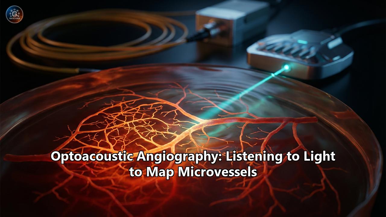
- Optical Excitation: A pulsed laser emits a burst of light (typically in the nanosecond range) into the tissue. The wavelength is carefully chosen to target a specific "chromophore"—a light-absorbing molecule. In angiography, the target is hemoglobin.
- Absorption and Heating: As photons travel through the tissue, they are absorbed by the hemoglobin within blood vessels. This absorption converts optical energy into thermal energy.
- Thermoelastic Expansion: The rapid heating (often just millidegrees Celsius) causes the blood vessel to expand. Because the laser pulse is ultra-short (satisfying the condition of "thermal confinement"), the heat doesn't have time to diffuse into the surrounding tissue. Similarly, the pulse is faster than the time it takes for the tissue to relax mechanically (satisfying "stress confinement").
- Acoustic Emission: This sudden, constrained expansion generates a pressure wave—an ultrasound wave.
- Detection: These waves propagate to the tissue surface, where they are detected by sensitive ultrasound transducers.
The result is an image where the contrast is optical (based on the chemical properties of blood) but the resolution is ultrasonic (based on the physics of sound, which scatters 1,000 times less than light in tissue). We are effectively "hearing" the color of the blood.
Part II: The Engine Room – Technology and Instrumentation
The transition from Bell’s primitive photophone to modern microvessel mapping relies on sophisticated engineering.
1. The Light Source:Modern systems typically use Q-switched Nd:YAG lasers coupled with Optical Parametric Oscillators (OPOs). These tunable lasers can sweep across the near-infrared (NIR) spectrum (680 nm to 980 nm). This tunability is crucial. Oxygenated hemoglobin ($HbO_2$) and deoxygenated hemoglobin ($HbR$) absorb light differently at different wavelengths. By pulsing multiple wavelengths rapidly, the system can perform spectral unmixing, calculating not just the presence of blood, but its oxygen saturation ($sO_2$). This turns a structural map into a functional metabolic readout.
2. The Transducer:The "ears" of the system are piezoelectric transducers, similar to those in standard ultrasound but optimized for broadband detection. For high-resolution skin imaging (Raster-Scan Optoacoustic Mesoscopy, or RSOM), high-frequency transducers (20–100 MHz) are used to resolve layers of the dermis. For deeper organ imaging (Multispectral Optoacoustic Tomography, or MSOT), lower frequencies (5–10 MHz) are employed to catch signals from centimeters deep. Newer "all-optical" ultrasound detectors using Fabry-Pérot interferometers are pushing detection sensitivity even further, eliminating electrical noise.
3. Image Reconstruction:Raw signals must be mathematically transformed into a 3D image. The classic algorithm is Filtered Back-Projection, similar to CT scanning. However, light fluence attenuation (the laser gets dimmer as it goes deeper) makes quantitative accuracy challenging. Advanced algorithms like Time Reversal and Model-Based Inversion account for the speed of sound variations and light distribution. The frontier is now Deep Learning (AI), where neural networks like U-Net are trained to reconstruct high-fidelity images from sparse data, removing artifacts and speeding up real-time processing.
Part III: Mapping the Microcosm – Clinical Applications
The ability to map microvessels—the tiny arterioles, venules, and capillaries—is a game-changer because disease often starts in the microcirculation.
1. Oncology: Visualizing the Engine of Cancer
Tumors are metabolically hungry. To survive, they hijack the body's vascular system, sprouting chaotic, leaky, and tortuous vessels—a process called angiogenesis. Optoacoustic angiography is uniquely suited to detect this.
- Breast Cancer: This is the most mature clinical application. The Imagio® Breast Imaging System (Seno Medical) was the first to receive FDA pre-market approval. It helps radiologists differentiate benign masses (fibroadenomas) from malignant tumors. Benign lesions tend to push vessels aside ("draping"), while malignant tumors show "feeding vessels" penetrating the core and a "claw sign" of irregular periphery vessels. Furthermore, because tumors consume vast amounts of oxygen, they often show a hypoxic (low oxygen) core. Optoacoustics can visualize this deoxygenation, providing a "functional biopsy" without a needle.
- Melanoma: In skin cancer, knowing the tumor depth (Breslow depth) is critical for prognosis. RSOM can profile the tumor's invasion into the dermis and visualize the surrounding vascular recruitment in 3D, potentially aiding in surgical planning.
2. Dermatology: Beneath the Surface
Skin diseases like psoriasis and eczema are inflammatory, driven by increased blood flow and dilated capillaries. RSOM provides a non-invasive look at these inflammatory biomarkers.
- Psoriasis: RSOM can quantify the vessel density and diameter in the psoriatic plaque. Studies have shown that these vascular metrics correlate closely with clinical severity scores (PASI) and can detect response to treatment weeks before the plaque visibly clears on the surface.
- Wound Healing: In chronic wounds or burns, assessing perfusion is vital. Optoacoustic angiography can map the revascularization of a wound bed, distinguishing between viable, oxygenated tissue and necrotic areas, guiding debridement.
3. Cardiology: The Vulnerable Plaque
Heart attacks are often caused by the rupture of "vulnerable plaques" in coronary arteries—plaques with a thin fibrous cap and a large lipid-rich necrotic core.
- Intravascular Photoacoustic (IVPA) Imaging: Researchers have developed catheter-based probes that combine IVUS with photoacoustics. While IVUS sees the structure, IVPA "sees" the chemistry. By targeting lipid absorption peaks (around 1200 nm or 1700 nm), IVPA can specifically highlight the fatty core of a plaque. This allows cardiologists to identify high-risk lesions that might otherwise look stable on standard angiography.
4. Neurology: Functional Brain Imaging
The brain is the most vascular-dependent organ. While fMRI relies on the BOLD signal (a complex mix of volume and oxygenation changes), optoacoustic imaging measures hemoglobin concentration and oxygenation directly.
- Mouse Models: In preclinical research, optoacoustic tomography has mapped "Resting-State Functional Connectivity" (RSFC) in the mouse brain with resolution superior to fMRI. It can visualize the hemodynamic waves propagating across hemispheres, offering a new tool to study neurovascular coupling in Alzheimer’s and stroke models.
- Human Potential: Transcranial imaging in humans is difficult due to the skull blocking light and sound. However, new techniques specifically targeting pediatric patients (thinner skulls) or using "photon recycler" probes are opening doors to monitoring neonatal brain injury and cerebral oxygenation in intensive care.
Part IV: Enhancing the Signal – Contrast Agents and Theranostics
While hemoglobin is a powerful endogenous contrast, sometimes we need to brighten the picture or target specific molecular markers.
- Gold Nanoparticles: Gold nanorods (GNRs) are the superstars of exogenous contrast. Their dimensions can be tuned to absorb light at any desired NIR wavelength. They have an absorption cross-section orders of magnitude higher than organic dyes.
- Organic Dyes: Indocyanine Green (ICG) and Methylene Blue are clinically approved dyes that fluoresce but also generate strong photoacoustic signals. They are used to map sentinel lymph nodes in cancer surgery.
- Theranostics: This portmanteau of "therapeutics" and "diagnostics" represents the future. Imagine gold nanorods targeted to a tumor. First, you use a low-energy laser pulse to image them (optoacoustic diagnosis). Once confirmed, you ramp up the laser power. The nanorods absorb the energy, heat up significantly, and cook the tumor from the inside (Photothermal Therapy or PTT), all while you monitor the temperature in real-time using the photoacoustic thermometer principle.
Part V: The AI Revolution and Future Horizons
Artificial Intelligence is the turbocharger for this technology.
- SenoGram®: Seno Medical’s AI tool assists radiologists by assigning a "likelihood of malignancy" score based on optoacoustic features, standardizing interpretation and reducing operator dependence.
- Sparse Sampling: Collecting a full 360-degree view of a patient is slow. Deep Learning algorithms are now enabling "sparse sampling"—reconstructing high-quality images from just a few laser shots, enabling real-time video-rate imaging of moving organs like the heart.
The "false positive" problem in breast cancer screening costs the healthcare system billions. Thousands of women undergo painful, anxiety-inducing biopsies for lesions that turn out to be benign. By providing a "functional rule-out," optoacoustic angiography could spare huge numbers of patients from unnecessary procedures, shifting the paradigm from "biopsy to diagnose" to "image to diagnose."
Conclusion
Optoacoustic angiography is more than just a new way to take a picture; it is a new way to sense the biology of the living body. It translates the molecular signature of hemoglobin into a high-definition map of life’s infrastructure. From the jagged feeding vessels of a malignant tumor to the rhythmic flushing of a healing wound or the subtle firing of a neural circuit, it allows us to see the unseen. As lasers become cheaper, compact, and faster, and as AI refines the "sound" of light, we are moving toward a future where the doctor’s stethoscope is replaced by a probe that listens not just to the heart, but to the very molecules that sustain it. The era of listening to light has only just begun.
Reference:
- https://pmc.ncbi.nlm.nih.gov/articles/PMC11297194/
- https://pmc.ncbi.nlm.nih.gov/articles/PMC9441264/
- https://pmc.ncbi.nlm.nih.gov/articles/PMC11685406/
- https://beonbrand.getbynder.com/m/d0616f35693d2397/original/Opto-acoustic-Imaging-of-the-Breast.pdf
- https://physicsworld.com/a/optoacoustic-imaging-identifies-breast-cancer-from-vascular-patterns/
- https://pmc.ncbi.nlm.nih.gov/articles/PMC5808678/
- https://validate.perfdrive.com/?ssa=7601d8e4-dedb-d312-a29b-8b6da04e38de&ssb=11646282132&ssc=https%3A%2F%2Fopg.optica.org&ssi=41706ed5-d3hy-89e7-10e4-ba83c735fdda&ssk=botmanager_support@radware.com&ssm=31022578822672636136647955513857&ssn=e02768c21fb2360c763b28b2e0666962972743295e87-76e3-285e-9b7053&sso=fe0a45e-91c6-3da2a841e6d5b1cc2ad5f40d0f010321274b97db13de547b&ssp=00514286791769873555176986471506673&ssq=93378401222391135914812222085551152870466&ssr=MTI0LjIxNy4xODkuMTE5&sst=python-requests%2F2.32.3&ssv=&ssw=
- https://www.diagnosticimaging.com/view/fda-gives-seno-medical-approval-for-opto-acoustic-breast-imaging-system