The human brain, an intricate web of nearly 100 billion neurons and trillions of synapses, has long been viewed through the lens of traditional "lock-and-key" biochemistry. For decades, neurobiologists conceptualized the cellular interior as a well-mixed soup where proteins diffused randomly until they docked onto specific membrane-bound organelles or receptors. This view, however, faced a fundamental paradox: how do neurons, with their sprawling arbors extending meters from the cell body, maintain the exquisite spatial organization required for millisecond-precision signaling? The answer, it appears, lies in a physical phenomenon more commonly associated with salad vinaigrettes than neuroscience: liquid-liquid phase separation (LLPS).
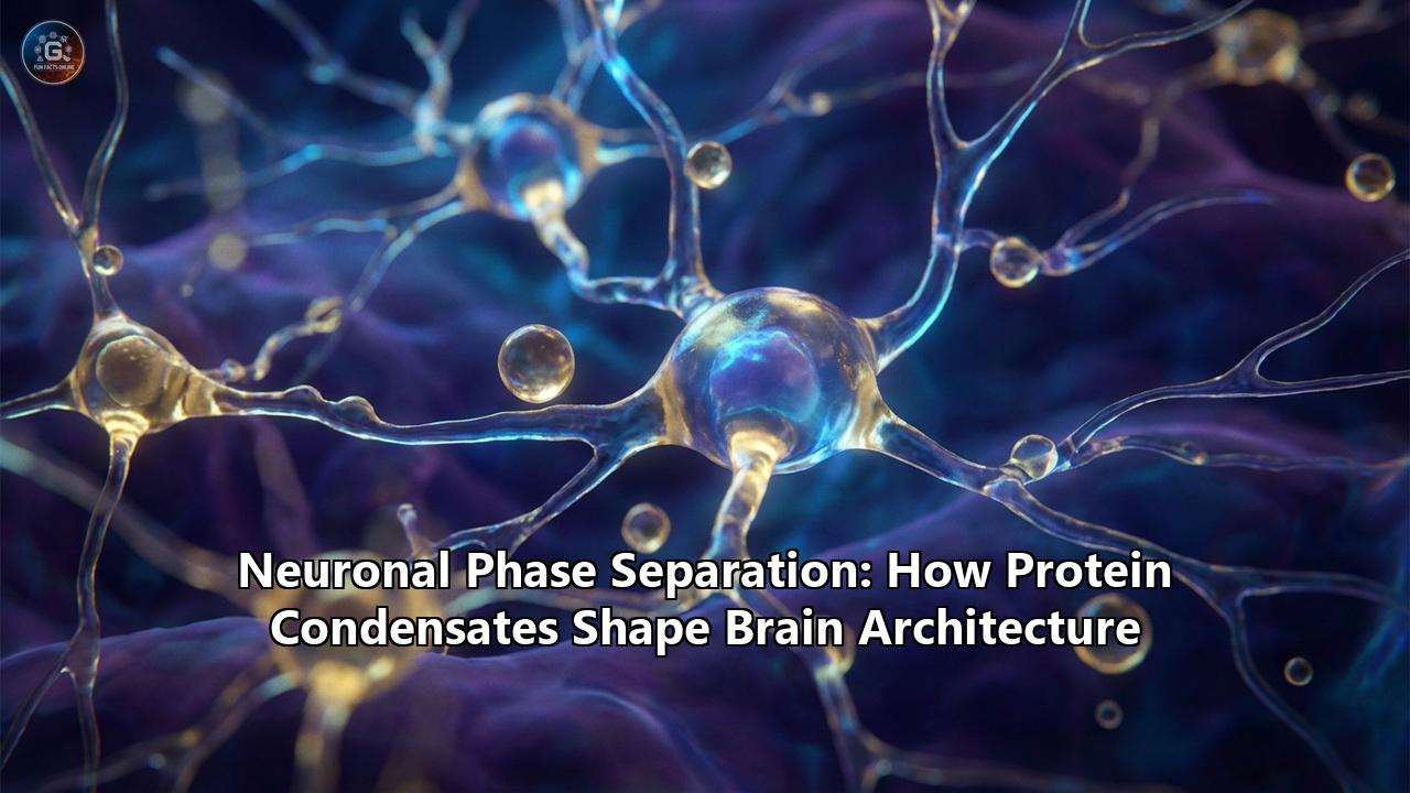
This paradigm-shifting discovery has revealed that the neuronal cytoplasm is not merely a soup, but an archipelago of dynamic, membrane-less organelles—droplets of protein and RNA that form, fuse, and dissolve in response to cellular cues. These "biomolecular condensates" are not random aggregates; they are sophisticated functional compartments that organize the synaptic machinery, regulate gene expression, and drive the morphological complexity of the brain. From the clustering of vesicles in the presynaptic terminal to the compaction of chromatin in the nucleus, phase separation is the invisible architect of neuronal identity.
This article explores the comprehensive role of neuronal phase separation, tracing its influence from the biophysical nanoscale to the macroscopic architecture of brain circuits, and examining how the solidification of these liquid droplets underlies neurodegenerative devastation.
Table of Contents
- The Physics of Thought: Introduction to Neuronal Phase Separation
Defining the "Membrane-less Organelle"
The Biophysics: Multivalency and Intrinsically Disordered Regions (IDRs)
Why Neurons Need Phase Separation: The Scale Problem
- The Presynaptic Terminal: Organizing the Release Machinery
The Vesicle Cluster: Synapsin as a Molecular Glue
The Active Zone: RIM, RIM-BP, and ELKS Condensates
Liprin-α: The Master Architect of Release Sites
Physiological Regulation: How Calcium Melts the Condensate
- The Postsynaptic Density: A Liquid Computer
The PSD-95/SynGAP Complex: Building the Excitatory Synapse
The "Phase-in-Phase" Architecture: Sub-compartmentalization
Synaptic Plasticity (LTP) and Condensate Expansion
The Inhibitory Synapse: Gephyrin and the "iPSD"
- Genomic Architecture: Chromatin Condensates and Gene Regulation
MeCP2 and Heterochromatin Compaction
Rett Syndrome: When the Condensate Fails
Activity-Dependent Transcription: The BDNF Story
Mitochondrial Nucleoids: TFAM Phase Separation
- Neuronal Logistics: RNA Transport Granules
The "Silent" State: Translational Repression in Transit
Key Players: FMRP, TDP-43, and G3BP1
KIF1C and the positioning of mRNA at Growth Cones
Local Translation: Reactivating the Granule at the Synapse
- Shaping the Neuron: Cytoskeleton and Polarity
Actin Polymerization via Condensates (Homer/Shank)
Tau Protein: Coating Microtubules via Phase Separation
Axon Guidance: How Condensates Steer Growth Cones
- The Solidification of Thought: Pathology and Neurodegeneration
The Liquid-to-Solid Transition: Aging of Condensates
ALS and FTD: The Case of TDP-43 and FUS
Alzheimer’s Disease: Tau Tangles as "Petrified" Droplets
Stress Granules: The Crucible of Aggregation
- Therapeutic Frontiers: Melting the Stone
Small Molecules: Lipoamide and 1,6-Hexanediol derivatives
PROTACs and Autophagy Activators
Future Directions: Gene Therapy for Condensate Regulation
1. The Physics of Thought: Introduction to Neuronal Phase Separation
To understand how phase separation shapes the brain, one must first grasp the physical principles that allow liquids to demix. Imagine a bottle of salad dressing containing oil and vinegar. When shaken, they mix, but given time, they separate into distinct layers. This is a phase transition driven by the minimization of free energy. In the cellular environment, a similar process occurs, but instead of oil and vinegar, the players are proteins and nucleic acids.
The "Membrane-less Organelle"Unlike mitochondria or lysosomes, which are enclosed by lipid bilayers, biomolecular condensates have no physical barrier separating them from the surrounding cytoplasm. They maintain their identity through a dense network of weak, transient interactions. This allows them to concentrate specific molecules—enhancing reaction rates by thousands of times—while excluding others. Crucially, because they are liquids, they allow for rapid exchange of components with the surroundings, enabling the cell to respond to signals in microseconds.
The Biophysics: Multivalency and IDRsWhat makes certain neuronal proteins "sticky" enough to phase separate? The secret lies in their structure—or lack thereof. Many key neuronal proteins contain large Intrinsically Disordered Regions (IDRs). Unlike the rigid lock-and-key structures of enzymes, IDRs are flexible, flopping molecular chains that do not fold into a stable 3D shape.
These IDRs are often enriched in specific amino acids (like arginine, glycine, and serine) that facilitate "multivalent" interactions. Think of these proteins as strands of Velcro. A single hook and loop are weak, but thousands of hooks and loops create a strong, yet adjustable, bond. Proteins like FUS, TDP-43, and Synapsin are essentially molecular Velcro, possessing multiple binding domains that allow them to cross-link with one another, reaching a critical concentration where they spontaneously condense into droplets.
Why Neurons Need Phase SeparationNeurons face a unique logistic challenge: size. A motor neuron axon can be a meter long, yet the nucleus is only 10 micrometers wide. Relying on simple diffusion to get neurotransmitter receptors to a synapse is like expecting a letter dropped in New York to drift on the wind to a specific mailbox in London.
Phase separation solves this by creating "local hubs" or depots. Instead of floating freely, mRNA encoding synaptic proteins is packed into high-density transport granules (condensates) that are hitched to motor proteins. At the synapse, phase separation creates a stable, high-density platform (the Postsynaptic Density) that traps receptors in the right place, ensuring they don't drift away.
2. The Presynaptic Terminal: Organizing the Release Machinery
The presynaptic terminal is a marvel of speed. When an action potential arrives, synaptic vesicles must fuse with the membrane in less than a millisecond. This requires vesicles to be clustered tightly near the "release site" (active zone) but kept mobile enough to move when needed.
The Vesicle Cluster: Synapsin as Molecular GlueFor years, electron microscopy revealed tight clusters of synaptic vesicles, but what held them together was a mystery. We now know that Synapsin, a protein coating these vesicles, contains a prominent IDR. In the resting state, Synapsin undergoes phase separation, forming a liquid droplet that engulfs the vesicles.
This "synapsin droplet" acts like a liquid cage. It keeps the reserve pool of vesicles clustered together, preventing them from wandering off. However, this cage has a key: phosphorylation. When a neuron fires, calcium enters and activates the enzyme CaMKII. CaMKII phosphorylates the IDR of Synapsin, disrupting the multivalent interactions that hold the droplet together. The condensate effectively "dissolves" or becomes more fluid, releasing the vesicles so they can move to the membrane and release their neurotransmitter payload. This is a prime example of how the brain uses phase transitions to switch between "storage" and "release" modes.
The Active Zone: RIM, RIM-BP, and ELKSCloser to the membrane lies the "Active Zone," the launchpad for vesicles. Recent structural biology has revealed that the core scaffold proteins here—RIM (Rab3-interacting molecule), RIM-BP (RIM-binding protein), and ELKS—also form condensates.
RIM and RIM-BP contain multiple SH3 and proline-rich domains that allow them to cross-link into a dense sheet on the presynaptic membrane. This condensate has a vital function: it recruits Voltage-Gated Calcium Channels (VGCCs). By concentrating calcium channels into a phase-separated droplet, the active zone ensures that the calcium influx is extremely localized and high-intensity, exactly where the vesicles are docked. This "nanodomain" of calcium is essential for the ultra-fast timing of neurotransmission.
Liprin-α: The Master ArchitectA recent breakthrough identified Liprin-α as a critical regulator of this system. Liprin-α forms a complex with RIM that undergoes phase separation. Intriguingly, Liprin-α appears to tune the material properties of the active zone condensate. Mutations in Liprin-α that disrupt this phase separation lead to "leaky" synapses where calcium channels are not properly clustered, resulting in sluggish or failed transmission. This suggests that the precise "viscosity" of the active zone droplet is tuned by evolution to optimize synaptic speed.
3. The Postsynaptic Density: A Liquid Computer
Across the synaptic cleft lies the Postsynaptic Density (PSD), a thick protein plaque that detects neurotransmitters. The PSD is essentially a biological computer that processes chemical inputs and turns them into electrical signals.
The PSD-95/SynGAP ComplexThe two most abundant proteins in the excitatory PSD are PSD-95 (a scaffold) and SynGAP (an enzyme). In isolation, they are soluble. But when mixed, they spontaneously separate into liquid droplets. This PSD condensate is the structural foundation of the synapse.
This liquid nature explains a long-standing puzzle: how synapses are stable for years (memory) yet plastic enough to change in seconds (learning). The PSD condensate is stable because of the high density of interactions, but individual molecules within it are constantly exchanging with the cytoplasm. This dynamic equilibrium allows the synapse to replace worn-out proteins without dismantling the whole structure.
The "Phase-in-Phase" ArchitectureHigh-resolution imaging has revealed that the PSD is not a uniform blob. It has layers. PSD-95 forms the "core" phase that sits directly under the membrane, trapping AMPA receptors via the auxiliary protein Stargazin. Homer and Shank proteins form a distinct, "pallium" phase further down. This "phase-in-phase" architecture (like a drop of oil inside a drop of water) allows the synapse to vertically stratify its functions: receptor trapping happens at the top, while cytoskeletal connection happens at the bottom.
Synaptic Plasticity (LTP) and Condensate ExpansionWhen we learn, synapses get stronger (Long-Term Potentiation, or LTP). This process involves the physical expansion of the PSD. During LTP, calcium influx triggers enzymes that modify SynGAP. This modification changes the "miscibility" of the SynGAP/PSD-95 phase, causing the droplet to effectively suck in more AMPA receptors from the surrounding membrane. The result is a larger, more sensitive synapse. Conversely, in Long-Term Depression (LTD), the phase properties change to expel receptors, weakening the connection.
The Inhibitory Synapse: Gephyrin and the "iPSD"While excitatory synapses use PSD-95, inhibitory synapses (which use GABA or Glycine) use a scaffold called Gephyrin. For a long time, the inhibitory PSD (iPSD) was thought to be a rigid lattice. New evidence shows that Gephyrin, when bound to GABA receptors, also undergoes phase separation.
The "iPSD" condensate is a thin, sheet-like liquid layer. Interestingly, the linker region of Gephyrin acts as an auto-inhibitor of this phase separation. Binding of other proteins (like Neuroligin-2) or phosphorylation releases this inhibition, triggering the formation of the condensate. This ensures that inhibitory synapses only form at the correct location, preventing "runaway" inhibition that could silence the brain.
4. Genomic Architecture: Chromatin Condensates and Gene Regulation
The brain's architecture isn't just about synapses; it starts in the nucleus with the regulation of gene expression. The 2-meter-long human genome must be packed into a 10-micron nucleus, yet specific genes must remain accessible for learning-induced transcription.
MeCP2 and Heterochromatin Compaction MeCP2 is a protein famous for its link to Rett Syndrome, a severe neurodevelopmental disorder. MeCP2 binds to methylated DNA (repressed genes) and condenses it into heterochromatin. It turns out that MeCP2 possesses massive IDRs and forms liquid condensates with DNA.These MeCP2 droplets act like oil droplets that pull distinct strands of DNA together, compacting them into a silenced state. This is crucial for "shutting off" genes that shouldn't be active in mature neurons, effectively locking in the neuronal identity.
Rett Syndrome: When the Condensate FailsMany mutations causing Rett Syndrome disrupt the ability of MeCP2 to phase separate. The R133C mutation, for example, prevents the IDR from interacting properly. The result is that heterochromatin does not compact into tight droplets; it remains "fluffy" and disorganized. This leads to "transcriptional noise"—genes that should be silent essentially leak expression, disrupting the precise symphony of proteins needed for brain function.
Activity-Dependent Transcription: The BDNF StoryWhen a neuron is highly active, it needs to produce BDNF (Brain-Derived Neurotrophic Factor) to reinforce its synapses. This requires rapidly opening the chromatin at the BDNF gene.
In the resting state, the BDNF promoter is buried inside a repressive MeCP2 condensate. Upon neuronal stimulation, calcium signaling leads to the phosphorylation of MeCP2. This phosphorylation acts as a "phase switch," reducing MeCP2's affinity for the condensate. MeCP2 exits the droplet, causing the local heterochromatin to dissolve (de-mix). This exposes the BDNF gene to transcription factors, allowing a burst of gene expression. This mechanism—phase transition as a gene switch—is a fundamental way the genome responds to experience.
Mitochondrial Nucleoids: TFAM Phase SeparationNeurons are energy-hungry, and their mitochondria contain their own DNA (mtDNA). The packaging protein TFAM organizes mtDNA into structures called "nucleoids." Recent studies show that TFAM and mtDNA undergo phase separation to form these nucleoids.
These droplets regulate mitochondrial transcription. When the TFAM condensate is "loose," transcription enzymes can enter, and the mitochondria produce energy. Under stress, the condensates can harden, shutting down mitochondrial gene expression to protect the DNA from damage. This highlights that phase separation is a universal organizational principle, extending even to endosymbiotic organelles.
5. Neuronal Logistics: RNA Transport Granules
A neuron's dendrites can be centimeters away from the nucleus. If the cell relied on proteins made in the soma to diffuse to the synapse, the delay would be prohibitive. Instead, neurons ship mRNA to the synapse and translate it locally "on demand." This shipping container is a phase-separated condensate.
The "Silent" State: Translational Repression in Transit RNA Transport Granules are essentially liquid droplets containing mRNA, ribosomes, and RNA-binding proteins (RBPs). Crucially, inside these granules, translation is paused. The high density of RBPs like FMRP (Fragile X Messenger Ribonucleotide Protein) creates an environment that sterically blocks the translation machinery. This prevents the neuron from making proteins in the wrong place (e.g., in the axon shaft) during transport. Key Players: FMRP, TDP-43, and G3BP1- FMRP: Loss of FMRP causes Fragile X Syndrome. FMRP is a master regulator of these granules. It uses its IDR to drive the phase separation of the granule. In Fragile X, without FMRP, these granules are unstable or "leaky," leading to excessive protein synthesis in dendrites, which results in immature, "spindly" dendritic spines and intellectual disability.
- TDP-43: Famous for its role in ALS, TDP-43 normally functions to shuttle mRNA for the cytoskeleton (like neurofilament mRNA) down the axon.
- G3BP1: A core component of stress granules, G3BP1 also helps scaffold neuronal transport granules.
The motor protein KIF1C, a kinesin, has been found to have a C-terminal IDR that forms condensates. These KIF1C condensates don't just move; they accumulate at the tips of growing axons (growth cones). By doing so, they drag specific mRNAs (like actin mRNA) to the very edge of the cell. This ensures that the building blocks for new cytoskeleton are synthesized exactly where the axon is trying to grow, providing the raw material for neural expansion.
Local Translation: Reactivating the GranuleWhen the granule reaches an active synapse, synaptic signaling (e.g., mGluR activation) triggers the phosphorylation of FMRP and other granule components. This causes the granule to "melt" or de-mix. The mRNA is released from the repressive liquid phase into the cytoplasm, where ribosomes immediately grab it and start synthesizing protein. This allows the synapse to remodel itself in real-time, strengthening the specific connection that was stimulated.
6. Shaping the Neuron: Cytoskeleton and Polarity
The shape of a neuron—its polarity, branching, and spine density—is determined by the cytoskeleton. Phase separation acts as a spatial template for cytoskeleton assembly.
Actin Polymerization via CondensatesIn dendritic spines, actin filaments must push against the membrane to make the spine grow. The PSD condensate, specifically the Homer/Shank layer, acts as a nucleation platform. These condensates can recruit actin-regulatory proteins and increase the local concentration of actin monomers (G-actin) by orders of magnitude. This high local concentration drives the rapid polymerization of F-actin bundles, providing the mechanical force to expand the spine head during LTP.
Tau Protein: Coating Microtubules Tau is an intrinsically disordered protein that stabilizes microtubules in the axon. We now know that Tau forms a liquid phase on the surface of the microtubule. This "liquid sheath" allows Tau to protect the microtubule from severing enzymes while still allowing motor proteins (like Kinesin) to slide through the liquid layer. It’s a protective lubricant. If Tau phase separates away* from the microtubule (due to hyperphosphorylation), the microtubule falls apart—a key event in Alzheimer’s pathology. Axon Guidance: Steering the Growth ConeAs an axon navigates the developing brain, its growth cone turns toward attractive cues (like Netrin) and away from repulsive ones. This turning is driven by asymmetric actin polymerization. Emerging evidence suggests that guidance cues trigger the local formation of signaling condensates on one side of the growth cone. These condensates trap actin regulators, causing the cytoskeleton to build up on that side and steering the axon. This effectively makes the growth cone a "phase separation engine" that steers based on where the droplets form.
7. The Solidification of Thought: Pathology and Neurodegeneration
The liquid nature of these condensates is their strength, but also their Achilles' heel. Liquids are metastable. Over time, or under stress, protein droplets can "age," becoming more viscous, then gel-like, and finally solid. This liquid-to-solid phase transition is the biophysical ground zero for many neurodegenerative diseases.
The "Aging" of CondensatesIn a healthy neuron, condensates form and dissolve constantly. However, genetic mutations or chronic stress can make these droplets "too sticky." They persist for too long. Over time, the disordered proteins inside them settle into a lower-energy state: the amyloid fibril. This is an irreversible solid aggregate. Once formed, it cannot be dissolved by the cell's machinery, and it often acts as a seed, converting other functional liquid proteins into solids.
ALS and FTD: TDP-43 and FUSIn Amyotrophic Lateral Sclerosis (ALS) and Frontotemporal Dementia (FTD), nearly 97% of cases involve the aggregation of TDP-43. Mutations in the IDR of TDP-43 (often in the alpha-helical region) disrupt its liquid properties, favoring the solid state.
Similarly, FUS (Fused in Sarcoma) mutations famously accelerate the liquid-to-solid transition. A "fresh" FUS droplet is liquid and functional. An "aged" FUS droplet (after a few hours in a test tube, or years in a patient) becomes a hardened gel that traps RNA and prevents it from being translated. This starves the axon of essential proteins, leading to motor neuron death.
Alzheimer’s Disease: Tau Tangles as "Petrified" DropletsThe neurofibrillary tangles of Alzheimer’s are made of solid Tau. As mentioned, Tau normally forms a liquid sheath on microtubules. In disease, hyperphosphorylation pushes Tau to detach and form droplets in the cytoplasm. These free-floating Tau droplets are highly concentrated. Inside this crowded environment, Tau molecules jam together, nucleating the formation of paired helical filaments (PHFs). The liquid droplet essentially "petrifies" into a tangle, killing the neuron from the inside.
Stress Granules: The Crucible of AggregationWhen a cell is stressed (e.g., by toxins or heat), it forms Stress Granules (SGs) to protect its mRNA. These are large condensates. Normally, when stress resolves, SGs disassemble. However, in aging brains or disease states, SGs fail to dissolve. They become "persistent SGs." Because SGs contain high concentrations of aggregation-prone proteins like TDP-43 and FUS, they act as a crucible. The longer they persist, the more likely the components inside will nucleate into pathogenic solids. This "intra-condensate demixing" is now believed to be the triggering event for protein aggregation in ALS.
8. Therapeutic Frontiers: Melting the Stone
If the problem is the transition from liquid to solid, the solution is to reverse it or prevent it. The field of "condensate therapeutics" is exploding.
Small MoleculesResearchers are screening for small molecules that act as "molecular solvents" for specific condensates.
- 1,6-Hexanediol is a chemical tool used in labs to dissolve weak condensates, though it is too toxic for drugs.
- Lipoamide and its derivatives have shown promise in restoring the liquidity of stress granules in ALS models.
- Planar aromatic compounds can intercalate into the amyloid structures or IDRs, preventing the "stacking" interactions that lead to solidification.
The goal is to find a drug that can cross the blood-brain barrier and subtly shift the phase diagram of proteins like TDP-43, keeping them in the functional liquid state without disrupting healthy condensates.
PROTACs and Autophagy ActivatorsAnother strategy is to simply remove the solid aggregates. PROTACs (Proteolysis Targeting Chimeras) are designed molecules that bind to a pathological protein (like aggregated Tau) and simultaneously bind to an E3 ubiquitin ligase, tagging the aggregate for destruction by the proteasome.
Similarly, drugs that boost autophagy (the cell's recycling system) can help the neuron "eat" the solidified condensates. Since the solid aggregates are often too large for the proteasome, autophagy is the only way to clear them.
Gene TherapyFor genetic forms of these diseases (like FUS-ALS), gene silencing (ASOs) can reduce the concentration of the mutant protein. Since phase separation is concentration-dependent, lowering the protein level below the "critical concentration" (Csat) can completely prevent the formation of pathological condensates, stopping the disease before it starts.
Conclusion
The discovery of neuronal phase separation has fundamentally rewritten the textbook on brain architecture. We now understand that the brain is not just a circuit of wires, but a landscape of shifting liquids. From the ephemeral flash of a thought (synaptic release) to the lifelong stability of a memory (chromatin compaction), the transition between liquid and solid states underpins our very existence. And in the tragic solidification of these vital fluids lies the root of neurodegeneration—a realization that has finally given us a clear target for curing the incurable. The future of neuroscience is, quite literally, fluid.
Reference:
- https://pmc.ncbi.nlm.nih.gov/articles/PMC12157082/
- https://www.researchgate.net/publication/229089723_The_Presynaptic_Active_Zone
- https://pmc.ncbi.nlm.nih.gov/articles/PMC12151379/
- https://www.biorxiv.org/content/10.1101/2024.08.29.610253v1.full-text
- https://pmc.ncbi.nlm.nih.gov/articles/PMC8026974/
- https://www.researchgate.net/publication/378873784_Thermodynamic_modulation_of_gephyrin_condensation_by_inhibitory_synapse_components
- https://www.pnas.org/doi/10.1073/pnas.2313236121
- https://www.researchgate.net/publication/358577139_MeCP2-induced_heterochromatin_organization_is_driven_by_oligomerization-based_liquid-liquid_phase_separation_and_restricted_by_DNA_methylation
- https://www.tandfonline.com/doi/full/10.1080/19491034.2021.2024691
- https://pubmed.ncbi.nlm.nih.gov/35156529/
- https://pubmed.ncbi.nlm.nih.gov/32698189/
- https://www.researchgate.net/publication/9030790_DNA_Methylation-Related_Chromatin_Remodeling_in_Activity-Dependent_Bdnf_Gene_Regulation
- https://www.researchgate.net/publication/236184288_Role_of_BDNF_epigenetics_in_activity-dependent_neuronal_plasticity
- https://link.springer.com/article/10.15252/embj.2020107165
- https://www.researchgate.net/figure/Phase-separation-of-TFAM-mtDNA-in-vitro-and-in-vivo-a-TEM-image-of-extracted-circular_fig3_355722973
- https://condensates.com/publications/self-assembly-of-multi-component-mitochondrial-nucleoids-via-phase-separation-5/
- https://www.biorxiv.org/content/10.1101/2021.12.30.474545.full
- https://www.biorxiv.org/content/10.1101/2021.12.30.474545v1.full
- https://pubmed.ncbi.nlm.nih.gov/33139925/
- https://pmc.ncbi.nlm.nih.gov/articles/PMC9510713/