For decades, the dawn of a new life was viewed through a lens of molecular chaos. Biologists peering into the microscopic world of a newly fertilized egg operated under a long-held assumption: the embryonic genome was a structural “blank slate.” It was envisioned as a disordered, tangled mass of DNA, waiting passively in the dark for the embryo to finally "wake up" and begin reading its genetic instructions. According to this prevailing dogma, biological order and architectural precision only emerged after the embryo flipped its master genetic switch—an event known as Zygotic Genome Activation (ZGA).
However, science is a discipline of perpetual revision, and this long-standing view has just been entirely overturned. In a landmark study published in February 2026 in the journal Nature Genetics, researchers have proven that the genetic blueprint of life is not born in chaos. Led by Professor Juanma Vaquerizas and his team at the MRC Laboratory of Medical Sciences (LMS), embedded within Imperial College London, scientists have mapped the earliest stages of genomic development to reveal a highly disciplined, intricately designed construction site. Well before the genome officially awakens, a sophisticated 3-dimensional scaffold of DNA is already being meticulously built.
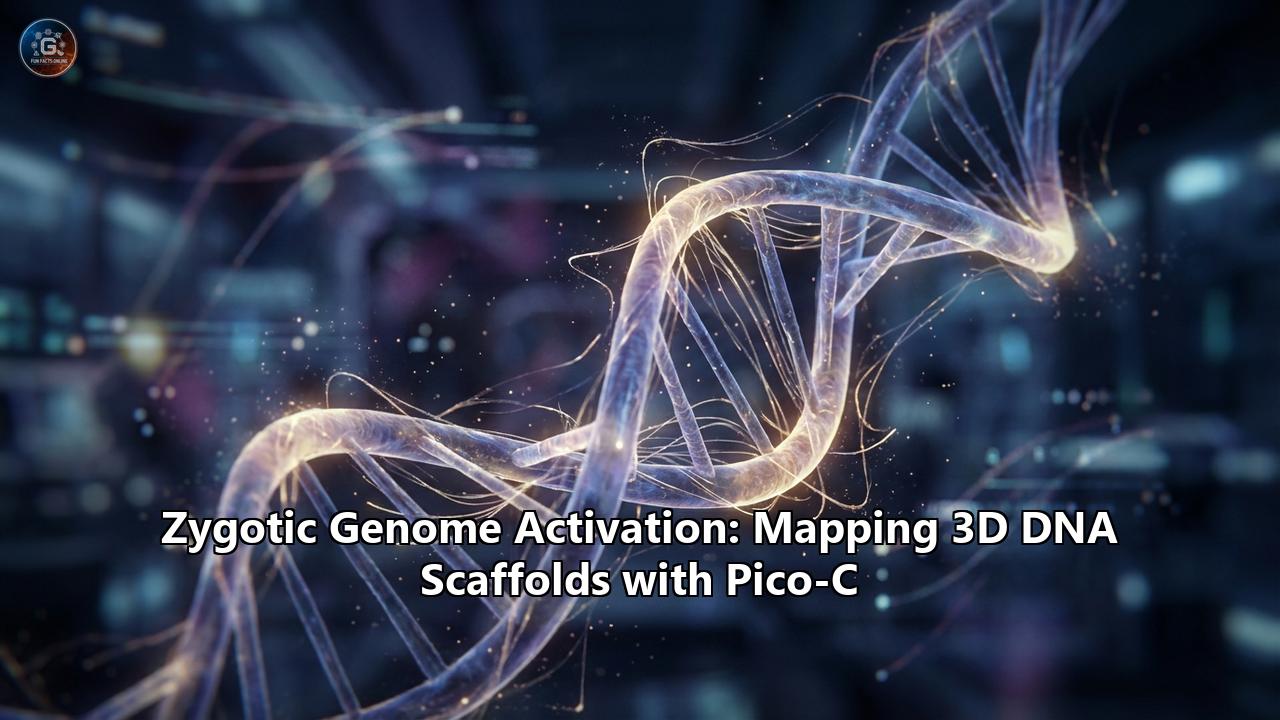
To achieve this breakthrough, the research team had to invent a pioneering, ultra-sensitive sequencing technology known as Pico-C. This technique allows scientists to visualize the 3D conformation of the genome with unprecedented resolution, using minute quantities of biological samples that are ten times smaller than those required by conventional methods. The implications of this discovery are profound, reshaping our fundamental understanding of early embryonic development, gene regulation, and the origins of developmental diseases.
To fully grasp the magnitude of this paradigm shift, we must journey into the microscopic, fast-paced world of early development, explore the revolutionary mechanics of Pico-C, and decode the modular scaffolding that anticipates the spark of life.
The Maternal-to-Zygotic Transition and the Spark of ZGA
When an egg is first fertilized, it does not immediately rely on its own DNA to survive and grow. Instead, it is coasting on a molecular inheritance. The mother packs the egg with a vast reservoir of maternal proteins and messenger RNAs (mRNAs). During the earliest hours of development, these maternal factors drive cellular division and basic metabolic functions. The embryo's own newly formed genome remains largely silent, tightly packed within the nucleus, acting merely as a passenger.
This period of maternal control is temporary. For the embryo to develop into a fully functioning, independent organism, it must eventually take control of its own destiny. This handover of power is called the Maternal-to-Zygotic Transition (MZT). The defining moment of this transition is Zygotic Genome Activation (ZGA)—the critical milestone where maternal mRNAs are degraded, and the embryo’s own DNA is transcribed into RNA for the first time.
The research team chose the fruit fly (Drosophila melanogaster) as their model organism to study this window. The fruit fly is a cornerstone of developmental genetics due to its incredibly rapid, synchronized early development. In the first few hours following fertilization, a fruit fly embryo undergoes a furious series of 13 nuclear divisions without fully dividing its cellular membranes, creating a single cell containing roughly 6,000 nuclei—a state known as a syncytium.
During these early pre-ZGA stages (Nuclear Cycles 1 through 13), the genome is mostly quiet, with only minor, sporadic transcription occurring around Nuclear Cycles 8 to 12. The major wave of ZGA erupts precisely at Nuclear Cycle 14. Because these divisions happen so rapidly—sometimes taking only eight minutes per cycle—scientists previously assumed there was simply no time for the DNA to organize itself into complex 3D structures. The assumption was that the fast-paced cellular dynamism would disrupt any attempt to build an organized genomic architecture.
The Blank Slate Myth and the Limits of Traditional Genomics
To understand why the "blank slate" myth persisted for so long, we must look at the limitations of previous genomic technologies. The human genome—and the fruit fly genome—is not just a linear string of A, C, T, and G letters. If you were to stretch out the DNA from a single human cell, it would be roughly two meters long. To fit inside a microscopic nucleus, this DNA must fold, loop, and coil in three-dimensional space.
This 3D architecture is not random. It is highly strategic. How DNA folds determines which genes are exposed to transcription machinery and which are hidden away. Distant regulatory elements, such as "enhancers," must physically loop through 3D space to touch the "promoters" of their target genes to turn them on. If the DNA misfolds, the wrong gene might be activated, or a crucial gene might remain silent, leading to severe developmental defects or diseases like cancer.
Over the last decade, scientists developed powerful techniques called chromosome conformation capture (such as Hi-C and Micro-C) to map these 3D interactions. These methods act like molecular glue, freezing the DNA in place, cutting it, and linking DNA strands that are physically close to each other in 3D space, even if they are millions of base pairs apart on the linear sequence. By sequencing these linked fragments, scientists can build a 3D map of the genome.
However, there was a massive catch: traditional Hi-C and Micro-C required millions of cells to generate a reliable map. This is relatively easy to achieve when studying a liver or a tumor, but it is impossible when studying the earliest moments of embryonic development, where an organism consists of only a handful of nuclei. Because of this technological blind spot, the pre-ZGA genome remained an unmapped territory. Without the tools to see it, the scientific community defaulted to the assumption that before ZGA, the genome was just a disordered tangle waiting for the 'on' switch.
Enter Pico-C: Seeing More with Less
To pierce the veil of early development, Professor Vaquerizas and his team had to push the boundaries of molecular biology. The result was Pico-C, an ultra-sensitive, low-input variant of Micro-C that acts as a high-resolution magnifying glass for the 3D genome.
Pico-C derives its name from the pico-scale samples it requires. It operates with about one-tenth of the biological input demanded by standard methods, allowing scientists to map genome structures from minute quantities of scarce material like early-stage embryos.
The innovation behind Pico-C lies in its exquisitely optimized biochemical workflow. Traditional Hi-C uses restriction enzymes to cut the DNA, which can leave blind spots and limit resolution. Pico-C, building on the principles of Micro-C, utilizes micrococcal nuclease (MNase), an enzyme that digests the DNA in a much finer, more controlled manner, yielding small fragments of about 150 base pairs. This preserves the finest, most delicate DNA-DNA interactions.
The Pico-C process unfolds in several meticulously choreographed steps:
- Isolation and Fixation: Nuclei are isolated from precisely staged Drosophila embryos (sorted via advanced fluorescence techniques like PCNA-GFP to isolate specific pre-ZGA nuclear cycles). The chromatin is then fixed with formaldehyde, effectively freezing the 3D architecture in place.
- Fine Digestion: MNase is deployed to carefully digest the chromatin into tiny, manageable fragments.
- Proximity Ligation and Biotinylation: The interacting, closely situated DNA ends are blunt-end ligated together, and the newly formed junctions are marked with biotin, a chemical tag.
- Enrichment and Sequencing: The DNA is sheared, and specialized capture probes combined with a biotin-streptavidin pulldown method are used to fish out only the informative, ligated junctions. These are then run through next-generation sequencers.
By applying Pico-C to the rapidly dividing Drosophila embryos in the hours before major ZGA, the research team finally had the data to see what the genome was doing in the dark.
A Highly Disciplined Construction Site
What the Pico-C data revealed was nothing short of revolutionary. As lead author Noura Maziak explained, “We used to think of the time before the genome awakens as a period of chaos. But by zooming in closer than ever before, we can see that it's actually a highly disciplined construction site. The scaffolding of the genome is being erected in a precise, modular way, long before the 'on' switch is fully flipped”.
The researchers found that well before Nuclear Cycle 14—the moment the genome officially "turns on"—an intricate, pre-wired 3D scaffold is actively forming. Contrary to the blank slate hypothesis, the genome was aggressively organizing itself into loops, topologically associating domains (TADs), and distinct spatial compartments.
Several key discoveries emerged from the Pico-C maps:
1. Modular Genomic Loops and Pre-WiringThe DNA was folding into specific, modular configurations. The team observed dynamic loops emerging in a stepwise, methodical fashion. At crucial developmental gene loci—such as zen, ftz, and Antp—the genome was already forming loops that anchored gene promoters to their respective distant enhancers. This means the physical connections necessary for gene activation were being established before the transcriptional machinery was even turned on. The genome was effectively pre-wiring itself, ensuring that the moment ZGA was initiated, the genetic instructions were primed and ready for immediate, rapid action.
2. The Role of Pioneer FactorsThis intricate architecture does not assemble itself by magic. The study identified that specific "pioneer factors"—proteins capable of accessing and opening condensed chromatin—are the master architects of this early scaffolding. The boundaries of these early structural domains were heavily enriched with binding motifs for factors like GAF (GAGA factor) and Zelda (Zld). These pioneer factors act like the construction foremen, laying down the structural boundaries and insulating different domains so that regulatory signals do not cross over and accidentally activate the wrong genes.
3. Resilience Amidst Cellular DynamismPerhaps most astonishingly, the researchers discovered that these early genomic loops were incredibly robust. Despite the fast-paced nuclear divisions occurring every few minutes in the Drosophila embryo, the 3D loops persisted through mitosis. The structure withstood the immense physical stress of cellular division, defying earlier assumptions that rapid mitosis would obliterate any complex genomic architecture.
4. Context-Dependent Regulation via Spatial AutocorrelationUsing advanced spatial autocorrelation analysis, the team found that the chromatin state and its 3D structure are intimately coupled long before widespread transcription. By inhibiting transcriptional elongation, they noted site-specific effects where some early loops were retained, pointing to a highly contextual, location-dependent regulatory logic governing the pre-ZGA genome.
Why Pre-Assemble the Genome?
From an evolutionary perspective, this pre-ZGA scaffolding makes perfect sense. Early development is a race against time. The embryo must rapidly transition from a single cell into a complex, multicellular organism capable of surviving in its environment.
If the genome were truly a disordered tangle at the onset of ZGA, the embryo would have to simultaneously untangle its DNA, find the correct genes, bring the regulatory elements into physical proximity, and begin transcription all at once. The potential for catastrophic errors—activating a leg-development gene in what should be the head, for instance—would be enormous.
By erecting a precise, modular 3D scaffold before the "on" switch is flipped, the embryo solves this logistical nightmare. The modularity of the loops allows the embryo to balance flexibility with tight control. Each architectural unit can be governed independently, ensuring that specific genomic regions can be activated or silenced with surgical precision the exact millisecond they are needed. It is the biological equivalent of an orchestra setting up their instruments, tuning their strings, and taking their seats, perfectly poised for the conductor's first downbeat.
Human Parallels: When the Scaffolding Collapses
While the initial discovery was made in the humble fruit fly, the principles of 3D genome organization are deeply conserved across the animal kingdom, including humans. The implications of the Pico-C findings extend far beyond basic developmental biology; they provide crucial insights into human fertility, genetic disorders, and cancer.
A companion paper published simultaneously in Nature Cell Biology by Renard Lewis and colleagues at ETH Zurich illustrated what happens in human cells when this vital 3D scaffolding is disrupted. The researchers utilized Pico-C on human cell lines and experimentally depleted specific tethering proteins (LBR and LAP2) that anchor the genome to the nuclear envelope.
The results were catastrophic. Without these tethers, the heterochromatin (the tightly packed, silent regions of DNA) detached and the 3D structure collapsed. This collapse did not just disrupt gene expression; the disorganized DNA mimicked the signature of a viral infection, triggering an intense cGAS-STING inflammatory immune response. As Professor Vaquerizas noted, "These two studies tell a complete story. The first shows us how the genome's 3D structure is carefully built at the start of life. The second shows us the disastrous consequences for human health if that structure is allowed to collapse".
In the realm of reproductive medicine, these findings shine a light on the heartbreaking mystery of in vitro fertilization (IVF) failures. It is estimated that 10-15% of human IVF failures occur during the critical pre-ZGA window. If the maternal factors fail to properly construct the pre-ZGA 3D scaffold, the subsequent activation of the embryonic genome will be chaotic, resulting in embryonic arrest. Understanding the architectural rules laid out by Pico-C may eventually lead to new diagnostics or interventions to support early embryonic viability in fertility clinics.
Furthermore, aberrant 3D genome looping is a known driver in oncology. In many cancers, the boundaries that separate different genomic compartments break down, allowing powerful enhancers to inappropriately contact and activate oncogenes (such as the MYC gene), driving explosive tumor growth. By studying how these loops are normally formed and insulated during the blank-slate-like state of early embryogenesis, cancer researchers can glean new strategies for identifying and potentially correcting pathogenic structural variants in tumors.
The Future of Precision Medicine and 3D Genomics
The introduction of Pico-C represents a watershed moment in the field of genomics. Because it bypasses the need for massive cell populations, Pico-C democratizes the study of the 3D genome. It opens entirely new frontiers for clinical research.
Moving forward, scientists can deploy Pico-C on scarce and precious clinical samples. This includes mapping the 3D genome of rare patient-derived stem cells, tracking architectural changes in circulating tumor cells isolated from liquid biopsies, or investigating tiny biopsies from patients with rare genetic or developmental diseases. Standard clinical diagnostic tests, which traditionally read DNA linearly, often miss complex structural variations that only manifest in 3D space. Pico-C, alongside other genomic proximity mapping techniques, promises to reveal hidden structural variants, bringing answers to patients with undiagnosed genetic conditions.
Additionally, the insights provided by Pico-C will fuel the development of predictive artificial intelligence models. Researchers are already training deep learning algorithms (such as Akita and Orca) to predict 3D genome folding directly from DNA sequences. With the high-resolution, temporally mapped data provided by Pico-C during the dynamic stages of ZGA, these computational models will become vastly more accurate, allowing scientists to simulate how genetic mutations might disrupt 3D architecture before a physical experiment is even performed.
The Symphony Before the Music Starts
For decades, we looked at the earliest moments of life and saw only chaos waiting for a spark. The development of Pico-C has handed us a new pair of glasses, and with them, the chaos has resolved into breathtaking order.
The discovery that the genome pre-assembles a complex, 3-dimensional scaffold before Zygotic Genome Activation is a testament to the elegant foresight of cellular biology. It reveals that life does not stumble into existence; it is carefully, architecturally prepared. The promoters and enhancers are brought together, the boundaries are drawn, and the stage is perfectly set.
As technology like Pico-C continues to refine our view of the genome's spatial organization, it solidifies the reality that DNA is much more than a linear code. It is a physical, dynamic structure—a microscopic origami whose folds dictate the blueprint of life itself. Unlocking the secrets of this hidden scaffolding not only rewrites the textbooks of developmental biology but also charts a promising new course for treating the diseases that arise when the architecture of life falls apart.
Reference:
- https://bioquicknews.com/new-technology-reveals-hidden-dna-scaffolding-built-before-life-switches-on/
- https://scitechdaily.com/scientists-discover-dna-is-already-organized-before-life-switches-on/
- https://www.news-medical.net/news/20260224/New-Pico-C-technology-maps-genome-structure-before-activation.aspx
- https://bioengineer.org/new-technology-uncovers-hidden-dna-scaffolding-formed-before-life-activates/
- https://distilinfo.com/2026/02/25/pico-c-technology-reveals-genome-structure-before-activation/
- https://www.academicjobs.com/research-publication-news/pico-c-reveals-pre-life-dna-scaffolding-or-mrc-lms-uk-research-6264
- https://www.news-medical.net/news/20251029/Novel-3D-chromosome-mapping-method-can-reliably-reveal-hidden-structural-variants.aspx
- https://www.researchgate.net/publication/360559715_Sequence-based_modeling_of_three-dimensional_genome_architecture_from_kilobase_to_chromosome_scale
- https://bioquicknews.com/