The human brain, a three-pound universe of gelatinous tissue, remains the most complex structure in the known cosmos. For centuries, explorers have sought to map its terrain, not with ships and compasses, but with scalpels, microscopes, and eventually, supercomputers. This is the story of Neuromapping and Brain Atlases—the centuries-long quest to draw a map of the human mind.
From the crude sketches of phrenologists to the digital "Google Earth" of the brain being built today, this field represents the intersection of anatomy, technology, and philosophy. It is a discipline where a difference of a few millimeters can mean the difference between moving your hand and being paralyzed, between speaking fluently and losing your voice forever.
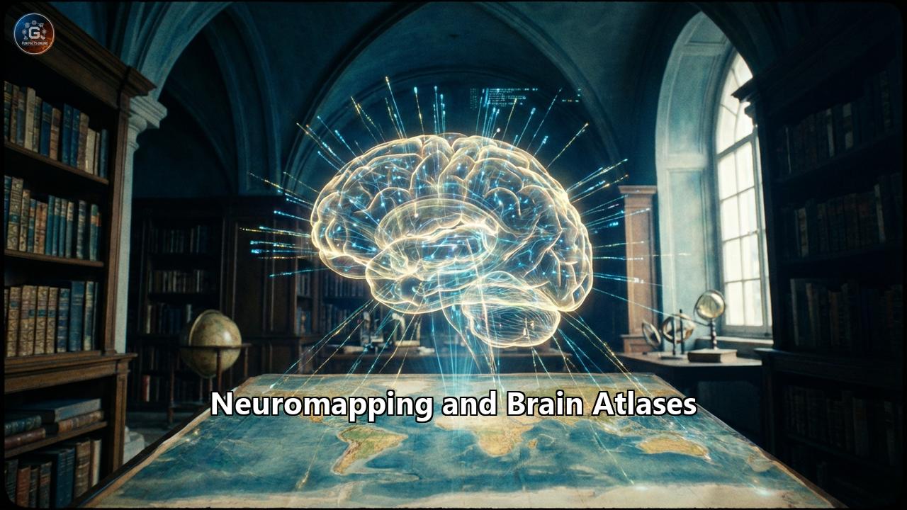
This comprehensive guide explores the history, technology, and future of brain mapping, designed to take you from the early days of rigorous dissection to the futuristic visions of digital twins and mind-reading interfaces.
Part I: The Cartographers of the Mind
The Early sketchers: From Phrenology to Cytoarchitecture
The desire to localize function in the brain is ancient, but the scientific journey truly began with a misstep: Phrenology. In the early 19th century, Franz Joseph Gall proposed that the brain was divided into 27 "organs," each responsible for a personality trait like "benevolence" or "destructiveness." He believed that the size of these organs would create bumps on the skull. While phrenology was scientifically flawed, it planted a crucial seed: the idea of functional localization—that specific parts of the brain do specific things.
The real breakthrough came not from feeling skulls, but from tragedy. In 1848, a railroad foreman named Phineas Gage had a tamping iron blasted through his left frontal lobe. Miraculously, he survived, but his personality shifted drastically. The polite, hardworking man became fitful and irreverent. This famous case study provided the first hard evidence that the frontal lobes were the seat of personality and social behavior.
Soon after, in 1861, the French physician Paul Broca met a patient named Louis Victor Leborgne, who could only say the syllable "tan." When Leborgne died, Broca autopsied his brain and found a specific lesion in the left frontal lobe. This area, now known as Broca’s Area, was the first anatomical proof of a language center in the brain.
Korbinian Brodmann and the Map That Lasted a Century
The true "Mercator Projection" of the brain arrived in 1909. A German anatomist named Korbinian Brodmann spent years hunched over a microscope, analyzing the cellular structure (cytoarchitecture) of brain slices. He noticed that the cortex—the brain's outer layer—was not uniform. In some places, the layers of cells were thick and dense; in others, they were thin and sparse.
Brodmann divided the cortex into 52 distinct areas based on these cellular patterns. Remarkably, his map was not just anatomical; it turned out to be functional. Brodmann Area 4 is the primary motor cortex, controlling voluntary movement. Area 17 is the primary visual cortex, processing what you see. Even today, more than 100 years later, neurosurgeons and researchers still refer to "Brodmann Areas" (BAs) as the standard coordinates of the cortex. It remains the most enduring map in neuroscience history.
Part II: The Tools of the Trade
Modern cartographers do not use quill pens; they use magnetic fields and quantum physics. The revolution in brain mapping is driven by imaging technology that allows us to peer inside the living skull without making a single cut.
MRI: The Structural Canvas
Magnetic Resonance Imaging (MRI) is the foundation of modern neuromapping. It uses a massive magnet (often 1.5 to 7 Tesla in strength) to align the protons in the water molecules of your body. When a radio frequency pulse knocks these protons out of alignment, they emit a signal as they spin back into place. Since different tissues (gray matter, white matter, fluid) have different water contents, they emit different signals.The result is a high-resolution, 3D structural map of the brain’s anatomy. An MRI scan is like a satellite photo of a city; it shows you the streets, the buildings, and the parks, but it doesn't tell you where the traffic is flowing or what the people inside the buildings are doing.
fMRI: Mapping the Traffic
To see the "traffic"—neural activity—scientists use functional MRI (fMRI). This technique relies on the BOLD signal (Blood-Oxygen-Level Dependent). When a specific brain area is active (e.g., when you tap your finger), it consumes more oxygen. The body rushes oxygenated blood to that spot. Oxygenated blood has different magnetic properties than deoxygenated blood, and the fMRI scanner can detect this change.
If an MRI is a map of the city, fMRI is the traffic report. It lights up the "roads" that are busy during specific tasks, allowing researchers to create functional maps for language, memory, and emotion.
DTI: Tracing the Wiring
The brain's gray matter (the computing centers) is connected by white matter (the cables). Diffusion Tensor Imaging (DTI) is a technique that tracks the movement of water molecules. In the brain's white matter tracts, water flows more easily along the nerve fibers than across them, much like water flowing through a hose.
By tracking this directional flow, DTI allows scientists to reconstruct the brain's wiring diagram. This process, called tractography, produces stunning, rainbow-colored images where red might represent fibers running left-to-right, blue for up-and-down, and green for front-to-back. It reveals the superhighways of information transfer, such as the corpus callosum (connecting the two hemispheres) and the arcuate fasciculus (connecting language areas).
Part III: The Great Atlases
A map is useless without a coordinate system. If a surgeon wants to target a tumor, they need a precise reference frame. This led to the creation of standardized brain atlases.
The Talairach Atlas: A Flawed Standard
For decades, the gold standard was the Talairach and Tournoux Atlas (1988). It was based on the post-mortem dissection of a single brain—that of a 60-year-old French woman. Researchers created a 3D coordinate grid (x, y, z) based on this one brain, and for years, every other brain was warped and stretched to fit this grid.
However, relying on a single, elderly brain had major drawbacks. It didn't account for the massive variability between individuals. A young man's brain and an elderly woman's brain can differ significantly in shape and size. The scientific community realized they needed a "population-based" map.
The MNI Templates: The Average Brain
Enter the Montreal Neurological Institute (MNI). Researchers there scanned hundreds of young, healthy brains and used supercomputers to average them together. The result, known as the MNI152 template, is a blurry, idealized brain that represents the "average" human anatomy.
Today, most neuroimaging software automatically registers individual brains to the MNI template. It is the standard reference space—the "Greenwich Mean Time" of neuroscience. If a paper says a specific activation was found at coordinate (34, -12, 50), they are almost certainly referring to MNI space.
The Julich-Brain: The Google Earth of Neuroscience
The most advanced frontier in atlasing comes from the Julich Research Centre in Germany. The Julich-Brain Atlas is a probabilistic, 3D atlas that goes far beyond the MNI template. It is based on the microscopic analysis of ten distinct post-mortem brains.
Because every person's brain is folded differently, a specific area (like Broca's area) might be a few millimeters to the left or right in different people. The Julich atlas captures this variability. Instead of drawing a hard line, it gives a probability: "There is a 90% chance this voxel is part of the visual cortex, and a 10% chance it is part of the association cortex." It is the first map to truly account for the biological diversity of the human mind.
Part IV: The Connectome and Big Data
We have moved from mapping regions to mapping connections. This is the era of Connectomics—the attempt to build a comprehensive wiring diagram of the entire nervous system.
The Human Connectome Project (HCP)
Launched in 2010, the Human Connectome Project is the "Human Genome Project" of neuroscience. Its goal was to map the macroscopic connections in the healthy human brain. Using custom-built MRI scanners with ultra-high magnetic gradients, the HCP scanned 1,200 healthy adults (including twins) to understand how brain wiring relates to genetics and behavior.
The HCP produced a new parcellation of the cortex, identifying 180 distinct areas per hemisphere—97 of which were previously unknown. They found that the brain is not just a collection of blobs, but a mosaic of highly specialized tiles, each with its own unique "fingerprint" of connectivity, function, and structure.
The Petabyte Challenge
Mapping the brain at a cellular level generates data on a scale that boggles the mind. A single cubic millimeter of brain tissue—about the size of a grain of sand—contains 50,000 neurons and 130 million synapses. Imaging this tiny speck at nanometer resolution produces 2 petabytes of data.
To map a whole human brain at this resolution would require zetabytes of storage—more than all the digital content in the world today. This "Big Data" challenge has turned neuroscience into a computational science. Researchers use AI and machine learning algorithms to automate the tracing of neurons, a task that would take a human thousands of years to do manually.
Part V: The Terra Incognita
Despite our advanced maps, vast territories of the brain remain "Terra Incognita" (unknown land).
The Claustrum: The Seat of Consciousness?
Deep beneath the cortex lies a thin, sheet-like structure called the claustrum. It is one of the most densely connected regions in the brain, receiving input from almost all cortical areas. Francis Crick (co-discoverer of DNA) and Christof Koch famously hypothesized that the claustrum acts as the "conductor" of the brain, integrating distinct sensory inputs (sight, sound, touch) into a single, unified conscious experience. Because it is so thin and irregular, it is notoriously difficult to map with standard MRI, making it one of the final frontiers in structural mapping.
The Subcortex and Brainstem
Most atlases focus on the cerebral cortex (the wrinkled surface) because it is large and easy to image. However, the subcortex and brainstem—evolutionarily older structures controlling breathing, heart rate, and basic arousal—are much harder to map. They are small, dense, and buried deep within the skull, where MRI signals are often distorted. Precision mapping of these "life-support" systems is a major focus of current research, particularly for understanding comas and vegetative states.
Part VI: Clinical Applications – Saving Lives
These maps are not just for textbooks; they are used daily in operating rooms to save lives.
Neurosurgery and the "Eloquent" Brain
Before removing a brain tumor, a surgeon must know exactly what the surrounding tissue does. If a tumor is located near the motor cortex or speech centers (so-called "eloquent cortex"), a wrong cut could leave the patient paralyzed or mute.
Surgeons use neuronavigation systems—essentially GPS for the brain—that overlay the patient’s MRI and DTI scans onto their real-world view of the open skull. They can see the "invisible" white matter tracts (like the corticospinal tract) on a screen and navigate around them, preserving the patient's function while removing the cancer.
Deep Brain Stimulation (DBS)
For patients with Parkinson’s disease, tremors are caused by misfiring circuits deep in the basal ganglia. Deep Brain Stimulation involves implanting a thin electrode into a specific millimeter-sized target, such as the Subthalamic Nucleus (STN).
Using high-precision atlases, surgeons can thread a needle through the brain to hit this tiny target with sub-millimeter accuracy. When the device is turned on, the tremors often vanish instantly—a "miracle" made possible by precise anatomical mapping.
Part VII: The Future – Digital Twins and BCI
The Virtual Brain
The future of neuromapping is simulation. Projects like The Virtual Brain are using atlas data to create personalized "digital twins" of patient brains. Doctors could one day simulate a surgery or a drug treatment on your digital twin to predict the outcome before ever touching your real brain. This computational modeling could revolutionize the treatment of epilepsy by predicting exactly where seizures originate and how to stop them.
Brain-Computer Interfaces (BCI)
Companies like Neuralink are pushing the boundaries of mapping by implanting thousands of microscopic electrodes directly into the cortex. To make these devices work, we need maps that are not just static, but dynamic—capable of decoding the "language" of neural firing in real-time.
As our maps improve, the line between mind and machine blurs. High-resolution functional mapping is the key to restoring sight to the blind, movement to the paralyzed, and potentially, allowing for direct brain-to-brain communication.
Conclusion
The map of the human brain is far from complete. We have sketched the continents and named the major cities, but the winding backstreets and the private interiors of the houses—the neurons and synapses—remain largely unexplored.
Neuromapping is the greatest cartographic challenge in human history. It is a journey inward, to understand the very organ that allows us to understand anything at all. As our atlases grow more detailed, we do not just see the brain more clearly; we see ourselves. The map is the territory, and the territory is us.Reference:
- https://www.psu.edu/news/research/story/new-detailed-brain-growth-atlas-mice-offers-insights-brain-development
- https://qbi.uq.edu.au/blog/2017/02/mapping-brain-scientists-define-180-distinct-regions-what-now
- https://www.sciencedaily.com/releases/2024/12/241206112108.htm
- https://obamawhitehouse.archives.gov/blog/2016/07/25/nih-funded-scientists-identify-97-previously-unknown-regions-brain
- https://www.medicaldaily.com/brain-map-100-regions-cerebral-cortex-392499
- https://www.riken.jp/en/news_pubs/research_news/rr/20200925_1/index.html
- https://en.wikipedia.org/wiki/Claustrum
- https://alleninstitute.org/news/a-mysterious-brain-region-the-claustrum/
- https://www.frontiersin.org/journals/systems-neuroscience/articles/10.3389/fnsys.2014.00048/full
- https://pmc.ncbi.nlm.nih.gov/articles/PMC3983483/