In the relentless pursuit of medical innovation, the field of biomedical engineering stands as a beacon of hope, consistently pushing the boundaries of what's possible. Among its most awe-inspiring frontiers is the realm of regenerative medicine, where scientists are not just treating diseases, but are striving to rebuild and restore the human body from its very foundations. Within this exciting landscape, a groundbreaking technology is emerging that sounds like it was plucked from the pages of a science fiction novel: a handheld device capable of printing bone tissue directly into the site of a complex fracture, healing devastating injuries with a precision and efficacy once thought unimaginable.
This is not a far-flung futuristic dream. It is the tangible result of pioneering research happening in laboratories across the globe, a testament to the convergence of materials science, cell biology, and 3D printing technology. This article delves deep into the world of handheld bioprinters, exploring how these remarkable devices are poised to revolutionize the treatment of complex fractures and offer new hope to patients with even the most severe orthopedic injuries.
The Unyielding Challenge of Complex Fractures
To fully appreciate the significance of this innovation, one must first understand the formidable challenge posed by complex fractures. These are not simple breaks that can be neatly set in a cast. A complex fracture, often the result of high-energy trauma such as a car accident, a significant fall, or a sports injury, involves the bone shattering into multiple pieces. These injuries are frequently accompanied by severe damage to the surrounding soft tissues, including muscles, nerves, and blood vessels, and can even involve bone loss, where fragments of bone are either missing or too small to be surgically repositioned.
The treatment of complex fractures is a multidisciplinary and often arduous journey for both the patient and the surgical team. The initial priority is to stabilize the patient, who may have sustained other life-threatening injuries. Surgeons must then piece the bone back together, a process akin to solving a three-dimensional jigsaw puzzle with missing pieces. Traditional methods of fixation involve the use of metal plates, screws, rods, and external fixators to hold the bone fragments in place while they heal. While often effective, these methods are not without their drawbacks. Metal implants can cause complications such as infection, irritation of surrounding tissues, and implant failure under mechanical stress. In some cases, a second surgery is required to remove the hardware after the bone has healed.
The Limitations of Nature's Scaffolds: Bone Grafts
When there is significant bone loss, surgeons often turn to bone grafts to fill the void and provide a scaffold for new bone to grow. The "gold standard" for bone grafting is the autograft, where bone is harvested from another part of the patient's own body, typically the pelvis, ribs, or wrist. Autografts have the significant advantage of being perfectly biocompatible, containing the patient's own living cells and growth factors, which actively promote bone formation and eliminate the risk of immune rejection.
However, autografts are not a perfect solution. The amount of bone that can be harvested is limited, which can be a significant issue in cases of extensive bone loss. The harvesting procedure itself is an additional surgery, which increases operative time, blood loss, and the risk of complications at the donor site, such as chronic pain and infection.
The alternative to autografts is the allograft, which uses bone tissue from a deceased donor. Allografts are available in larger quantities and eliminate the need for a second surgical site on the patient. However, they come with their own set of challenges. The donated bone must be rigorously screened and sterilized to minimize the risk of disease transmission. This processing strips the allograft of its living cells, rendering it less effective at stimulating new bone growth compared to an autograft. There is also a small but persistent risk of an immune response from the recipient's body.
Synthetic bone graft substitutes, typically made from ceramic materials like hydroxyapatite and tricalcium phosphate, have also been developed to circumvent the limitations of autografts and allografts. These materials are osteoconductive, meaning they provide a scaffold for new bone to grow upon, but they generally lack the osteoinductive properties of natural bone, which is the ability to actively stimulate the formation of new bone.
The Dawn of a New Era: 3D Bioprinting in Bone Regeneration
It is against this backdrop of clinical challenges and the limitations of existing treatments that the revolutionary potential of 3D bioprinting comes into sharp focus. 3D printing, also known as additive manufacturing, has been transforming industries for decades, but its application in medicine, and specifically in the printing of living tissues, is a more recent and profoundly exciting development.
The history of 3D bioprinting can be traced back to the early 2000s, building on the foundations of conventional 3D printing developed in the 1980s. Early pioneers in regenerative medicine demonstrated that it was possible to build scaffolds and seed them with cells to grow new tissues. The first hand-built urinary bladders, created by layering scaffolds and patient cells, were a landmark achievement. This success spurred researchers to find ways to automate and refine the process, leading to the birth of 3D bioprinting. This technology uses a "bioink"—a printable material that can contain living cells, growth factors, and other biological components—to construct complex three-dimensional structures, layer by layer.
In the realm of orthopedics, 3D bioprinting has already made significant inroads. Surgeons are now able to use patient-specific 3D printed models of complex fractures to plan and rehearse surgeries, leading to more precise and efficient procedures. But the ultimate goal has always been to move beyond simply creating models to printing the living tissue itself. The dream is to fabricate patient-specific, living bone grafts that can be implanted into the body to regenerate lost or damaged tissue.
The Game-Changer: Handheld Bioprinters for In-Situ Repair
While the concept of lab-grown, 3D printed bone grafts is a significant leap forward, it still presents logistical challenges. The process of creating a custom graft typically involves taking a CT scan of the patient's injury, designing a 3D model of the required implant, printing the implant in a specialized lab, and then sterilizing and transporting it to the operating room for implantation. This multi-step process can be time-consuming and expensive.
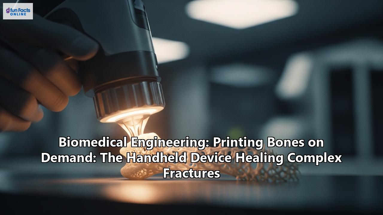
This is where the next evolution of the technology, the handheld bioprinter, emerges as a true game-changer. These portable devices are designed to be used directly in the operating room, allowing the surgeon to print a customized bone scaffold in-situ, or directly into the fracture site. This approach eliminates the need for pre-operative imaging and fabrication, dramatically reducing surgical time and complexity. The surgeon can see the defect in real-time and, with the handheld device, fill it precisely, layer by layer, with a material that promotes healing.
This in-situ approach offers several profound advantages over traditional methods and even lab-based bioprinting:
- Unprecedented Precision and Customization: The surgeon can perfectly match the contours of the fracture, no matter how irregular, ensuring a seamless fit that is impossible to achieve with pre-fabricated implants.
- Reduced Surgical Time and Complexity: By eliminating the need for pre-operative CT scans and off-site fabrication, the entire process can be completed in a single surgical session, potentially reducing a procedure that could take hours down to a fraction of the time.
- Improved Healing and Integration: The ability to print directly into the defect allows for intimate contact between the bioink and the surrounding healthy bone, which can enhance the integration of the graft and promote more rapid healing.
- Minimally Invasive Potential: As the technology advances, it is envisioned that some procedures could be performed through minimally invasive approaches, further reducing patient trauma and recovery time.
The 'Bone Pen' and the 'Glue Gun': A Closer Look at the Technology
The development of handheld bioprinters is being driven by innovative research groups around the world. Two of the most prominent and illustrative examples are the "BioPen," developed in Australia, and a device ingeniously modified from a hot glue gun by researchers in South Korea and the United States.
The BioPen: Drawing a Path to Regeneration
The BioPen, a collaborative effort between the University of Wollongong and St. Vincent's Hospital in Melbourne, is a handheld device that allows a surgeon to "draw" a scaffold directly into a cartilage or bone defect. This remarkable device works by extruding a bioink that is protected by an outer layer of gel. The two materials are combined in the pen's nozzle as the surgeon applies the scaffold layer by layer. A low-powered ultraviolet light source attached to the pen solidifies the bioink as it is deposited, protecting the cells within and giving the scaffold its shape.
The bioink used in the BioPen is a hydrogel, a water-based polymer network that can be designed to mimic the extracellular matrix of natural tissue. For bone and cartilage repair, the bioink is typically composed of materials like methacrylamide-modified gelatin (GelMA) and methacrylated hyaluronic acid (HAMA), and is laden with the patient's own stem cells, harvested during the same surgical procedure. The hydrogel provides a supportive environment for the cells, and the addition of growth factors can encourage them to differentiate into the desired cell type—in this case, chondrocytes for cartilage or osteoblasts for bone.
The promise of the BioPen has been demonstrated in preclinical studies. In one key study, the device was used to repair full-thickness chondral (cartilage) defects in the knees of sheep. The results showed that the in-situ printed scaffolds promoted early cartilage regeneration, demonstrating the feasibility and potential of this approach in a large animal model. The ultimate goal of this technology is to repair cartilage injuries that are currently very difficult to treat, and in doing so, potentially prevent the onset of osteoarthritis, a debilitating condition that often follows such injuries.
The 'Glue Gun' Printer: A Simple Solution to a Complex Problem
In a striking example of innovative thinking, researchers from Sungkyunkwan University and Korea University in South Korea, in collaboration with institutions like MIT and Harvard-affiliated hospitals in the US, have developed a handheld bone printer by modifying a commercially available hot glue gun. This approach highlights a key goal in the development of these devices: to create a tool that is not only effective but also user-friendly, portable, and cost-effective.
Instead of a glue stick, this device uses a specially formulated filament made from a composite of polycaprolactone (PCL) and hydroxyapatite (HA). PCL is a biodegradable and biocompatible polymer that has a low melting point (around 60 degrees Celsius), making it safe to use in close proximity to living tissues. HA is a ceramic material that is a major component of natural bone and is known for its osteoconductive properties. The combination of these two materials creates a filament that is both strong and flexible, and which can be melted and extruded directly into a bone defect.
The researchers also incorporated antibiotics into the filament, allowing for a sustained release of the drugs at the injury site to prevent post-operative infections, a significant risk in complex fracture surgery. The device itself was modified to operate at a lower temperature than a standard glue gun and was fitted with a specialized nozzle for more precise application.
The efficacy of this "glue gun" printer has been tested in rabbits with critical-sized defects in their femurs. The results were impressive. The in-situ printed scaffolds promoted rapid bone regeneration without causing infection or tissue damage. Micro-CT scans revealed that the new bone growth in the rabbits treated with the handheld printer was significantly better than in those treated with traditional bone cement. The biodegradable PCL scaffold gradually dissolved over time, allowing the rabbit's own newly formed bone to take its place.
The Science of the Scaffold: Inside the Bioink
The success of any bioprinting technology, and especially a handheld device for in-situ repair, hinges on the properties of the bioink. This is not just a passive material; it is an active participant in the healing process, a carefully engineered microenvironment designed to support and guide the body's own regenerative capabilities. The ideal bioink for bone regeneration must possess a unique combination of properties:
- Printability: The material must be able to be smoothly extruded from the printer nozzle and hold its shape after being deposited. This often involves the use of hydrogels with specific rheological properties, such as shear-thinning, where the material becomes less viscous under the stress of printing but quickly solidifies once in place.
- Biocompatibility: The bioink must not be toxic to the surrounding tissues or elicit a harmful inflammatory or immune response. This is why materials like gelatin, hyaluronic acid, and alginate, which are derived from natural sources, are often used.
- Mechanical Strength: The printed scaffold must be strong enough to provide structural support to the fracture site, especially in load-bearing bones. This is a significant challenge for hydrogels, which are often mechanically weak. To address this, researchers are developing composite bioinks, incorporating materials like ceramics or stronger polymers to enhance their mechanical properties. The PCL/HA composite used in the "glue gun" printer is a prime example of this approach.
- Biodegradability: The scaffold is intended to be a temporary structure. It must degrade at a rate that matches the rate of new bone formation, gradually being replaced by the patient's own tissue. The degradation rate of polymers like PCL can be tuned by altering their molecular weight.
- Osteoinductivity and Osteoconductivity: As mentioned earlier, osteoconductivity is the ability to act as a scaffold for bone growth, while osteoinductivity is the ability to actively stimulate the differentiation of stem cells into bone-forming cells. To achieve osteoinductivity, bioinks can be loaded with growth factors, which are signaling molecules that play a crucial role in the natural bone healing process.
The Power of Growth Factors: Orchestrating Regeneration
Growth factors are the conductors of the cellular orchestra that is tissue regeneration. In the context of bone healing, two of the most important are Vascular Endothelial Growth Factor (VEGF) and Bone Morphogenetic Proteins (BMPs).
- VEGF is a key player in angiogenesis, the formation of new blood vessels. A robust blood supply is essential for bone healing, as it delivers the oxygen, nutrients, and cells needed for tissue regeneration.
- BMPs, particularly BMP-2, are potent osteoinductive agents. They signal to mesenchymal stem cells, which are present in the surrounding tissues, to migrate to the injury site and differentiate into osteoblasts, the cells that build new bone.
The challenge with using growth factors therapeutically has been delivering them to the right place, at the right time, and in the right concentration. Simply injecting them into the body is often ineffective, as they have a short half-life and can cause side effects if they spread to other areas.
This is another area where 3D bioprinting, and by extension handheld bioprinters, offers a transformative solution. By incorporating growth factors directly into the bioink, it is possible to create a system for their sustained and localized release. Advanced bioprinting techniques even allow for the creation of spatial gradients of growth factors within a scaffold. For example, a scaffold could be printed with a higher concentration of VEGF in one area to promote the ingrowth of blood vessels, and a higher concentration of BMP-2 in another area to stimulate bone formation. This ability to spatiotemporally control the delivery of growth factors allows for a more biomimetic approach to healing, one that more closely replicates the natural processes of the body.
The Surgeon's New Tool: The Operating Room of the Future
The introduction of handheld bioprinters has the potential to profoundly change the way orthopedic surgery is performed. Surgeons who have been involved in the development and testing of these devices speak of a new paradigm in treatment. Instead of being limited to a selection of pre-sized metal implants or having to perform a separate procedure to harvest a bone graft, the surgeon will be empowered to create a truly personalized solution in real-time.
The user-friendly design of devices like the BioPen and the "glue gun" printer means that they could be adopted by surgeons without extensive additional training. The intuitive, manual operation allows the surgeon to use their skill and judgment to fill the defect precisely. This technology could also democratize access to advanced regenerative therapies. While a full-scale, laboratory-based bioprinting setup is a major investment, a portable, cost-effective handheld device could be more widely adopted in hospitals around the world.
In a survey of orthopedic surgeons regarding their perspectives on bioprinted cartilage grafts, the need for better solutions for cartilage injuries was a recurring theme. The surgeons expressed hope that this technology could offer a better quality of life for their patients, for whom current treatments are often inadequate. While they also emphasized the need for strong clinical evidence of safety and efficacy, the potential benefits were seen as a powerful driver for adoption.
Navigating the Road Ahead: Challenges and Future Directions
Despite the remarkable progress and immense promise of handheld bioprinters, there are still significant hurdles to overcome before this technology becomes a standard of care.
- Vascularization: Creating a blood supply for larger printed constructs remains one of the biggest challenges in tissue engineering. While the incorporation of growth factors like VEGF is a promising strategy, ensuring the rapid formation of a functional vascular network throughout a large scaffold is complex.
- Regulatory Approval: Any new medical device and material must go through a rigorous regulatory approval process, such as that overseen by the FDA in the United States. This involves demonstrating the safety and efficacy of the device through extensive preclinical and clinical trials. The personalized nature of in-situ bioprinting presents unique regulatory challenges, as each "implant" is created at the point of care.
- Long-Term Studies: While early preclinical studies have been very encouraging, long-term studies are needed to assess the durability of the regenerated tissue and to ensure that there are no unforeseen adverse effects.
- Cost and Accessibility: While the goal is to create a cost-effective solution, the development and initial implementation of this technology will likely be expensive. Ensuring equitable access to this advanced form of treatment will be an important consideration.
Looking to the future, the field is ripe with possibilities. The integration of artificial intelligence (AI) and robotics with handheld bioprinting could lead to even more sophisticated systems. AI could be used to analyze the fracture in real-time and guide the surgeon in the optimal deposition of the bioink. Robotic arms could be used to hold and manipulate the printer with a level of precision and stability that surpasses that of the human hand.
The development of "4D bioprinting" is another exciting frontier. This involves printing with "smart" materials that can change their shape or properties over time in response to stimuli in the body. This could allow for the creation of scaffolds that can adapt and remodel themselves as the tissue heals.
Ethical Considerations in a Brave New World
As with any technology that has the power to fundamentally alter the human body, the rise of bioprinting brings with it a host of ethical considerations that must be carefully navigated. Issues of safety are paramount; the long-term effects of implanting living, bioprinted constructs into the human body are still being studied, and the risk of complications such as tumor formation, however small, must be thoroughly investigated.
The principle of informed consent takes on a new level of complexity. Patients will need to understand not only the surgical procedure itself but also the novel biological materials and processes being used to heal their bodies.
Questions of equity and access are also critical. Will this revolutionary technology be available to all who need it, or will it be a luxury reserved for the wealthy? The goal of many researchers to create cost-effective, accessible devices is a crucial step in addressing this concern.
A Future Forged in the Lab, Realized in the Operating Room
The journey of the handheld bioprinter from a concept to a clinical reality is a powerful illustration of the transformative potential of biomedical engineering. What begins as a creative solution in a research lab—the idea to turn a glue gun into a bone printer or to draw new cartilage with a "pen"—is steadily making its way toward the operating room, promising to change the lives of patients with devastating injuries.
The ability to print bone on demand, directly at the site of a complex fracture, represents a paradigm shift in orthopedic medicine. It is a move away from off-the-shelf solutions and toward a future of truly personalized, regenerative healing. While the road ahead is still long, with significant scientific, regulatory, and ethical challenges to navigate, the progress made so far is nothing short of extraordinary. The day may not be far off when a surgeon, faced with a shattered bone, reaches not for a metal plate and screws, but for a handheld device that can rebuild what was broken, cell by cell, layer by layer, ushering in a new era of healing.
Reference:
- https://pubmed.ncbi.nlm.nih.gov/28512850/
- https://pmc.ncbi.nlm.nih.gov/articles/PMC2922269/
- https://today.uconn.edu/2020/02/3d-printers-developed-treat-musculoskeletal-injuries/
- https://pmc.ncbi.nlm.nih.gov/articles/PMC8287509/
- https://pmc.ncbi.nlm.nih.gov/articles/PMC6540724/
- https://www.frontiersin.org/journals/bioengineering-and-biotechnology/articles/10.3389/fbioe.2021.630488/full
- https://www.researchgate.net/figure/Mechanical-Properties-of-the-bioinks-A-Youngs-Modulus-and-B-Equilibrium-Modulus-for_fig3_321439671
- https://quasa.io/media/korean-innovators-transform-glue-gun-into-breakthrough-bone-repair-device
- https://newatlas.com/medical-tech/3d-printed-bone-grafts-directly/
- https://www.researchgate.net/figure/a-Design-of-the-Biopen-which-shows-the-two-separate-chambers-with-a-motor-control-The_fig1_317005635
- https://www.bohrium.com/paper-details/3d-bioprinting-spatiotemporally-defined-patterns-of-growth-factors-to-tightly-control-tissue-regeneration/812578887471988737-6329
- https://www.researchgate.net/figure/The-Biopen-is-used-for-cartilage-regeneration-a-Schematic-of-the-two-chambers-inside_fig5_370240608
- https://www.thenews.com.pk/latest/1342363-scientists-develop-3d-printing-glue-gun-to-repair-bone-fractures
- https://pubmed.ncbi.nlm.nih.gov/40896112/
- https://3dprint.com/175766/3d-stem-cell-biopen-cartilage/
- https://www.regenhealthsolutions.info/2020/04/13/handheld-bioprinter-to-treat-skeletal-muscle-injuries/
- https://www.regmednet.com/in-situ-3d-bioprinting-of-musculoskeletal-tissues-in-orthopedic-surgery/
- https://pmc.ncbi.nlm.nih.gov/articles/PMC7831065/
- https://www.youtube.com/watch?v=oggI-2NfuWA
- https://ris.utwente.nl/ws/files/6833014/Barradas11osteo.pdf
- https://aoa.org.au/for-patients/celebrating-orthopaedics/innovations---the-biopen
- https://3dprint.com/320454/handheld-3d-printer-repairs-bone-like-a-glue-gun-tested-in-rabbits-planned-for-or-use/
- https://pmc.ncbi.nlm.nih.gov/articles/PMC9472017/
- https://www.sciencenews.org/article/handheld-bone-printer
- https://pmc.ncbi.nlm.nih.gov/articles/PMC5798025/
- https://www.mdpi.com/2313-7673/10/8/491
- https://portlandpress.com/essaysbiochem/article/65/3/569/229077/Strategies-for-inclusion-of-growth-factors-into-3D
- https://pmc.ncbi.nlm.nih.gov/articles/PMC7428335/
- https://biomedeng.jmir.org/2019/1/e12148
- https://accscience.com/journal/IJB/10/2/10.36922/ijb.1752
- https://publications.waset.org/10011703/optimization-of-mechanical-properties-of-alginate-hydrogel-for-3d-bio-printing-self-standing-scaffold-architecture-for-tissue-engineering-applications
- https://pmc.ncbi.nlm.nih.gov/articles/PMC8720383/
- https://www.voxelmatters.com/ai-turns-bioprinter-into-a-partner-for-tissue-engineering/
- https://accscience.com/journal/IJB/11/4/10.36922/IJB025170164
- https://www.medicaldesignbriefs.com/component/content/article/36927-handheld-3d-printers-treat-musculoskeletal-injuries