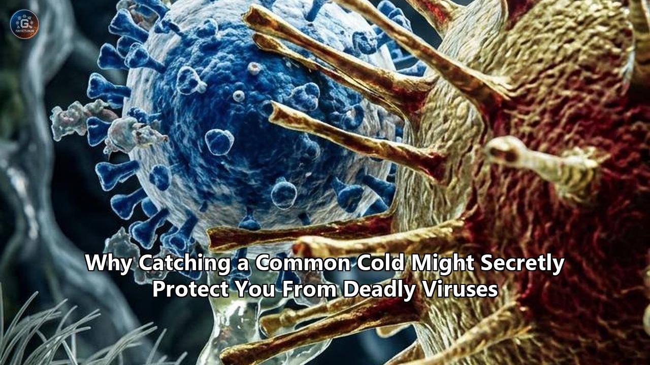
When seasonal respiratory viruses collide in human airway tissues, the outcome of the battle is rarely determined by the incoming pathogen alone. Data emerging from multi-institution molecular virology teams at Yale University, University College London (UCL), and the MRC-University of Glasgow Centre for Virus Research demonstrate that an active infection with a mild upper-respiratory pathogen can halt the invasion of far more lethal viruses, including Influenza A and SARS-CoV-2.
Through two distinct biological processes—innate mucosal viral interference and cross-reactive heterologous T-cell memory—the mild viruses responsible for seasonal sniffling create short- and medium-term biological defenses that systematically disrupt the replication kinetics of dangerous pathogens.
Virologists and immunologists have pinpointed the precise signaling pathways, cellular organelle interactions, and sequence-level cross-reactivities that drive this interaction. As public health agencies and biotech labs race to develop pan-respiratory countermeasures, these discoveries are forcing a re-evaluation of how mucosal barriers defend the host, uncovering the technical mechanisms behind common cold immunity that standard single-pathogen models missed for decades.
The Molecular Alarm: How Rhinovirus Closes the Epithelial Gate
The primary line of defense is an innate, non-specific biological blockade triggered by human rhinoviruses (HRVs), the single-stranded RNA enteroviruses responsible for more than half of all common colds.
               [ INCOMING RHINOVIRUS (HRV) ]
                            │
                            ▼
          Binds Epithelial Receptors (ICAM-1 / CDHR3)
                            │
                            ▼
            Endosomal / Cytoplasmic Replication
                            │
              Uncoated Viral dsRNA Intermediates
                            │
            ┌───────────────┴───────────────┐
            ▼                               ▼
       RIG-I Sensor                   MDA-5 Sensor
            │                               │
            └───────────────┬───────────────┘
                            ▼
              MAVS Adapter Protein (Mitochondria)
                            │
                            ▼
           TBK1 / IKK-ε Phosphorylation Complex
                            │
            ┌───────────────┴───────────────┐
            ▼                               ▼
      IRF3 Activation                 IRF7 Activation
            │                               │
            └───────────────┬───────────────┘
                            ▼
     Nuclear Translocation & Transcriptional Induction
                            │
                            ▼
           Release of Type I (IFN-α/β) & Type III (IFN-λ)
                            │
            ┌───────────────┴───────────────┐
            ▼                               ▼
  Autocrine Loop (Infected Cell)  Paracrine Loop (Neighbor Cells)
            │                               │
            └───────────────┬───────────────┘
                            ▼
               JAK-STAT Signaling Cascade
       (JAK1 / TYK2 Phosphorylation → STAT1/STAT2 Dimers)
                            │
                            ▼
                 ISGF3 Transcription Complex
                            │
                            ▼
              Broad Induction of ~300+ ISGs
  (OAS1, MX1/MxA, PKR, IFITM3, Viperin, RNASEL Activation)
                            │
                            ▼
     [ HOST AIRWAYS SEALED: SECONDARY PATHOGENS BLOCKED ]
       • SARS-CoV-2 entry down-regulated / replication halted
       • Influenza A viral polymerase & transcription blocked
When an HRV particle invades human ciliated bronchial or nasal epithelial cells via intercellular adhesion molecule 1 (ICAM-1) or cadherin-related family member 3 (CDHR3), it undergoes rapid translation and replication. During this process, viral replication intermediates generate double-stranded RNA (dsRNA) motifs inside the host cytoplasm.
Airway epithelial cells continuously scan their intracellular space using pattern recognition receptors (PRRs), predominantly retinoic acid-inducible gene I (RIG-I) and melanoma differentiation-associated protein 5 (MDA5). The moment these sensors bind foreign viral RNA loops, they recruit mitochondrial antiviral signaling proteins (MAVS).
This trigger activates TANK-binding kinase 1 (TBK1) and IκB kinase-epsilon (IKKε), phosphorylating interferon regulatory factors 3 and 7 (IRF3 and IRF7). Within hours of an HRV invasion, the infected cells transcribe and export vast concentrations of:
- Type I Interferons: Primarily IFN-α and IFN-β.
- Type III Interferons: Specifically IFN-λ1, IFN-λ2, and IFN-λ3, which act locally on mucosal epithelial surfaces.
This burst of local interferon functions as a regional alarm. The secreted interferons bind in both an autocrine and paracrine fashion to nearby, uninfected airway cells via the heterodimeric IFN-α/β receptor (IFNAR1/IFNAR2) and the IFN-λ receptor (IFNLR1/IL10Rβ).
This binding sets off the Janus kinase-signal transducer and activator of transcription (JAK-STAT) cascade. JAK1 and TYK2 phosphorylate STAT1 and STAT2, which then assemble with interferon regulatory factor 9 (IRF9) to form the ISGF3 transcription factor complex.
ISGF3 translocates into the nucleus, binding interferon-stimulated response elements (ISRE) across the genome to rapidly activate hundreds of interferon-stimulated genes (ISGs). The airway epithelium becomes an inhospitable zone for secondary viral replication:
- 2'-5'-Oligoadenylate Synthetase 1 (OAS1): Synthesizes 2',5'-oligoadenylates that activate latent Endoribonuclease L (RNase L), cutting both host and incoming viral single-stranded RNA to destroy viral genomes on arrival.
- Myxovirus Resistance Protein A (MxA / MX1): Forms high-affinity oligomeric rings around foreign viral nucleocapsids, trapping viral replication machinery and blocking nuclear translocation.
- Protein Kinase R (PKR / EIF2AK2): Autophosphorylates upon detecting residual viral RNA, subsequent to which it phosphorylates the eukaryotic initiation factor eIF2α, shutting down host cell translation to prevent secondary invaders from producing viral proteins.
- Interferon-Induced Transmembrane Protein 3 (IFITM3): Embeds within endosomal and plasma membranes, altering local membrane fluidity to prevent viral-envelope fusion and endosomal egress.
+------------------+-----------------------------------------------------------+
| Effector Gene    | Primary Antiviral Mechanism                               |
+------------------+-----------------------------------------------------------+
| OAS1 / RNase L   | Catalyzes targeted degradation of single-stranded         |
|                  | incoming viral genomic and messenger RNA.                 |
+------------------+-----------------------------------------------------------+
| MxA (MX1)        | Assembles into tight multimeric rings around viral        |
|                  | ribonucleoprotein complexes, blocking transcription.      |
+------------------+-----------------------------------------------------------+
| PKR (EIF2AK2)    | Inactivates translation factor eIF2α via phosphorylation, |
|                  | shutting down the protein synthesis machinery.            |
+------------------+-----------------------------------------------------------+
| IFITM3           | Rigidifies host cell membranes, preventing viral envelope |
|                  | fusion and blocking cytosolic entry.                      |
+------------------+-----------------------------------------------------------+
Bench Analysis: How Rhinovirus Halts Lethal Pathogens
In experiments conducted at the Yale School of Medicine by a research team led by immunobiologist Dr. Ellen Foxman, human respiratory tract tissue was cultured at the air-liquid interface to create differentiated airway organoids containing ciliated, basal, and goblet cells.
When these airway models were infected exclusively with SARS-CoV-2, the pathogen's viral load surged rapidly, doubling approximately every six hours across the initial 72-hour window. SARS-CoV-2 achieves this early runaway replication because its non-structural proteins (specifically nsp1, nsp6, and ORF6) delay the host's interferon response.
           VIRAL KINETIC DIVERGENCE (ORGANOID MODEL)

  Log10 Viral RNA
  Copies / mL
       ▲
   8 ──│                                    / [SARS-CoV-2 Alone]
       │                                   /  (Doubles every 6 hrs;
   6 ──│                                  /    IFN suppressed by ORF6/nsp1)
       │                                 /
   4 ──│                                /
       │                               /
   2 ──│───────────────────────────────   [HRV Pre-Exposure + SARS-CoV-2]
       │                                  (ISGs Pre-Activated; OAS1/MxA/PKR
   0 ──└─────┬──────────────┬──────────────┬──────────────► Active; 0 Replication)
            0 hr          24 hr          48 hr          72 hr
However, when tissues were exposed to human rhinovirus prior to SARS-CoV-2 challenge, the dynamic reversed. Because rhinovirus triggers immediate RIG-I/MDA5-mediated interferon induction, the tissue was flooded with ISG effectors before the coronavirus entered.
The viral load of SARS-CoV-2 in tissues pre-treated with rhinovirus flatlined. The replication complex was unable to gain traction in an intracellular environment full of active RNase L and phosphorylated eIF2α.
To confirm that interferon signaling was the exact variable controlling this interference, Foxman's team introduced BX795, a synthetic small-molecule inhibitor of the TBK1/IKKε complex that blocks downstream interferon transcription.
When interferon signaling was pharmacologically shut off, rhinovirus lost its protective effect, and SARS-CoV-2 replicated unimpeded. The experiment proved that the protective effect was not direct physical competition for host cell receptors, but host-mediated innate immune priming.
Similar interference mechanics occur between rhinovirus and Influenza A virus (IAV). Clinical data from over 13,000 patients analyzed across three years at Yale New Haven Hospital revealed that simultaneous dual infections with active rhinovirus and influenza were statistically rare. Laboratory-engineered primary human airway models showed that rhinovirus exposure produced an antiviral state that completely prevented influenza replication for up to five days.
The UCL Discovery: Abortive Infections and Non-Structural T-Cell Targets
Beyond the short-lived, broad-spectrum protection provided by rhinovirus-induced interferons, a long-term protective mechanism operates through seasonal coronavirus exposures.
The human population is perpetually reinfected with four endemic human coronaviruses (HCoVs):
- Alphacoronaviruses: HCoV-229E and HCoV-NL63
- Betacoronaviruses: HCoV-OC43 and HCoV-HKU1
These endemic strains cause an estimated 15% to 30% of standard annual common colds. While conventional immunology long maintained that immunity against seasonal HCoVs offered little cross-protection against lethal betacoronaviruses due to structural divergences in the Spike (S) glycoprotein, work by T-cell immunologists Dr. Leo Swadling and Professor Mala Maini at University College London overturned this assumption.
                THE CORONAVIRUS GENOMIC ARCHITECTURE

  5' [────────────────── ORF1a / ORF1ab (75%) ──────────────────] [── Structural (25%) ──] 3'
                                 │                                         │
                                 ▼                                         ▼
            Encodes Replication-Transcription Complex (RTC)        Encodes Spike (S),
            Proteolytically cleaved into nsps (nsp1 - nsp16)       Envelope (E), Membrane (M),
                                 │                                 Nucleocapsid (N)
            ┌────────────────────┴────────────────────┐                    │
            ▼                                         ▼                    ▼
     nsp12 (Polymerase / RdRp)                nsp13 (Helicase)       High Mutational Drift;
     • 85–96% Amino Acid Homology             • Deeply Conserved     Neutralizing Antibodies
       Across All Coronaviridae                 Across All HCoVs     Evaded by Variant Evolution
            │                                         │                    │
            └────────────────────┬────────────────────┘                    ▼
                                 ▼                              [ Strain-Specific Protection ]
             Cross-Reactive CD4+ / CD8+ Memory T Cells
              Primed by Prior HCoV Common Colds
                                 │
                                 ▼
         Rapid Destruction of Newly Infected Host Cells
               During Earliest Intracellular Phase
                                 │
                                 ▼
             [ ABORTIVE INFECTION / ZERO SEROCONVERSION ]
During the initial pandemic wave in early 2020, Swadling and Maini monitored an intensively characterized cohort of frontline healthcare workers who were repeatedly exposed to high viral loads in hospital COVID-19 wards, yet consistently tested negative via RT-PCR and never developed anti-SARS-CoV-2 spike or nucleocapsid antibodies.
Standard clinical diagnostic criteria categorized these individuals as entirely "uninfected". However, deep-phenotyping immunoassays tell a different story.
By tracking blood transcriptomics, the UCL investigators found a selective spike in IFI27 (Interferon Alpha-Inducible Protein 27), an early and specific host innate biomarker of coronavirus entry. The virus had entered their cells, but the infection was rapidly terminated before productive viral shedding or humoral seroconversion could occur—an outcome termed an abortive infection.
+--------------------------+-----------------------+-----------------------+
| Diagnostic Metric        | Abortive Infection    | Productive Infection  |
+--------------------------+-----------------------+-----------------------+
| Nasopharyngeal RT-PCR    | Negative              | Positive (High Ct)    |
+--------------------------+-----------------------+-----------------------+
| Anti-Spike IgG Serology  | Negative              | Positive (High Titers)|
+--------------------------+-----------------------+-----------------------+
| IFI27 Transcript Marker  | Transiently Elevated  | Sustained Elevation   |
+--------------------------+-----------------------+-----------------------+
| RTC-Specific Memory T    | Rapid, Robust         | Delayed / Variable    |
| Cells (nsp12 / nsp13)    | Clonal Expansion      | Kinetics              |
+--------------------------+-----------------------+-----------------------+
| Clinical Presentation    | Asymptomatic          | Mild to Critical      |
+--------------------------+-----------------------+-----------------------+
When the team profiled the cellular immune repertoires of these resistant individuals, they found they harbored high baseline frequencies of cross-reactive, tissue-tropic CD4+ and CD8+ memory T cells targeting the internal Replication-Transcription Complex (RTC).
The RTC: An Invariable Viral Target
Unlike the external Spike protein, which undergoes rapid evolutionary drift and immune evasion mutations, the internal non-structural replication enzymes of coronaviruses are constrained by biochemical requirements. The enzymatic core of the virus cannot alter its fundamental geometry without destroying its ability to replicate RNA.
- nsp12 (RNA-dependent RNA Polymerase, RdRp): Catalyzes the synthesis of viral RNA, exhibiting up to 90% amino acid sequence homology across endemic common cold coronaviruses and SARS-CoV-2.
- nsp13 (RNA 5'-Triphosphatase / Helicase): Drives the ATP-dependent unwinding of double-stranded RNA intermediates, preserving conserved peptide epitopes across diverse betacoronavirus lineages.
The underlying mechanism of this cross-reactive common cold immunity operates on timing.
Structural proteins like Spike and Nucleocapsid are synthesized later in the viral cycle through subgenomic transcription. In contrast, open reading frames ORF1a and ORF1ab are translated immediately upon viral uncapping in the host cytosol to assemble the RTC.
Host major histocompatibility complex (MHC) Class I and II pathways process and present these newly synthesized RTC peptides on the cell surface within two to four hours of infection.
Because these individuals had encountered endemic common cold strains like HCoV-OC43 and HCoV-HKU1 in previous years, they maintained long-lived memory T cells primed against conserved nsp12 and nsp13 epitopes.
These memory T cells recognized the presented RTC peptides on the respiratory mucosa, activated, and eliminated the infected host cell before the incoming virus could assemble progeny virions. The host aborted the disease process entirely, bypassing systemic illness without needing neutralizing antibodies.
Epidemiological Footprints: When Respiratory Viruses Compete in Human Populations
The clinical and cellular reality of viral interference matches historical epidemiological anomalies that long puzzled public health modelers.
                     THE 2009 EUROPEAN INTERFERENCE ANOMALY

  Viral Prevalence
       ▲
       │             /────────\   [Rhinovirus Autumn Surge]
       │            /          \  (Triggers widespread mucosal IFN)
       │           /            \
       │          /              \
       │         /                \
       │  ──────/                  \────────
       │  .................................. [H1N1 Pandemic Flu Suppressed]
       │                                     \
       │                                      \─────────/ [H1N1 Delayed Wave]
       │                                                 \ (Rebounds only after
       │                                                  \ HRV levels decline)
       0 ──┴───────────┬───────────┬───────────┬───────────┬───────────► Time
                      Aug         Sep         Oct         Nov
The 2009 H1N1 Pandemic Delay in Europe
In late summer 2009, mathematical models predicted that the emergent H1N1 swine flu pandemic would cause severe infection waves across Western Europe by early September as schools reopened.
In France, Norway, Sweden, and the UK, however, the primary flu surge failed to appear on schedule. National respiratory surveillance networks documented that H1N1 was held at bay throughout September and October, only surging later in November and December.
Epidemiological post-mortems combined with multiplex PCR mapping resolved the anomaly. A massive, early-autumn seasonal rhinovirus epidemic had swept through European school systems.
The widespread circulation of rhinovirus acted as a population-level firewall, flooding human airways with interferon and transiently rendering millions of individuals refractory to secondary Influenza A infection. Only when the rhinovirus wave subsided did the population become susceptible, allowing H1N1 transmission to accelerate.
Post-Lockdown Perturbations and Competitive Niches
The implementation of Non-Pharmaceutical Interventions (NPIs)—including isolation policies, mask mandates, and border closures during 2020–2022—disrupted normal human respiratory virome dynamics.
While enveloped viruses like Influenza and Respiratory Syncytial Virus (RSV) saw transmission rates plunge, non-enveloped rhinoviruses continued to circulate widely due to greater environmental durability and higher resistance to ethanol hand sanitizers.
+------------------+------------------+-------------------+--------------------+
| Pathogen Class   | Enveloped Status | NPI Susceptibility| Primary Protective |
|                  |                  | (Disinfectants)   | Mechanism Induced  |
+------------------+------------------+-------------------+--------------------+
| Human            | Non-enveloped    | Low (Resistant to | Innate Mucosal     |
| Rhinovirus       | (Capsid only)    | alcohol rubs)     | Interferon Surge   |
+------------------+------------------+-------------------+--------------------+
| Endemic HCoVs    | Enveloped        | Moderate          | Cross-Reactive RTC |
| (OC43, HKU1, etc)| (Lipid Bilayer)  |                   | Memory T Cells     |
+------------------+------------------+-------------------+--------------------+
| Influenza A      | Enveloped        | High              | Secondary Target   |
| (IAV)            | (Lipid Bilayer)  |                   | of Mucosal ISGs    |
+------------------+------------------+-------------------+--------------------+
| SARS-CoV-2       | Enveloped        | Moderate-High     | Secondary Target   |
|                  | (Lipid Bilayer)  |                   | of ISGs & T Cells  |
+------------------+------------------+-------------------+--------------------+
When broad-scale NPI measures were abruptly lifted, public health surveillance recorded severe surges of RSV and influenza, frequently termed the "tripledemic".
Beyond waning adaptive immunity, mathematical virology teams noted that the altered seasonal timing of cold-virus peaks meant that normal background viral interference was misaligned, leaving mucosal barriers unprimed during vulnerable winter transmission windows.
The Biological Constraints: The Timing and Kinetic Dilemma
Despite the protective biology behind common colds, this defense cannot simply be generalized into an uncontrolled preventative strategy. The clinical outcome of viral coinfection is governed by strict chronological windows, host baseline genetics, and localized tissue compartmentalization.
                 THE IMMUNE PRIMING TIMING WINDOW

  Interferon-Driven Protection
       ▲
       │                  /─────────────────\
       │                 /                   \
       │                /  PROTECTIVE ZONE    \
       │               /  (Optimal Mucosal     \
       │              /    Antiviral Defense)   \
       │             /                           \
       │            /                             \
       │  ─────────/                               \─────────────────
       │  [CONCURRENT / DELAYED]                     [DECAYED EFFECT]
       │  Compounding Pathology                      ISG expression returns
       │  Exhausts airway cilia                      to baseline levels
       0 ──┴───────────────────────┬───────────────────────┬─────────►
         -12 hr                  +24 to +72 hr           +120 hr
                             Time of Secondary Exposure
The Kinetic Sweet Spot
For viral interference to confer protection, the primary, mild infection must precede the secondary lethal challenge by roughly 24 to 72 hours.
- The 24–72 Hour Window: The host epithelium has fully upregulated its ISG cascades (OAS1, MxA, PKR), establishing a defensive mucosal barrier.
- The Simultaneous Exposure Failure: If an individual is exposed to both rhinovirus and influenza or SARS-CoV-2 simultaneously, the viruses replicate in parallel without an established interferon shield. The dual burden can overwhelm epithelial repair systems, leading to severe dual-pathogen pneumonia.
- The Post-5-Day Decay: Type I and III interferon expressions naturally decline after five to seven days to prevent cytotoxic damage to the airway lining. Once baseline homeostasis returns, the window of innate protection closes.
The Late-Stage Paradox
While early mucosal interferon expression halts virus progression, delayed or persistent systemic interferon expression is a known driver of immunopathology.
In late-stage, severe SARS-CoV-2 infections, high concentrations of systemic Type I interferons drive hyper-inflammatory signaling, fuel macrophage activation syndrome, and worsen alveolar damage.
A transient, localized, early burst of interferon in the upper nasopharynx is protective, whereas a prolonged, systemic release deep in the lung parenchyma is destructive.
Furthermore, over-activation of cellular stress pathways can disrupt this protective interaction. Research at Yale revealed a cellular balancing act between the transcription factor NRF2 (which regulates host antioxidant defense against oxidative stress) and IRF3-mediated interferon signaling.
Upregulating the NRF2 antioxidant pathway dampens the cellular interferon response, revealing how environmental stressors, pollutants, and metabolic state can shut down the antiviral defenses that rhinovirus usually triggers.
Translational Viromics: Turning Common Cold Mechanisms into Next-Generation Therapeutics
Instead of relying on natural, variable, and symptom-inducing infections to establish common cold immunity, translational immunology teams are engineering therapeutic modalities that isolate the exact molecular mechanisms driving these defenses.
+------------------------------------+------------------------------------+
| Therapeutic Strategy               | Underlying Cold-Derived Mechanism  |
+------------------------------------+------------------------------------+
| Targeted Mucosal Type III          | Recreates the non-inflammatory     |
| Interferon-Lambda (IFN-λ)          | protective ISG expression of       |
| Inhalation Formulations            | Rhinovirus at mucosal entry points.|
+------------------------------------+------------------------------------+
| Conserved RTC Core                 | Directs memory CD4+/CD8+ T-cell    |
| Pan-Coronavirus Vaccines           | responses toward invariant nsp12   |
| (Targeting nsp12 / nsp13)          | RdRp sequences discovered in HCoVs.|
+------------------------------------+------------------------------------+
| Synthetic Nucleic Acid Agonists    | Safely triggers cellular RIG-I     |
| (Engineered TLR3/MDA-5 Inhalers)   | and MDA-5 sensors to establish a   |
|                                    | 72-hour viral-free mucosal state.  |
+------------------------------------+------------------------------------+
1. Inhaled Mucosal Type III Interferons (IFN-Lambda)
Pharmaceutical development has largely avoided systemic Type I interferons (IFN-α/β) because systemic injection induces severe flu-like side effects, including high fever, debilitating fatigue, and systemic myalgia.
Biotechnology platforms are focusing instead on recombinant Type III Interferon-Lambda (IFN-λ).
Because IFNLR1 receptors are predominantly restricted to mucosal epithelial tissues and absent on hematopoietic and endothelial cells, intranasally administered IFN-λ triggers the same localized ISG upregulation (OAS1, MxA, IFITM3) as human rhinovirus—sealing the upper respiratory tract against incoming pathogens without inducing systemic inflammation.
2. Universal Pan-Coronavirus Vaccines Targeting the RTC
First-generation COVID-19 vaccines focused on the surface Spike glycoprotein to generate circulating neutralizing antibodies.
However, antigenic drift in the receptor-binding domain (RBD) continually undermines neutralizing antibody titers.
Building on the findings from Swadling and Maini, next-generation vaccine consortia are designing multivalent mRNA and viral-vectored formulations that integrate conserved non-structural replication sequences alongside structural targets.
       NEXT-GEN PAN-CORONAVIRUS VACCINE DESIGN

  [ Conventional Vaccine ] ──► [ Spike (S) Antigen Only ]
                                      │
                                      ▼
                        (Susceptible to Antigenic Drift)

  [ Next-Gen Design ]     ──► [ Mucosal Delivery Vehicle ]
                                      │
           ┌──────────────────────────┴──────────────────────────┐
           ▼                                                     ▼
  [ Stabilized S2 Subunit ]                           [ Conserved RTC Core ]
  (Broadly Neutralizing Surface Sites)               (nsp12 RdRp / nsp13 Helicase)
           │                                                     │
           ▼                                                     ▼
  Induces Mucosal sIgA                               Induces Airway Resident
  & Broad Neutralization                             CD4+/CD8+ Memory T Cells
                                                                 │
                                                                 ▼
                                                  [ RAPID ABORTIVE CLEARANCE ]
By encoding high-homology epitopes from nsp12 (RdRp) and nsp13 (helicase), these vaccines train the adaptive immune system to maintain a permanent cohort of lung-resident memory T cells ($T_{\text{RM}}$).
These $T_{\text{RM}}$ cells remain stationed in the respiratory mucosa, ready to destroy host cells displaying early non-structural proteins during the first hours of infection, recreating the abortive clearance seen in healthcare workers with broad cross-reactive common cold immunity.
3. Broad-Spectrum Synthetic PRR Agonists
Another clinical pathway focuses on transient, non-replicating pathogen-associated molecular pattern (PAMP) mimetics.
Aerosolized small-molecule agonists and synthetic double-stranded RNA formulations (such as optimized Poly(I:C) derivatives) interact with toll-like receptor 3 (TLR3) and RIG-I/MDA5 in the nasopharynx.
Administered via an inhaler or nasal spray during high-risk exposure windows, these agents trigger an autocrine and paracrine interferon cascade without introducing a live virus.
This treatment gives the recipient a temporary 48-to-72-hour window of mucosal resistance, providing broad-spectrum protection against structurally unrelated respiratory pathogens.
Institutional Hurdles: The Regulatory Shift Toward Mucosal and Innate Surrogates
Translating these biological insights into standard medical interventions requires overcoming regulatory frameworks built around simple antibody metrics.
For decades, international regulatory agencies such as the U.S. FDA, the European Medicines Agency (EMA), and the WHO used serum-neutralizing antibody titers as the primary surrogate endpoint for respiratory vaccines.
Serum antibodies are easy to extract, measure via standard in vitro microneutralization or ELISA assays, and standardize across clinical trial phases.
However, serum antibodies correlate poorly with abortive infection, broad viral interference, or mucosal-level clearance.
Assaying tissue-resident memory T cells requires mucosal brushings or bronchoalveolar lavages, which are harder to standardize across global trials.
Similarly, proving the real-world clinical efficacy of an innate mucosal priming agent requires event-driven human challenge trials or broad multi-pathogen surveillance studies rather than single-target efficacy studies.
The growing scientific consensus around viral interference is accelerating this regulatory evolution.
Public health institutions are moving away from studying single viral threats in isolation, turning instead toward a multi-pathogen framework that views the human respiratory mucosa as a dynamic, interactive ecosystem.
The Path Forward: Unresolved Questions and Clinical Milestones
As research continues, several structural questions remain at the center of ongoing respiratory virology programs:
- Durability of Mucosal $T_{\text{RM}}$ Populations: While systemic central memory T cells ($T_{\text{CM}}$) circulating in blood can persist for decades, airway-resident memory T cells ($T_{\text{RM}}$) positioned in the upper respiratory epithelium naturally decline over 6 to 18 months. Determining how to maintain durable mucosal protection without requiring seasonal booster formulations is an active area of investigation.
- The Structural Limits of Epithelial Memory: Beyond classical adaptive immunology, researchers are investigating trained immunity in respiratory epithelial stem cells. Early evidence suggests that repeated viral encounters epigenetically reprogram basal cells to maintain altered chromatin accessibility, permanently enhancing baseline innate sensor expression.
- Predictive Multipathogen Transmission Modeling: Epidemic forecasting platforms are integrating real-time multiplex PCR pathogen arrays. Instead of modeling influenza or SARS-CoV-2 spread in isolation, modern public health platforms are incorporating the background prevalence of rhinovirus and seasonal coronaviruses into computational transmission models to improve accuracy.
The biological insights uncovered behind the scenes of common cold infections have rewritten core assumptions in respiratory immunology.
The ubiquitous common cold is far from a minor seasonal inconvenience. It is an active shaper of host resistance, capable of closing cellular entry pathways and priming the human immune system to abort deadly viral challenges long before they can take hold.
Reference:
- https://tools.cdc.gov/podcasts/media/pdf/EID_2_22_InterferenceRespiratoryViruses.pdf
- https://pmc.ncbi.nlm.nih.gov/articles/PMC8798701/
- https://www.sciencemediacentre.org/expert-reaction-to-lab-study-looking-at-respiratory-cells-infected-with-sars-cov-2-in-the-presence-or-absence-of-rhinovirus-infection/
- https://medicine.yale.edu/lab/foxman/research/viral-interference/
- https://afludiary.blogspot.com/2021/03/study-human-rhinovirus-infection-blocks.html
- https://www.sciencemediacentre.org/expert-reaction-to-study-looking-at-memory-t-cells-and-the-immune-response-to-sars-cov-2/
- https://medicine.yale.edu/profile/ellen-foxman/
- https://www.frontiersin.org/journals/immunology/articles/10.3389/fimmu.2025.1596135/full
- https://www.ucl.ac.uk/news/2021/nov/new-target-covid-19-vaccines-identified
- https://news.yale.edu/2021/06/15/common-cold-combats-covid-19
- https://www.mdpi.com/2076-393X/14/6/498
- https://globalbiodefense.com/2020/09/04/rhinovirus-assist-common-cold-jump-starts-bodys-antiviral-defense-against-seasonal-influenzas/
- https://www.facebook.com/medicalxpress/posts/exposure-to-the-rhinovirus-the-most-frequent-cause-of-the-common-cold-can-protec/4322243374488179/
- https://www.the-scientist.com/nasal-cells-fight-the-common-cold-via-interferons-73952
- https://swarajyamag.com/news-brief/exposure-to-common-cold-virus-likely-to-protect-against-covid-19-infection-finds-study
- https://www.researchgate.net/publication/403456623_Abortive_infection_T_cells_as_early_antibody-independent_defenders
- https://www.mdpi.com/1422-0067/24/5/4371
- https://pmc.ncbi.nlm.nih.gov/articles/PMC8647507/
- https://www.pnas.org/doi/10.1073/pnas.2532920123
- https://www.emjreviews.com/respiratory/news/flu-symptoms-return-as-winter-viruses-rebound/
- https://journals.asm.org/doi/10.1128/spectrum.03187-24