The crisis of dental decay is a silent pandemic. For millennia, humanity has resigned itself to a biological tragedy: once tooth enamel is lost, it is gone forever. Unlike bone, which knits itself back together after a fracture, or skin, which seals over a wound with fresh tissue, dental enamel—the hardest substance in the human body—is biologically dead. It has no cells, no blood supply, and no capacity for self-repair. When the acid byproducts of bacteria erode this crystalline shield, the damage is permanent. The standard of care for the last century has been remarkably primitive: drill out the rot and patch the hole with inert metals or plastics. It is a philosophy of "repair and patch," not "regenerate and heal."
But a paradigm shift has emerged from an unlikely source, promising to rewrite the rules of dentistry. In the laboratories of King’s College London, researchers have unlocked a method to regenerate dental enamel using one of the most abundant biological waste products on Earth: human hair.
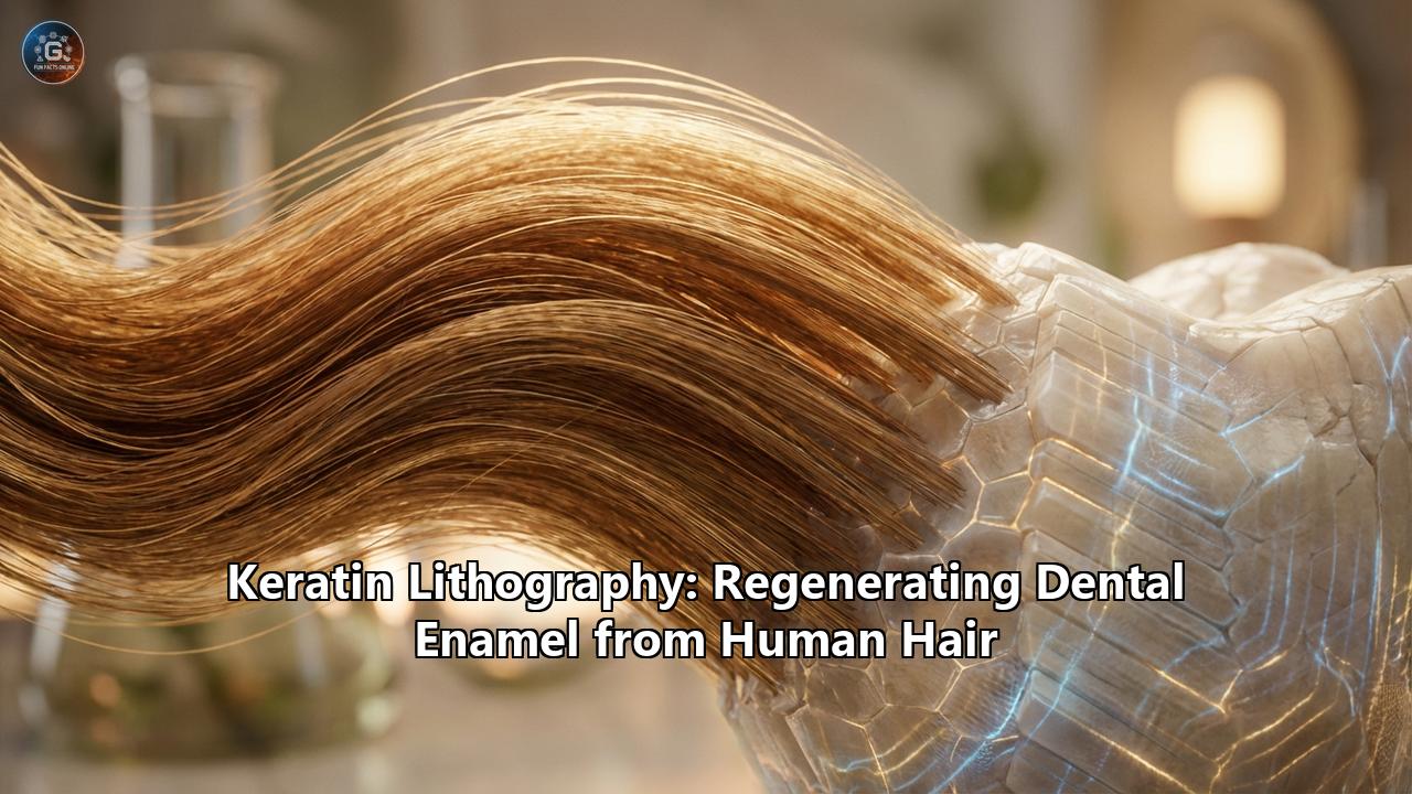
This breakthrough, often described in scientific circles as a form of "biomimetic mineralization" or "keratin lithography," represents more than just a new toothpaste ingredient. It is the dawn of a new era in materials science, where the discarded proteins of the barber shop are engineered to rebuild the crystalline fortifications of the human smile. This is the comprehensive story of how hair is being turned into stone, and how the future of dentistry lies in the trash bin of the salon.
Part I: The Unforgiving Shield
To understand the magnitude of this breakthrough, one must first appreciate the architectural marvel that is dental enamel. To the naked eye, it appears as a simple, smooth, white coating. But under the magnification of a scanning electron microscope, enamel reveals itself as a landscape of staggering complexity.
Enamel is composed of approximately 96% mineral, specifically calcium hydroxyapatite, arranged in long, tightly packed prisms or rods. These rods are woven together in a microscopic basket-weave pattern that provides immense structural resilience, allowing our teeth to withstand the crushing forces of mastication—up to 70 kilograms of pressure—thousands of times a day for decades.
The tragedy of enamel lies in its developmental history. The cells that create enamel, known as ameloblasts, are active only during the formation of the tooth within the gum. Once the tooth erupts into the oral cavity, the ameloblasts die and are shed. The tooth enters the harsh environment of the mouth—a chemical warzone of acidic foods, abrasive chewing, and bacterial fermentation—completely orphaned from its creators. It is a castle built once, with no masons left to repair the walls.
For centuries, dentistry has been a battle to delay the inevitable. Fluoride was the first great revolution, a chemical that hardens the existing mineral structure and makes it more resistant to acid. But fluoride acts as a shield reinforcement; it cannot rebuild a collapsed wall. When a cavity (caries) forms, the structural integrity is breached. The dentist’s drill removes the decayed tissue, and the void is filled with amalgam (silver mercury) or composite resin (plastic).
These fillings are imperfect substitutes. Amalgams require the removal of healthy tooth structure to secure a mechanical hold. Composite resins, while aesthetically pleasing, are prone to shrinkage, leakage, and secondary decay. They are distinct materials, foreign bodies glued into a biological hole. They do not mimic the intricate, prismatic structure of natural enamel. They are patches on a tire, destined to fail and be replaced in a cycle that often ends with the loss of the tooth.
Part II: The Biomimetic Turn
The solution to this stalemate was never going to be found in better plastics or stronger metals. It had to be found in biology. This is the realm of biomimetics—the abstraction of good design from nature. If the ameloblast cells are gone, can we create a synthetic process that mimics what they used to do?
The ameloblast does not simply dump calcium and phosphate onto the tooth. It secretes an organic protein matrix—a scaffold. This protein scaffold acts as a blueprint. It captures mineral ions from the fluid environment and guides them into specific alignment, forcing them to crystallize into the long, hard rods of hydroxyapatite. Once the crystals are grown, the protein scaffold degrades, leaving behind the 96% mineral rock.
For years, scientists have hunted for a protein that could act as a surrogate scaffold. They tried amelogenin, the original protein used by the body, but it is expensive and unstable. They tried synthetic peptides and DNA. But the answer, it turned out, was hiding in plain sight, in a protein responsible for toughness and protection across the animal kingdom: keratin.
Part III: The Keratin Revolution
Keratin is the structural protein of the integumentary system. It is the stuff of hair, nails, hooves, horns, and wool. It is incredibly tough, insoluble in water, and resistant to chemical degradation. It is also one of the most abundant biopolymers on the planet. Every year, millions of tons of hair and wool are sheared, cut, and discarded as waste.
The research team at King’s College London, led by Dr. Sherif Elsharkawy and PhD researcher Sara Gamea, hypothesized that keratin could function as the missing scaffold for enamel regeneration. Their logic was rooted in the protein’s structure. Keratin possesses a specific molecular architecture—rich in cysteine and capable of forming disulfide bridges—that allows it to self-assemble into fibrous networks.
The process they developed, which we can call "Keratin Lithography," transforms this waste product into a sophisticated architectural tool. The methodology is elegantly simple yet chemically profound:
- Extraction: Keratin is extracted from organic sources like sheep’s wool or human hair. Through a chemical process, the robust fibers are broken down into liquid keratin proteins.
- Application: This liquid keratin is applied to the tooth surface. This can be done via a gel, a varnish, or even a toothpaste.
- Self-Assembly: Upon contact with the tooth, the keratin proteins begin to self-assemble. They link hands, forming a microscopic "mesh" or scaffold over the enamel. This mesh is not just a passive net; it has a specific electromagnetic charge and chemical affinity for calcium and phosphate.
- Mineralization: The oral environment is naturally rich in calcium and phosphate ions from saliva. Under normal conditions, these ions float aimlessly. However, the keratin scaffold acts as a magnet and a mold. It captures these ions and organizes them.
- Crystallization: The ions trapped in the keratin mesh begin to nucleate and grow into crystals. Because they are confined by the keratin structure, they cannot grow into chaotic, random shapes. They are forced to grow into ordered, aligned prisms that mimic the structure of natural enamel.
The result is not a glued-on patch, but a grown-on layer. The keratin acts as a transient template, guiding the minerals to rebuild the lost architecture of the tooth.
IV. The Mechanism: How Hair Grows Stone
The genius of this approach lies in the "epitaxial growth." In materials science, epitaxy refers to the deposition of a crystalline overlayer on a crystalline substrate. The keratin allows the new mineral to grow directly out of the old mineral.
When the keratin gel is applied to an early-stage cavity (a white spot lesion where the enamel has softened but not yet cavitated), it infiltrates the microscopic pores of the damaged tissue. As the scaffold sets, it recruits minerals from saliva. The study published in Advanced Healthcare Materials confirmed that the resulting layer is not merely a calcium deposit. It is a highly organized, crystal-like structure.
Microscopic analysis revealed that the keratin directs the formation of "spherulites"—clusters of crystals—that eventually fuse and align to form a continuous, hard shell. This new layer fuses seamlessly with the underlying native enamel. There is no "glue line" or weak interface where bacteria can sneak in. The repair is chemically identical to the original tooth.
Furthermore, the keratin-mediated mineralization does something current fillings cannot: it seals the dentinal tubules. Beneath the enamel lies the dentin, a porous layer filled with microscopic tubes that lead directly to the nerve pulp. When enamel wears away, these tubes are exposed, causing the sharp, shooting pain of tooth sensitivity (dentin hypersensitivity). By growing a new mineral layer over these exposed tubes, the keratin treatment eliminates the root cause of sensitivity, rather than just numbing the nerve.
V. Strength and Durability
One of the most startling findings of the King’s College study was the mechanical property of the regenerated layer. In comparative tests, the keratin-mineralized enamel was significantly harder and more durable than conventional treatments.
Standard composite resins are plastics. Over time, they wear down, absorb stains, and release microplastics. They are softer than natural enamel. The keratin-grown layer, however, is essentially stone reinforced by a protein matrix. It exhibits a "graded" structure—hard on the outside, slightly more elastic on the inside—which mimics the shock-absorbing properties of natural teeth.
This durability suggests that keratin treatments could be used not just for filling cavities, but for preventing them. Imagine a world where, instead of waiting for a hole to form, you apply a keratin varnish every six months that adds a fresh micron of sacrificial enamel to your teeth, constantly replenishing what daily life wears away.
VI. The "Waste to Wealth" Paradigm
The implications of this technology extend far beyond the dentist’s chair. We live in an age of resource scarcity and waste management crises. The dental industry is currently a significant contributor to pollution, relying on single-use plastics and synthetic resins that persist in the environment.
Keratin lithography flips this script. It utilizes a biological waste product. Hair clippings from salons and low-grade wool that is unsuitable for the textile industry are currently burned or dumped in landfills. This technology monetizes that waste, turning it into a high-value biomedical resource.
It is a closed-loop system: biological waste (hair) is used to repair biological damage (teeth). The extraction process is water-based and solvent-free, avoiding the harsh petrochemicals required to manufacture synthetic dental resins. It is biodegradable, biocompatible, and non-toxic. For a world striving for sustainability, "hair toothpaste" is a poster child for the circular economy.
VII. Clinical Applications: The End of the Drill?
How will this technology manifest in our daily lives? The researchers envision a two-tiered approach.
1. The Consumer Tier:Within the next few years, we may see "Keratin-Enhanced Toothpastes" on supermarket shelves. Unlike current "enamel repair" toothpastes, which largely rely on depositing amorphous calcium phosphate (a soft, unorganized mineral), these pastes would contain the keratin precursors. Daily brushing would leave behind a microscopic scaffold, allowing the minerals in your saliva to constantly rebuild the enamel surface at a molecular level. It would be a daily "re-setting" of the tooth’s structural integrity.
2. The Professional Tier:For more significant damage—visible white spots, early decay, or severe sensitivity—a dentist would apply a concentrated keratin gel. This would be a painless procedure. No needles, no drilling. The dentist would clean the tooth, paint on the keratin varnish, and perhaps cure it with a light or simply let it set. Over the next few days, the patient’s own saliva would mineralize the scaffold, healing the lesion.
This non-invasive approach is particularly revolutionary for pediatric dentistry. The "drill and fill" experience is traumatic for children and often requires sedation. A "paint and heal" approach could eliminate dental phobia for an entire generation.
VIII. Challenges and the Road Ahead
While the laboratory results are groundbreaking, the path to the clinic is paved with regulatory hurdles. The oral environment is more chaotic than a petri dish. The pH fluctuates wildly; bacteria are constantly attacking surfaces; enzymes in saliva (like proteases) can degrade proteins.
The researchers must ensure that the keratin scaffold remains stable long enough for mineralization to occur before the mouth’s enzymes break it down. However, keratin’s natural insolubility and toughness give it a massive advantage over other proteins like collagen or amelogenin. It is naturally designed to survive harsh conditions.
Furthermore, clinical trials must demonstrate long-term stability. Does the new enamel layer stay attached for 10 or 20 years? Does it stain? These questions will be answered in the coming phase of human trials. The team at King’s College London is optimistic, projecting that products could be available to the public within a relatively short timeframe—perhaps as soon as three to five years.
IX. Broader Implications for Regenerative Medicine
The success of keratin lithography in dentistry serves as a proof-of-concept for a broader field: protein-mediated tissue engineering. If we can use keratin to guide the mineralization of bone-like structures (teeth), can we use it for bone grafts? Can we use it to coat orthopedic implants to ensure better integration with the skeleton?
The study highlights the power of "disordered proteins"—proteins that don't have a rigid shape until they interact with their target. This flexibility allows them to adapt to the complex, irregular shapes of a cavity or a bone fracture, providing a custom-fit scaffold every time.
X. Conclusion
For decades, we have accepted that our teeth are finite resources, destined to erode and decay. We have accepted that the only solution is to replace biology with plastic. The discovery of keratin-mediated enamel regeneration shatters this assumption.
By looking at the piles of hair sweeping across the barbershop floor, scientists have found the key to immortality for our teeth. It is a triumph of lateral thinking—connecting the keratin of the hair to the hydroxyapatite of the tooth. It promises a future where dentistry is regenerative rather than restorative, where the drill is replaced by a brush, and where the human body is healed by its own discarded blueprints.
As we move forward, the phrase "by the skin of your teeth" takes on a literal new meaning. We will save our teeth not with gold or silver, but with the very stuff of hair and skin, closing the loop of biology to maintain the hardest substance we possess. The era of the living filling has arrived.
Reference:
- https://scitechdaily.com/toothpaste-made-from-hair-could-regrow-tooth-enamel/
- https://www.kcl.ac.uk/news/toothpaste-made-from-hair-provides-natural-root-to-repair-teeth
- https://adanews.ada.org/huddles/toothpaste-made-from-hair-may-help-repair-tooth-enamel/
- https://www.thehealthy.com/news/kings-college-london-keratin-tooth-enamel-study-december-2025/
- https://www.cosmeticsdesign-europe.com/Article/2025/08/21/keratin-breakthrough-promises-sustainable-tooth-enamel-repair/
- https://www.specialchem.com/cosmetics/news/scientists-discover-keratin-from-hair-skin-and-wool-can-repair-tooth-enamel
- https://news.sky.com/story/toothpaste-made-with-hair-naturally-repairs-tooth-enamel-scientists-discover-13414542
- https://www.researchgate.net/publication/397186484_Keratin_protein_mediated_bio-remineralisation_of_early_enamel_erosion_A_pilot_study
- https://www.dental-tribune.com/news/hair-derived-keratin-toothpaste-a-natural-enamel-repair-revolution/
- https://www.youtube.com/shorts/j7z7HKC3FVc