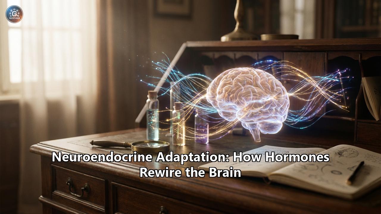
The human brain was once thought to be a static organ—a biological computer whose hardware was finalized in childhood, destined only to decline with age. We now know this to be a fundamental misconception. The brain is a dynamic, shifting landscape, capable of profound physical reorganization throughout the entire lifespan. This quality, known as neuroplasticity, is not merely a response to learning or trauma; it is a relentless, daily adaptation driven by a powerful internal chemical orchestra: the endocrine system.
Neuroendocrine adaptation is the scientific frontier where hormones—typically viewed as regulators of metabolism, reproduction, and stress—reveal themselves as master architects of neural circuitry. From the distinct reshaping of the maternal mind to the synaptic pruning caused by chronic stress, hormones do not just signal the brain; they rewire it. They alter dendritic spine density, modulate the birth of new neurons, and resize entire brain regions to suit the environmental demands placed upon the organism. This is the story of how the chemical self constructs the electrical self.
The Chemical Architects: Mechanisms of Remodeling
To understand how a molecule floating in the bloodstream can restructure a thought, we must look at the cellular machinery. Hormones like cortisol, estradiol, and insulin do not simply knock on the door of a neuron; they often possess keys to the very genetic control room of the cell.
The influence of hormones on the brain occurs through two primary pathways: genomic and non-genomic actions. In the genomic pathway, steroid hormones (such as estrogen and cortisol) are lipophilic; they slip effortlessly through the neuron's fatty outer membrane, bind to receptors in the cytoplasm, and travel into the nucleus. There, they act as transcription factors, literally reading sections of DNA to increase or decrease the production of specific proteins. These proteins might be the building blocks of new synaptic terminals or the receptors that make a neuron more sensitive to electrical signals.
The non-genomic pathway is faster, occurring in seconds or minutes. Here, hormones bind to receptors on the cell surface, triggering rapid signaling cascades—kinase pathways that can instantaneously alter the excitability of a neuron, making it more or less likely to fire.
At the structural level, the most visible impact of these mechanisms is on dendritic spines. These tiny, mushroom-shaped protrusions on the branches of neurons are the receiving docks for synaptic signals. A single neuron may have thousands of spines, each one a potential connection point. Hormones regulate the density and shape of these spines. Under the influence of estrogen, for instance, the hippocampus (the seat of memory) blooms with new dendritic spines, creating a dense forest of connectivity. Conversely, under the siege of chronic stress hormones, these spines wither and retract, thinning the forest and severing the lines of communication.
The HPA Axis: The double-Edged Sword of Stress
The Hypothalamic-Pituitary-Adrenal (HPA) axis is the body’s primary alarm system, and its end-product, cortisol, is perhaps the most potent rewiring agent in the modern human experience.
In the short term, cortisol is a performance enhancer for the brain. During an acute crisis—a near-miss car accident or a high-stakes presentation—cortisol floods the system, sharpening attention and mobilizing glucose for immediate neural energy. It acts on the amygdala, the brain's threat-detection center, to etch the memory of the danger deeply into the neural fabric. This is an adaptive survival mechanism; the brain prioritizes remembering what could kill you so you can avoid it next time. This "flashbulb memory" effect is why you can remember exactly where you were during a traumatic event but might forget what you had for lunch yesterday.
However, the system was designed for sprints, not marathons. When stress becomes chronic—relentless work pressure, financial insecurity, or systemic trauma—neuroendocrine adaptation turns maladaptive. The constant bath of cortisol begins to poison the very structures it once protected.
The Hippocampal Shrinkage: The hippocampus contains a high density of glucocorticoid (cortisol) receptors. Chronic overstimulation of these receptors triggers a process of excitotoxicity and dendritic retraction. The neurons in the CA3 region of the hippocampus begin to shed their dendritic spines and eventually die off. MRI studies have consistently shown that individuals with Cushing’s disease (who have excess cortisol) or severe, untreated PTSD possess significantly smaller hippocampal volumes. The result is a fragmentation of narrative memory and a loss of context. The brain loses its ability to distinguish between a past threat and a present safety, trapping the individual in a loop of reactive anxiety. The Amygdala Hypertrophy: While the hippocampus withers, the amygdala does the opposite. Chronic stress causes the amygdala to grow larger and more reactive. Dendritic trees in the basolateral amygdala expand, creating more connections. The result is a brain that is physically wired for fear—a "hyper-vigilant" state where neutral stimuli (a loud noise, a stern look) are misinterpreted as severe threats. The Prefrontal Disconnect: The prefrontal cortex (PFC) is the CEO of the brain, responsible for logic, impulse control, and emotional regulation. It normally keeps the primitive amygdala in check. Chronic stress severs the synaptic connections between the PFC and the emotional centers. The "cables" are cut. The CEO loses control of the security guard. This neuroendocrine remodeling explains the brain fog, impulsivity, and emotional volatility often seen in burnout and chronic anxiety disorders.The HPG Axis: Sex Hormones as Neural Sculptors
For decades, science relegated sex hormones—estrogen, progesterone, and testosterone—to the realm of reproduction, assuming their influence stopped at the neck. We now know that the brain is a sexual organ, at least in terms of its receptor density. These hormones act as potent neurotrophic factors, protecting neurons from death and stimulating the growth of new connections. The Hypothalamic-Pituitary-Gonadal (HPG) axis drives massive remodeling events at key life stages: puberty, pregnancy, and menopause.
Puberty: The Great Pruning: During puberty, a tidal wave of gonadal hormones triggers a massive reorganization of the brain. The brain actually loses gray matter volume during this time, a process known as synaptic pruning. This is not a loss of function, but a gain in efficiency. The hormones signal the brain to cut away weak, unused connections to strengthen the major highways of communication. It is a refinement process, transitioning the brain from a child’s sponge-like absorption state to an adult’s focused, executive state. The Estrous Cycle and Micro-Remodeling: In females, the brain is not static from month to month. Research on the hippocampus has shown that dendritic spine density fluctuates in rhythm with the menstrual cycle. When estrogen levels peak (around ovulation), spine density in the CA1 region of the hippocampus increases significantly, enhancing verbal memory and spatial reasoning. As progesterone rises and estrogen falls, these spines are pruned back. This cyclical "breathing" of the brain suggests a flexibility in cognitive strategies across the month, optimizing the brain for different types of tasks at different hormonal phases. Pregnancy: The Matrescence: Perhaps the most dramatic example of neuroendocrine adaptation is pregnancy. The flood of hormones during gestation—estradiol levels rise hundreds of times above normal—orchestrates a radical restructuring of the maternal brain. MRI studies reveal that pregnancy leads to a substantial reduction in gray matter volume in regions associated with social cognition, such as the Theory of Mind network. Far from being a deficit (the "mommy brain" myth), this volume loss is a focused specialization. The brain is streamlining its circuits to become hyper-efficient at reading the infant's cues, predicting needs, and detecting threats. This restructuring predicts the quality of maternal attachment; the more the brain changes, the deeper the bond. These changes can last for at least two years postpartum, and some evidence suggests they may be permanent, leaving a "fingerprint" of motherhood on the brain's architecture. Menopause and the Neuroprotective Withdrawal: As women enter menopause, the precipitous drop in estrogen represents a bioenergetic crisis for the brain. Estrogen is a master regulator of the brain's glucose metabolism. When it fades, neurons in the hippocampus and cortex can struggle to generate energy, leading to temporary cognitive instability—hot flashes, brain fog, and memory lapses. This is a critical window of vulnerability. However, the brain is resilient; it eventually undergoes a compensatory adaptation, shifting its metabolic machinery to utilize ketone bodies and other fuel sources, settling into a new equilibrium.Metabolic Hormones: The Gut-Brain Intelligence
The brain consumes 20% of the body's energy despite being only 2% of its weight. It is ravenous. Therefore, it listens intently to the hormones that regulate energy balance: insulin, leptin, and ghrelin.
Insulin and "Type 3 Diabetes": Insulin receptors are densely packed in the hippocampus. Insulin does not just regulate blood sugar; it regulates synaptic plasticity. It supports the survival of neurons and the formation of memories. A growing body of research suggests that Alzheimer’s disease may be, in part, a form of insulin resistance in the brain, often termed "Type 3 Diabetes." When neurons become deaf to insulin's signal due to chronic metabolic dysfunction (like a high-sugar diet), they can no longer repair their synapses, leading to cognitive decline. Leptin and Ghrelin: Leptin (the satiety hormone) and ghrelin (the hunger hormone) have receptors throughout the brain's reward centers. They do more than tell you when to put down the fork; they alter the "value" of external stimuli. High levels of ghrelin (hunger) actually rewire the hippocampus to be better at spatial learning—an evolutionary adaptation to help a starving hunter remember where the food is. Conversely, leptin promotes long-term potentiation (LTP) in the hippocampus. Obesity, which often involves leptin resistance, can impair this plasticity, creating a link between metabolic health and cognitive flexibility.Social Hormones: The Chemistry of Connection
Oxytocin and vasopressin are unique in that they act as both hormones in the blood and neurotransmitters in the brain. They are the architects of social circuitry.Oxytocin, released during touch, breastfeeding, and orgasm, quiets the amygdala. It inhibits the fear response and strengthens the connection between the emotional centers and the prefrontal cortex, promoting trust and "social synchronization." In animal models, oxytocin can change the auditory cortex of a mother mouse to be specifically tuned to the frequency of her pup's distress call. It literally rewires the sensory processing centers to prioritize the social bond above all other noise.
Vasopressin, closely related, is vital for social memory and territoriality. In the famous studies of voles, the difference between a monogamous, pair-bonding prairie vole and a promiscuous meadow vole comes down to the distribution of vasopressin receptors in the brain's reward system. By genetically altering these receptors, scientists can rewrite the social behavior of the animal. This implies that our capacity for loyalty, monogamy, and tribalism is deeply rooted in the neuroendocrine tuning of our reward circuits.
Epigenetics: The Ghost in the Machine
The story of neuroendocrine adaptation gets even deeper when we consider epigenetics. Hormones can permanently tag DNA, switching genes on or off for the long term.
This is the mechanism behind transgenerational trauma. If a parent experiences severe, chronic stress, their HPA axis dysregulation can lead to epigenetic changes in their sperm or egg cells (or the uterine environment). High levels of glucocorticoids can methylate genes related to stress response in the developing fetus. The result is an offspring with a brain already wired for hyper-vigilance and high anxiety, before they have taken their first breath. The hormone rewires the brain of the next generation, preparing them for a dangerous world that the parent experienced.
Harnessing the Plasticity
The implications of this science are profound. If the brain is plastic and responsive to hormones, it means we have agency. We can influence the hormonal bath our brains soak in.
- Exercise as Endocrine Therapy: Physical activity is the most potent natural modulator of neuroendocrine function. Muscle contraction releases myokines, which cross the blood-brain barrier and stimulate the release of BDNF (Brain-Derived Neurotrophic Factor). BDNF is "Miracle-Gro" for the brain, triggering neurogenesis in the hippocampus and protecting against cortisol-induced atrophy.
- Sleep Architecture: Sleep is when the neuroendocrine system resets. Growth hormone acts during deep sleep to repair tissue, while the "glymphatic" system flushes out metabolic waste. Protecting sleep is protecting the hormonal regulation of brain structure.
- Social Regulation: Positive social interaction releases oxytocin, which buffers the corrosive effects of cortisol. We can literally protect our brain structure through the maintenance of healthy relationships.
Conclusion
We are not fixed entities. We are a fluid collaboration between our genetic blueprint and our chemical environment. Neuroendocrine adaptation is the mechanism of this fluidity. It is the bridge between our lived experience and our biological structure.
From the pruning fires of puberty to the protective rewiring of motherhood, and from the ravages of chronic stress to the healing power of social bonding, hormones are the silent sculptors of the self. Understanding this interplay offers us a new map for mental health—one that treats the brain not just as a machine of electrical impulses, but as a garden dependent on the chemical soil in which it grows. By tending to our endocrine health—through diet, stress management, connection, and movement—we are effectively acting as the architects of our own brains.
Reference:
- https://pmc.ncbi.nlm.nih.gov/articles/PMC10043452/
- https://pmc.ncbi.nlm.nih.gov/articles/PMC10741468/
- https://www.openaccessjournals.com/articles/the-impact-of-chronic-stress-on-brain-function-and-structure-18223.html
- https://www.jneurosci.org/content/16/13/4059
- https://pubmed.ncbi.nlm.nih.gov/35863332/
- https://pmc.ncbi.nlm.nih.gov/articles/PMC9337272/
- https://www.researchgate.net/publication/362188695_Oxytocin_vasopressin_and_social_behavior_from_neural_circuits_to_clinical_opportunities
- https://www.youtube.com/watch?v=UQiXXw42AdE