In the microscopic theater of war that plays out within the human respiratory tract, few adversaries are as cunning, adaptable, and mechanically sophisticated as the influenza virus. To the naked eye, the "flu" is merely a seasonal nuisance—a week of fever, aches, and fatigue. But at the molecular level, an influenza infection is a masterclass in biological engineering. It is a sequence of precise, machine-like operations where a nanoscopic capsule hijacks the complex machinery of a human cell to turn it into a viral factory.
This is not a simple story of infection; it is a story of lock-and-key espionage, acid-triggered shape-shifting, molecular theft, and evolutionary arms races. To understand how influenza invades, we must look under the hood of this viral machine and watch its gears turn.
Part I: The Anatomy of a Shapeshifter
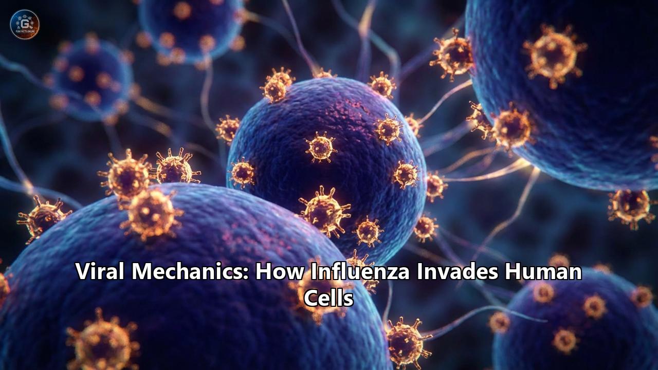
Before understanding the invasion, one must understand the invader. The influenza A virus is an enveloped virus, meaning it is wrapped in a lipid membrane stolen from the host cell it previously destroyed. Studding this membrane are the virus’s primary weapons—glycoprotein spikes that give the virus its jagged appearance and its family name, Orthomyxoviridae (from the Greek myxa, meaning mucus).
The Surface Spikes
Two major proteins dominate the surface, acting as the hands and the scissors of the virus:
- Hemagglutinin (HA): This trimeric protein is the key to entry. It accounts for about 80% of the surface proteins and looks like a mushroom. The "head" of the mushroom binds to receptors on human cells, while the "stalk" hides a deadly secret—a fusion peptide capable of merging viral and cellular membranes.
- Neuraminidase (NA): If HA is the grapnel used to board the ship, NA is the sword used to cut the lines upon departure. This tetrameric enzyme cleaves sialic acid residues, ensuring that newly formed viruses don't get stuck to the dying cell or to each other.
The Internal Machinery
Beneath the lipid envelope lies a shell of Matrix Protein 1 (M1), which gives the virus its structure and rigidity. Piercing through the envelope is the M2 Ion Channel, a small but vital pore that acts as a pH-gated proton pump—a mechanism we will see is crucial for the virus’s "unboxing" process.
Deep inside the core lies the viral genome. Unlike many viruses that carry a single strand of genetic material, influenza carries a segmented genome of eight separate negative-sense RNA strands. Each segment is wrapped in a protective coat of Nucleoprotein (NP) and capped with its own RNA polymerase complex (consisting of the subunits PB1, PB2, and PA). This segmented nature is the virus's "evolutionary superpower," allowing it to shuffle genes with other flu strains in a process called reassortment, leading to pandemics.
Part II: The Breach – Binding and Entry
The invasion begins with a case of mistaken identity. The epithelial cells lining the human upper respiratory tract are coated with a forest of glycoproteins. These proteins end in a specific sugar molecule called sialic acid. To the cell, sialic acid is a harmless surface marker. To the influenza virus, it is a landing pad.
The Sialic Acid Lock
Not all sialic acids are the same. The chemical linkage between the sialic acid and the underlying galactose sugar determines who gets infected.
- Alpha-2,3 linkage: This configuration looks like a straight cone and is found abundantly in the intestines of wild birds (ducks, gulls). Avian influenza viruses have HA proteins shaped specifically to bind this structure.
- Alpha-2,6 linkage: This configuration looks like a bent umbrella and is dominant in the human upper respiratory tract.
For an avian virus (like H5N1) to become a human pandemic, it must mutate its HA receptor-binding pocket to fit the Alpha-2,6 "umbrella." This often requires as few as two specific amino acid changes—such as the Q226L (Glutamine to Leucine) and G228S (Glycine to Serine) mutations seen in H2 and H3 subtypes. Without these mutations, an avian virus struggles to latch onto human throat cells, explaining why bird flu doesn't easily spread between people.
The Trojan Horse
Once the HA protein binds to a sialic acid receptor, the virus surfs along the cell surface until it finds a receptor-rich patch. The cell, sensing a ligand bound to its surface, initiates receptor-mediated endocytosis. The cell membrane invaginates, wrapping around the virus and pinching off to form a bubble called an endosome.
The virus is now inside the citadel, but it is trapped within a bubble. To replicate, it must release its genetic payload into the cell's cytoplasm. It does this by turning the cell's own defenses against it.
Part III: The Acid Trigger and the Harpoon
The cell, thinking it has captured a piece of food or debris, begins to acidify the endosome. It pumps protons (H+) into the bubble to lower the pH, aiming to break down the contents. This drop in pH is exactly what the virus is waiting for. It triggers a two-step mechanical sequence that is one of the most dramatic events in structural biology.
Step 1: The M2 Proton Pump
As the endosome becomes acidic (around pH 6.0), the M2 ion channels on the viral surface open. These channels allow protons to flood inside the virus particle itself. The acidification of the viral interior disrupts the interactions between the internal M1 matrix protein and the viral RNA coils (vRNPs). This "loosens" the genetic core, preparing it to be released.
Step 2: The HA "Spring-Loaded" Fusion
Simultaneously, the lower pH (around pH 5.0 to 5.5) acts on the external Hemagglutinin spikes. The HA protein is metastable; it is like a loaded spring held in place by a safety pin. The protons bind to specific histidine residues (such as His184), acting as a switch that releases the safety pin.
In a split second, the HA protein undergoes a massive conformational change. The "stalk" of the protein shoots upward, thrusting a hydrophobic sequence called the fusion peptide into the endosomal membrane. This peptide acts like a harpoon, anchoring the virus to the endosome wall.
The HA protein then collapses back on itself (a "jackknife" motion), pulling the two membranes together with immense force. The viral envelope and the endosomal membrane merge, opening a pore. The viral contents—the eight RNA segments and their polymerase enzymes—spill out into the cytoplasm. The Trojan Horse has opened.
Part IV: The Hijack – Nuclear Entry and Cap-Snatching
Most RNA viruses replicate in the cytoplasm (the cell's main room), but influenza is an aristocrat; it demands the VIP suite—the nucleus.
The viral ribonucleoproteins (vRNPs) contain nuclear localization signals (NLS) that mimic the cell’s own cargo tags. They bind to the host's importin-alpha transport machinery, which unknowingly ferries the viral genome through the nuclear pore complex and into the nucleus.
The Crime of "Cap-Snatching"
Once inside the nucleus, the virus faces a problem. To make viral proteins, it needs to produce messenger RNA (mRNA) that the host's ribosomes will recognize. Human mRNA has a special chemical "cap" at the 5' end that acts as a verification code. The influenza virus does not have the enzyme to make this cap. So, it steals it.
The viral polymerase complex executes a molecular heist known as "cap-snatching":
- The PB2 subunit of the viral polymerase binds to a newly forming host mRNA transcript.
- The PA subunit, which acts as an endonuclease (a molecular guillotine), slices the host mRNA about 10–13 nucleotides downstream of the cap.
- The virus uses this stolen capped fragment as a primer to start transcribing its own viral RNA.
This mechanism serves a dual purpose: it ensures viral mRNAs are translated efficiently by the host ribosomes, and it sabotages the host cell by destroying its own messenger RNAs, effectively shutting down host gene expression.
Part V: The Assembly Line
The nucleus now becomes a factory producing two things:
- Viral mRNA: These exit to the cytoplasm to be translated into viral proteins (HA, NA, M1, M2, etc.).
- vRNA (Genomic RNA): The viral polymerase switches modes to replicate the full genome. These new negative-sense RNA copies are wrapped in newly synthesized Nucleoprotein (NP) and form new vRNPs.
The Traffic Jam
The newly made viral proteins have different destinations. HA, NA, and M2 are processed through the Endoplasmic Reticulum and Golgi apparatus, where they are glycosylated (coated in sugars) and sent to the cell surface. They cluster in specific regions of the cell membrane called lipid rafts—thickened patches of the membrane rich in cholesterol.
Meanwhile, the new genomic vRNPs must leave the nucleus. The viral M1 and NEP (Nuclear Export Protein) bind to the vRNPs and guide them out of the nucleus and to the cell membrane.
The Packaging Mystery
One of the greatest marvels of influenza is how it ensures that exactly one copy of each of the eight segments gets into the new virus particle. If a virus is missing Segment 4 (HA), it is a dud. Research suggests that inter-segment interactions allow the eight RNAs to form a specific "daisy chain" or "bundle" configuration, ensuring a complete set is packaged.
Part VI: Escape – Budding and Release
At the lipid raft sites on the cell membrane, the components converge. The HA and NA spikes push outward, curving the membrane. The M1 protein lines the inside, pulling the membrane around the vRNP bundle.
The virus begins to bud off, but it remains tethered to the cell by the very same sialic acid receptors that allowed it to enter. The new HA spikes on the emerging virus bind to sialic acids on the dying cell's surface. If this bond is not broken, the new viruses will clump together and die on the surface of the cell.
This is where Neuraminidase (NA) plays its critical role. It acts as a molecular lawnmower, slicing off the sialic acid residues on the cell surface and on the nascent virion itself. This releases the virus into the extracellular space, free to infect the next cell. This step is the target of our most common flu drugs, neuraminidase inhibitors like Oseltamivir (Tamiflu), which gum up the NA blades, trapping the virus on the cell surface.
Part VII: The Host Strikes Back (and Why It Often Fails)
The human body does not take this invasion lying down.
- The Innate Response: Sensors like RIG-I detect the viral RNA and trigger the release of Interferons. These signaling proteins warn neighboring cells to shut down protein synthesis and activate antiviral genes.
- The Viral Countermeasure: The influenza NS1 (Non-Structural Protein 1) is a dedicated immune saboteur. It binds to the viral RNA to hide it from RIG-I, and it interacts with cellular machinery to block the processing of interferon mRNA.
- The Cytokine Storm: In highly pathogenic strains (like the 1918 flu or H5N1), the virus replicates so fast that the immune system panics. It floods the lungs with pro-inflammatory cytokines (IL-6, TNF-alpha), causing massive fluid buildup and tissue damage. Often, it is this "friendly fire"—the cytokine storm—that kills the patient, not the virus itself.
Conclusion: The Perpetual War
Influenza is not a static enemy. Through Antigenic Drift (slow mutations in HA/NA) and Antigenic Shift (swapping gene segments with avian or swine viruses to create hybrid pandemics), it constantly changes its appearance to evade our immune memory.
Understanding these viral mechanics—the alpha-2,6 lock, the pH trigger, the cap-snatching heist—is our only hope for defense. It has led to the development of endonuclease inhibitors (like Baloxavir) that jam the cap-snatching machinery and is driving the search for a Universal Flu Vaccine that targets the non-mutating "stalk" of the HA protein. Until then, the dance between the shapeshifting invader and the human cell continues, a deadly and beautiful display of biological mechanics.