For centuries, the map of the human body was considered complete. We had cataloged the continents of the organs—the heart, the liver, the lungs—and mapped the rivers of the veins and arteries. We had identified the microscopic cities of cells and the neural telegraph lines that connected them. Anatomy was a finished science, a subject of memorization rather than exploration.
But imagine walking through a dense forest for decades, studying every tree, every leaf, and every stone, only to realize one day that you had completely ignored the air itself. You had missed the medium that held it all together.
In 2018, a team of researchers stumbled upon something that had been hiding in plain sight for the entire history of medicine. It wasn’t a microscopic particle or a rare genetic mutation. It was a massive, body-wide structure—a fluid-filled superhighway functioning beneath our skin, surrounding our gut, encircling our lungs, and sheathing our muscles. They called it the interstitium.
This discovery did not just add a footnote to medical textbooks; it threatened to rewrite them. It suggested that what we previously dismissed as dense "connective tissue" was actually a dynamic, living network—a shock-absorbing, fluid-transporting, immune-regulating system that might just be the largest organ in the human body.
This is the story of that discovery, the anatomy of this hidden web, and how it fundamentally changes our understanding of cancer, healing, and the ancient mysteries of the human form.
Part I: The Accidental Discovery
The Phantom in the Bile Duct
The year was 2015. Dr. David Carr-Locke and Dr. Petros Benias, endoscopists at Mount Sinai Beth Israel in New York, were performing a routine procedure. They were using a cutting-edge piece of technology called Probe-based Confocal Laser Endomicroscopy (pCLE). Unlike traditional microscopes that require tissue to be sliced, stained, and placed on a slide, pCLE allows doctors to peer into living human tissue in real-time, inserting a microscopic camera probe directly into the body.
They were examining a patient’s bile duct, looking for signs of cancer. On the screen, they saw something strange.
According to standard anatomy, the bile duct’s wall should have been a dense, solid wall of connective tissue. But the live feed showed something else entirely. They saw a bizarre pattern of open, fluid-filled cavities. It looked like a honeycomb, or a lattice of dark spaces supported by a mesh of thick, bright bands.
Confused, they checked the equipment. They checked the literature. There was no record of such a structure.
They took a sample of the tissue and sent it to Dr. Neil Theise, a prominent liver pathologist at NYU Langone Health. When Theise looked at the sample under a standard microscope—the kind used in pathology labs for over a century—the open spaces were gone. The tissue looked exactly like the dense, solid wall described in textbooks.
The Great Collapse
This discrepancy sparked a detective story that unraveled a 100-year-old error in medical observation.
Dr. Theise realized that the standard method of preparing tissue for study was destroying the evidence. For decades, pathologists have "fixed" tissue samples by dehydrating them with chemicals and embedding them in wax. This process creates a snapshot of the cells, but it drains all the fluid.
Theise hypothesized that the "dense connective tissue" seen in textbooks was actually a collapsed building. In the living body, these tissues were open, fluid-filled skyscrapers. But when the fluid was drained during the fixation process, the floors (collagen bundles) collapsed on top of each other, appearing as a solid, flat pancake of tissue.
The "cracks" visible in old slides, which pathologists had long dismissed as artificial tears from the slicing process, were not accidents. They were the footprints of the vanished fluid. The open spaces were real. They had just never survived the journey to the microscope slide before.
To prove this, the team froze tissue samples immediately after removal, preserving the fluid structure before the collapse could occur. Under the microscope, the honeycomb structure remained. The "phantom" was real.
The Announcement
In March 2018, the team published their findings in Scientific Reports. They proposed that this network of fluid-filled spaces was not just a local quirk of the bile duct. It was everywhere. They found it in the dermis of the skin, the lining of the digestive tract, the lungs, the urinary system, and the fascia between muscles.
They argued that because this network possessed a unitary structure and function, it should be classified as a distinct organ: the interstitium.
The announcement sent shockwaves through the scientific community. Headlines blared: "Scientists Discover New Organ." While some anatomists debated the semantics of the word "organ," no one could deny the physical reality of what Theise and his colleagues had illuminated. The body was not a collection of solid parts packed in inert filler. It was a fluid continuum.
Part II: Anatomy of the Hidden Web
To understand the interstitium is to understand the architecture of life itself. It is the "space between," but it is far from empty.
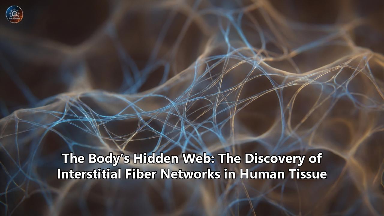
The Lattice: Collagen and Elastin
The structural skeleton of the interstitium is built from two primary proteins, creating a lattice that is both strong and flexible.
- Collagen Bundles: These are the pillars of the system. Thick, strong bundles of collagen (Types I, III, and V) form the walls of the fluid compartments. They provide tensile strength, ensuring that our tissues don’t simply dissolve into a puddle.
- Elastin Fibers: Interwoven with the collagen are fibers of elastin. As the name suggests, these provide elasticity. They allow the lattice to snap back into shape after being compressed or stretched.
Imagine a sponge. The solid material of the sponge is the collagen/elastin lattice. The holes in the sponge are the interstitial spaces. But unlike a kitchen sponge, this biological sponge is incredibly organized and spans the entire body.
The Fluid: A Pre-Lymphatic Ocean
Filling the spaces between these protein bundles is interstitial fluid. For a long time, scientists knew this fluid existed—it’s the nutrient-rich broth that bathes our cells—but they thought it merely seeped passively through dense tissue like water through soil.
The new discovery revealed that this fluid actually flows through open highways. It is a dynamic river system. The interstitium contains roughly 20% of the body's total fluid volume—more than the amount of blood in your veins.
This fluid is not stagnant. It is in constant motion, generated by the leakage of blood plasma from capillaries. It flows through the interstitial lattice, washing over cells, delivering nutrients, and collecting waste. Eventually, it drains into the lymphatic system.
This led to a crucial realization: The interstitium is the source of lymph. It is the "pre-lymphatic" system. Before immune fluid enters the lymph nodes to be scrubbed of bacteria, it travels through the open roads of the interstitium.
The Shock Absorber
Why does this structure exist? The primary evolutionary function appears to be mechanical protection.
The interstitium is found in tissues that undergo constant rhythmic movement:
- The pulsing of arteries.
- The peristalsis of the gut.
- The expansion of the lungs.
- The compression of the skin.
If these tissues were solid and rigid, the constant friction and pressure would tear them apart. The fluid-filled interstitium acts as a body-wide shock absorber. Because water is incompressible, the fluid-filled spaces prevent the tissues from collapsing under pressure, while the collagen lattice prevents them from over-expanding. It is a perfect hydraulic cushioning system, protecting the delicate machinery of our organs from the wear and tear of daily life.
Part III: The Superhighway of Disease
The discovery of the interstitium was not just an anatomical curiosity; it provided the "missing link" for understanding how diseases—particularly cancer—spread with such terrifying speed.
The Metastatic Waterslide
For decades, oncologists were puzzled by the behavior of certain cancers. A tumor might start in the stomach, but metastatic cells would appear in lymph nodes far away, much faster than expected. If the connective tissue was a dense wall, how were these large cancer cells tunneling through it so quickly?
The interstitium provided the answer. The connective tissue wasn't a wall; it was a highway.
When cancer cells break away from a primary tumor, they don't have to hack their way through dense muscle or fibrous tissue. They simply slip into the interstitial space. Once inside, they are in a fluid-filled channel with little resistance.
Dr. Theise described it chillingly: "Once they get in, it's like they're on a water slide."
The fluid flow within the interstitium carries these malignant cells directly toward the lymphatic system. This explains why the presence of cancer in the "stroma" (the connective tissue layer) is such a critical prognostic factor. It marks the moment the enemy breaches the castle walls and enters the river system that connects the entire kingdom.
New Mechanisms of Migration
Recent research (2020-2025) has deepened this understanding. Scientists found that cancer cells don't just passively float; they actively navigate this space.
- Downstream Flow: Cells can ride the natural flow of interstitial fluid toward lymph nodes (autologous chemotaxis).
- Upstream Struggle: Surprisingly, some aggressive cancer cells can crawl against the flow, using the stress of the fluid on their membranes to guide them upstream to other tissues.
This knowledge is revolutionizing cancer staging. It suggests that sampling the interstitial fluid (ISF) could provide an early warning system, detecting metastatic cells before they even reach the lymph nodes or the blood.
Fibrosis and Edema
The interstitium also explains the mechanics of other diseases.
- Edema (Swelling): Edema is essentially the flooding of the interstitium. When the balance of fluid entering and leaving these spaces is broken (e.g., heart failure or kidney disease), the "rooms" of the interstitium overfill, leading to visible swelling.
- Fibrosis (Scarring): In diseases like scleroderma or liver cirrhosis, the delicate collagen lattice becomes corrupted. The fluid spaces collapse, and the collagen bundles thicken and fuse. The shock absorber turns into a stiff, unyielding scar, causing the organ to fail.
Part IV: The Eastern Connection
Perhaps the most fascinating ripple effect of this discovery lies not in Western hospitals, but in the ancient clinics of Traditional Chinese Medicine (TCM).
For over 2,000 years, acupuncturists have treated the body based on a map of Meridians—channels through which Qi (energy) flows. Western anatomy has notoriously failed to find physical evidence of these meridians. Dissect a human arm, and you find nerves, veins, and muscles, but no "energy tubes" corresponding to the acupuncture charts.
Until now.
The San Jiao (Triple Burner)
In TCM, there is an organ system called the San Jiao, or "Triple Burner." Unlike the heart or stomach, the San Jiao has no distinct physical sac. Ancient texts describe it as "having a name but no form." It is described as a system of spaces and waterways responsible for the movement of fluids and energy throughout the entire body.
Sound familiar?
The description of the San Jiao aligns with startling precision to the description of the interstitium.
- TCM: A reticular network connecting all organs.
- Science: A body-wide network of connective tissue.
- TCM: Controls the metabolism of fluids.
- Science: Transports interstitial fluid and lymph.
- TCM: Located between the skin and muscles, and around the organs.
- Science: Located in the dermis, fascia, and organ linings.
Many integrative medicine researchers now propose that the interstitium is the anatomical basis of the San Jiao.
Explaining Acupuncture
This connection may also explain how acupuncture works.
Critics of acupuncture often ask: "How can inserting a needle into the toe affect the liver?" If the body is a collection of isolated parts, it makes no sense. But if the body is interconnected by a continuous, fluid-filled web, it becomes plausible.
- Mechanical Transduction: When an acupuncture needle is rotated, it winds the collagen fibers of the interstitium around it, like spaghetti on a fork. This mechanical stress signal is transmitted through the collagen lattice (fascia) at high speeds, potentially reaching distant organs.
- Electrical Conductivity: The interstitial fluid is an electrolyte-rich solution. It conducts electricity better than the surrounding cells. The "meridians" might be the paths of least electrical resistance flowing through the interstitial channels. Stimulation of these points could modulate the bioelectric signals passing through the "hidden web" to the internal organs.
While this theory is still being debated, the discovery of the interstitium has provided the first viable anatomical bridge between the reductionist view of Western anatomy and the holistic view of Eastern medicine.
Part V: The Future of Medicine
We are currently in the infancy of "Interstitial Science." The recognition of this network as a functional unit opens up frontiers that were previously unimaginable.
1. A New Diagnostic Window
Currently, we diagnose disease primarily through blood (hematology) or tissue biopsy (histology). The interstitium offers a third window.
Interstitial Fluid (ISF) Sampling is poised to become the next big diagnostic tool. ISF contains a different biomarker profile than blood. It is the direct environment of the cells. While blood tells us what is happening system-wide, ISF tells us what is happening in the local neighborhood of the tissue.Wearable microneedle patches are already being developed to continuously monitor glucose, lactate, and cancer markers in the interstitial fluid without drawing blood.
2. Drug Delivery Systems
Getting drugs into solid tumors is notoriously difficult. The high pressure within a tumor often pushes drugs away. Understanding the fluid dynamics of the interstitium could allow engineers to design nano-particles that ride the interstitial currents directly into the heart of a tumor, or use the network to distribute therapies more evenly across a fibrotic organ.
3. Immunology and Healing
We now know that the interstitium is a highway for macrophages and other immune cells. Understanding how these cells navigate the lattice could lead to new treatments for autoimmune diseases. If we can "close the road" to inflammatory cells in conditions like Crohn's disease or Rheumatoid Arthritis, we could stop the damage without suppressing the entire immune system.
Part VI: The Philosophical Shift
The discovery of the interstitium is more than just a triumph of microscopy; it is a triumph of perspective.
For centuries, Western medicine has been dominated by a "part-list" mentality. We break the body down into smaller and smaller pieces—organs, tissues, cells, organelles, molecules—assuming that if we understand the parts, we understand the whole.
But the interstitium reminds us that connection is as important as composition.
You cannot understand a forest just by studying the trees; you must understand the air, the soil, and the water that flows between them. The human body is not a machine made of distinct parts bolted together. It is a fluid continuum. The skin is connected to the fascia, which is connected to the lung lining, which is connected to the heart sac.
When you take a breath, the pressure wave travels through this web. When you move your arm, the fascia slides and the fluid shifts, rippling through the lattice. We are a singular, unified, hydro-dynamic organism.
The Unseen Space
It is humbling to realize that in the 21st century, in an age where we can map the genome and photograph black holes, we can still discover a new organ inside our own bodies. It begs the question: What else is hiding in the "empty" spaces?
The interstitium was invisible not because it was too small to see, but because we didn't know how to look. We were looking at the bricks and ignoring the mortar. We were looking at the dried ruins and missing the flowing river.
As we move forward, the "Hidden Web" will likely become a central focus of medical research. It bridges the gap between structure and function, between the solid and the liquid, and potentially, between the ancient wisdom of the past and the high-tech medicine of the future.
The map of the human body has changed. The empty spaces have been filled. And we are just beginning to explore the depths of this inner ocean.
Reference:
- https://www.sci.news/medicine/interstitium-new-organ-human-body-05860.html
- https://www.24hourfitness.com/24life/recover/2018/what-is-the-interstitium-and-why-should-we-care-about-it
- https://nyulangone.org/news/researchers-find-new-organ-missed-gold-standard-methods-visualizing-anatomy-disease
- https://www.wkyc.com/article/news/nation-world/scientists-say-theyve-discovered-a-new-human-organ-the-interstitium/507-532777731
- https://www.webmd.com/a-to-z-guides/what-is-the-interstitium
- https://newatlas.com/interstitium-new-organ-discovery/53981/
- https://www.the-scientist.com/cancer-cells-could-travel-through-the-interstitium-study-68680
- https://www.fasciatraininginstitute.com/fascia/fascia-and-interstitium-understanding-the-bodys-architectural-framework/
- https://www.weforum.org/stories/2018/04/there-s-a-previously-undiscovered-organ-in-your-body-and-it-could-explain-how-cancer-spreads/
- https://www.todaysrdh.com/the-interstitium-new-organ-discovery-and-its-potential-influence-on-the-oral-systemic-connection/
- https://www.dailysabah.com/health/2018/03/28/scientists-discover-biggest-human-organ-interstitium-hiding-in-plain-sight
- https://fasciaguide.com/research/fascia-is-the-largest-organ-in-the-body/
- https://www.researchgate.net/publication/325090406_NEWS_ALERT_-_The_Discovery_of_Interstitium_How_it_Relates_to_Energy_Medicine
- https://en.wikipedia.org/wiki/Interstitium
- https://www.ncbi.nlm.nih.gov/search/research-news/6489/
- https://www.researchgate.net/publication/265175041_Interstitial_fluid_flow_in_cancer_implications_for_disease_progression_and_treatment
- https://www.meer.com/en/80621-fascia-and-interstitiums-role-in-health-and-movement
- https://pmc.ncbi.nlm.nih.gov/articles/PMC6816778/
- https://www.seedacupuncture.com/the-seed-blog/bioelectric-interstitium-acupuncture-meridian-science-connective-tissue
- https://www.acupuncturenorthclinic.com/the-interstitium-and-how-it-s-relevant-to-your-acupuncture-treatments