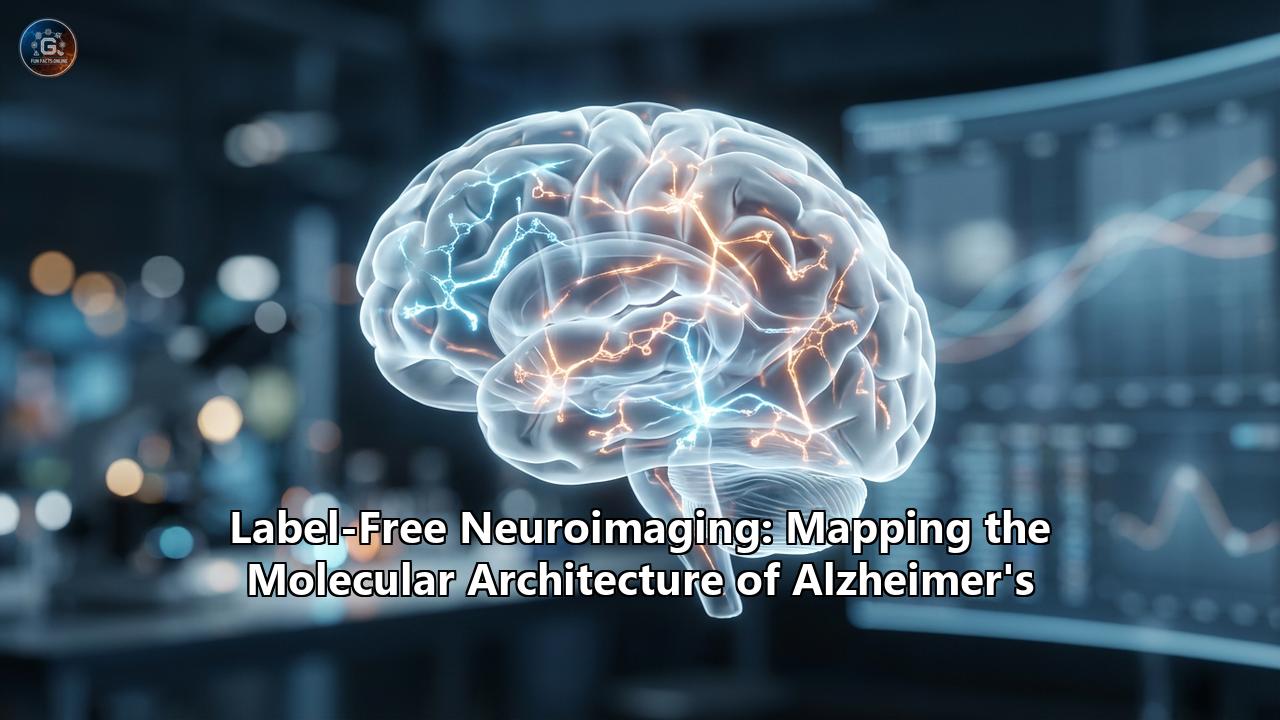
The human brain is arguably the most complex and intricate architecture in the known universe, a staggering network of billions of neurons and trillions of synapses. Yet, for all its structural majesty, it is acutely vulnerable to microscopic errors in its own molecular machinery. Chief among these devastating errors is Alzheimer’s disease, an insidious neurodegenerative disorder that systematically dismantles memory, cognition, and identity. For over a century, since Dr. Alois Alzheimer first peered through a microscope in 1906 and observed the strange "plaques" and "tangles" in a patient’s brain, scientists have relied on chemical stains and dyes to map this pathology.
However, a quiet revolution is currently rewriting the rules of neurobiology. Driven by breakthroughs in optics, quantum physics, and artificial intelligence, researchers are abandoning the dyes, the radioactive tracers, and the fluorescent tags. Welcome to the era of label-free neuroimaging—a paradigm shift that allows us to map the molecular architecture of Alzheimer’s disease in its pure, unaltered, and native state.
To truly appreciate the magnitude of this shift, we must first understand the "observer effect" in biological imaging. For decades, the gold standard for diagnosing and studying Alzheimer’s disease at the tissue level has been histological staining using exogenous reagents. Pathologists utilize fluorescent dyes like Thioflavin S and Congo Red, or complex immunohistochemical cocktails containing targeted antibodies, to force the brain’s hidden pathology to reveal itself. While these methods have been foundational, they come with a profound scientific cost. Exogenous dyes can induce unspecific binding, creating false-positive signals or uneven labeling. The very act of attaching a bulky fluorophore to a delicate lipid or protein molecule can alter its natural conformation, obscure its interactions, and disrupt the native tissue environment. In clinical diagnostics, techniques like Positron Emission Tomography (PET) rely on injecting radioactive tracers into the patient—a process that is not only expensive and invasive but also offers limited spatial resolution.
Label-free neuroimaging sidesteps these limitations by interrogating the intrinsic physical and chemical properties of the brain tissue itself. Rather than introducing a foreign molecule to light up a target, these advanced optical modalities listen to the innate "songs" of the native molecules—their natural vibrations, their interaction with light, their mass, and their structural density.
At the vanguard of this revolution is a suite of techniques based on Raman scattering. First described by the Indian physicist C.V. Raman in the early 20th century, the Raman effect occurs when light interacts with the chemical bonds of a molecule, resulting in a tiny fraction of the light being scattered at a different energy level. This shift in energy corresponds precisely to the vibrational modes of specific molecular bonds—acting as a unique "fingerprint" for every chemical substance. In the context of Alzheimer's disease, advanced nonlinear Raman techniques—specifically Coherent Anti-Stokes Raman Scattering (CARS) and Stimulated Raman Scattering (SRS)—have become extraordinary tools for mapping brain pathology.
Consider the amyloid-beta (Aβ) plaque, one of the primary pathological hallmarks of Alzheimer's. Traditional biology tells us that these plaques are simply aggregates of a misfolded protein. However, when researchers aim an SRS microscope at these plaques, an entirely new dimension of data emerges. By tuning the laser to the Amide I band—a vibrational mode corresponding to the carbon-oxygen double bonds in the backbone of proteins—scientists can detect the exact secondary structure of the tissue. Functional, healthy proteins in the brain are largely composed of alpha-helices. But when amyloid-beta misfolds and aggregates, it adopts a rigid, flat structure known as a beta-sheet. SRS microscopy can detect a distinct "blue shift" (a shift of approximately 10 inverse centimeters) in the Amide I spectrum that occurs precisely when proteins transition from healthy alpha-helices to toxic beta-sheets. For the first time, researchers can rapidly and directly image the spatial distribution of misfolded proteins in a living or fresh brain slice without a single drop of dye, distinguishing diseased tissue from healthy tissue purely by the vibrational frequency of its chemical bonds.
But the story told by label-free imaging does not end with proteins. For a long time, the dominant narrative of Alzheimer's disease has been the "amyloid hypothesis," which heavily focuses on protein aggregation. However, label-free CARS microscopy, which is uniquely suited to imaging lipids by detecting the symmetric stretching vibrations of carbon-hydrogen (CH2) bonds at 2840-2850 cm⁻¹, has brought lipids back into the spotlight. When imaging Alzheimer's brain tissue, CARS reveals that senile plaques are frequently surrounded by dense "halos" of abnormal lipid accumulations. By analyzing the intensity ratio between the CH2 stretching (at 2850 cm⁻¹) and the CH3 stretching (at 2930 cm⁻¹), label-free vibrational imaging can easily distinguish lesion areas from healthy neural real estate. This direct, undisturbed observation strongly supports the emerging view that localized lipid metabolism dysregulation is not merely a byproduct of Alzheimer's, but a core component of its architectural collapse.
As we dive deeper into the microscopic world, Fourier-Transform Infrared (FTIR) spectroscopy paired with Raman mapping has allowed scientists to reconstruct the actual life-cycle and chronological development of an amyloid plaque. According to recent label-free infrared imaging studies on human brain tissue, amyloid plaques undergo a distinct maturation sequence. In their infancy, "diffuse plaques" consist of short, antiparallel beta-sheets, which indicate the presence of toxic amyloid oligomers. As the disease progresses, these diffuse structures condense into "compact plaques," eventually maturing into "classic cored plaques" characterized by large, highly organized parallel beta-sheets indicative of dense fibrillar networks. Seeing this progression mapped out spatially, without the bias of antibody affinity, provides an unparalleled window into the biophysics of neurodegeneration.
Fascinatingly, the application of label-free Raman mapping (using 532 nm excitation) to human brain tissue has also unmasked a hidden player in the Alzheimer's microenvironment: carotenoids. In non-plaque areas of the brain, these molecules are virtually absent or randomly dispersed. Yet, around the dense cores of mature amyloid plaques, label-free imaging detects intense spikes in carotenoid signatures. Because carotenoids are intimately linked to oxidative stress and antioxidant defense mechanisms, their localized presence suggests a highly specific, localized neuroinflammatory response to the misfolded protein accumulations. This kind of micro-environmental mapping is nearly impossible to achieve with traditional staining, which requires researchers to know exactly what they are looking for in advance. Label-free techniques are fundamentally unbiased; they illuminate the entire chemical landscape, allowing the tissue to speak for itself.
While vibrational spectroscopy unveils the chemical composition of the brain, another marvel of label-free technology—Cryo-Micro-Optical Sectioning Tomography (Cryo-MOST)—has cracked the code of whole-brain, 3D structural mapping. Traditional histology requires slicing the brain into incredibly thin sections, staining them, and attempting to digitally stitch them back together—a painstaking process fraught with mechanical distortion. Cryo-MOST circumvents this by utilizing the natural autofluorescence of amyloid plaques. Researchers discovered that chilling the brain tissue to temperatures below -100 degrees Celsius dramatically enhances the natural fluorescence of senile plaques, while simultaneously preserving the tissue's structural integrity. An automated system then incrementally shaves the frozen brain block, capturing high-resolution optical images at every micron. The result is a breathtaking, three-dimensional, micron-resolution map of the entire brain’s amyloid burden, captured in its native anatomical context without a single exogenous label. This holistic view is vital for evaluating how experimental Alzheimer's drugs distribute and clear plaques across different regions of the brain over time.
Moving beyond photons, Mass Spectrometry Imaging (MSI), particularly utilizing high-resolution platforms like Fourier Transform Ion Cyclotron Resonance (FTICR), provides a different but highly complementary label-free perspective. Instead of bouncing light off the tissue, MALDI-MSI uses a precise laser to vaporize microscopic grid points across a frozen brain section. The vaporized molecules are then weighed in a mass spectrometer based on their mass-to-charge ratio. This allows for the simultaneous spatial mapping of hundreds of specific lipids, metabolites, and peptides across an Alzheimer's brain section. While standard immunohistochemistry can only target one or two proteins at a time based on antibody availability, mass spectrometry imaging acts as a broadband molecular radar, identifying complex overlapping pathologies—such as the exact spatial co-localization of specific gangliosides, myelin lipids, and amyloid fragments in the degenerating cortex.
Furthermore, the physical architecture of the brain tissue—its density, refractive index, and the way it bends light—holds untapped diagnostic potential. The accumulation of tangles and plaques fundamentally alters the microscopic optical properties of the tissue. Techniques exploiting phase and polarization, such as Quantitative Label-Free Imaging with Phase and Polarization (QLIPP), measure the retardance and anisotropy of brain slices. Normal white matter tracts have a highly ordered, directional architecture due to the alignment of myelin sheaths and axons. When Alzheimer's disease ravages these tracts, the structural entropy increases. Recent studies using learning-based quantum sensing and polarization metrics have shown that the physical structural differences caused by intracellular Tau tangles versus extracellular amyloid plaques yield distinct "linear entropy" signatures. The sheer physical density and disorder of these protein aggregates scatter light differently than healthy tissue, allowing researchers to discern the severity of pathology simply by analyzing how the tissue warps passing light.
This influx of complex, multidimensional data from label-free optics has paved the way for the ultimate convergence: the integration of Artificial Intelligence. When a human looks at a label-free phase image or an autofluorescence scan, the image often appears as a grayscale landscape of varying contrasts, lacking the bright, color-coded certainty of fluorescent dyes. Enter Deep Learning. Researchers have begun training sophisticated convolutional neural networks, such as the 2.5D U-Net architecture, to translate label-free physical measurements directly into molecular descriptions. By training the AI on thousands of paired images (where the tissue was first imaged label-free, and then chemically stained to reveal the "ground truth"), the neural network learns to recognize the invisible physical signatures of specific organelles and protein aggregates. Once trained, the AI can take a raw, label-free image and perfectly "predict" or computationally generate the fluorescent stain. This concept of "virtual staining" represents the holy grail of histopathology: achieving the chemical specificity of a targeted dye using only the non-destructive, label-free physical measurement of the tissue.
The implications of this label-free revolution extend far beyond the laboratory bench; they are charting a direct course toward clinical, in vivo applications. Today, diagnosing Alzheimer's definitively still requires either a costly PET scan, a painful lumbar puncture to assess cerebrospinal fluid (CSF), or, ultimately, a post-mortem brain autopsy. The dream of neurobiology is to detect the disease non-invasively, decades before cognitive decline manifests. Label-free optical coherence tomography (OCT) and Raman spectroscopy are currently being adapted for retinal imaging. The retina is fundamentally an extension of the central nervous system, sharing embryonic origins with the brain. Studies indicate that amyloid accumulation and vascular changes occur in the retina parallel to their occurrence in the cerebral cortex. Because the eye is optically clear, label-free techniques can be deployed through the pupil to measure the molecular and structural health of the retinal layers in living patients, potentially offering a routine, non-invasive screening tool for early-stage Alzheimer's.
Furthermore, the miniaturization of multiphoton and nonlinear optical systems into coherent anti-Stokes Raman scattering (CARS) microendoscopes is paving the way for revolutionary intraoperative tools. While primarily explored for brain tumor margin detection, the ability to maneuver a fiber-optic probe into living tissue to obtain real-time, label-free histological images could transform how neurodegenerative diseases are monitored. A surgeon or clinician equipped with a stimulated Raman scattering probe could instantly determine the chemical composition—differentiating lipids, amyloid fibers, and collagen—of the tissue in real-time, without waiting days for a pathology lab to fix, section, and stain a biopsy.
The transition to label-free neuroimaging is not merely a technological upgrade; it is a profound philosophical evolution in the life sciences. For centuries, our understanding of biology has been inherently destructive—we kill, fix, freeze, slice, and stain life to understand it. In doing so, we often study an artifact of our own creation rather than the biological reality. Alzheimer's disease, with its delicate interplay of misfolded proteins, shifting lipid metabolism, and localized neuroinflammation, demands to be understood in its native, unperturbed context.
By harnessing the subtle physics of light—the inelastic scattering of photons, the generation of harmonic frequencies, the measurement of quantum anisotropy, and the translation of these physical phenomena through artificial intelligence—we are finally learning to observe the brain without altering it. We are listening to the vibrational frequencies of the amide bonds; we are mapping the lipid halos of senile plaques; we are tracking the structural evolution of amyloid from soluble oligomer to intractable fibril. We are mapping the molecular architecture of Alzheimer's disease in its purest form. And in this pristine, label-free clarity, we may finally find the insights needed to dismantle the architecture of the disease itself.
Reference:
- https://pmc.ncbi.nlm.nih.gov/articles/PMC8341907/
- https://pubs.acs.org/doi/10.1021/acsphotonics.5c01192
- https://neurosciencenews.com/brain-mapping-alzheimers-7749/
- https://www.mdpi.com/2079-6374/13/1/27
- https://www.mdpi.com/2079-6374/11/8/255
- https://www.spiedigitallibrary.org/journals/journal-of-biomedical-optics/volume-20/issue-05/056013/Label-free-imaging-and-quantitative-chemical-analysis-of-Alzheimers-disease/10.1117/1.JBO.20.5.056013.full
- https://pmc.ncbi.nlm.nih.gov/articles/PMC6239428/
- https://www.researchgate.net/publication/355013832_Distinguishing_Amyloid_b-Protein_in_a_Mouse_Model_of_Alzheimer's_Disease_by_Label-Free_Vibrational_Imaging
- https://www.researchgate.net/publication/346962626_Label-free_vibrational_imaging_of_different_Ab_plaque_types_in_Alzheimer's_disease_reveals_sequential_events_in_plaque_development
- https://research.vu.nl/en/publications/multimodal-label-free-fluorescence-and-raman-imaging-of-amyloid-d/
- https://www.frontiersin.org/journals/neuroscience/articles/10.3389/fnins.2024.1301107/full
- https://repositum.tuwien.at/bitstream/20.500.12708/221820/1/Jansky%20Raphaela%20-%202025%20-%20Label-free%20characterization%20of%20alpha-synuclein...pdf
- https://www.researchgate.net/publication/343710618_Revealing_architectural_order_with_quantitative_label-free_imaging_and_deep_learning