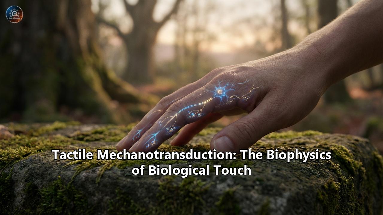
The sensation of a gentle breeze, the reassuring grasp of a loved one's hand, the sharp prick of a thorn, and the complex texture of woven silk—our interaction with the physical world is fundamentally mediated by the sense of touch. Unlike vision or hearing, which rely on the localized capture of light and sound waves, touch is uniquely intimate and distributed across the entirety of our largest organ: the skin. At the heart of this sensory experience lies one of the most elegant and complex processes in biology: tactile mechanotransduction.
Tactile mechanotransduction is the biophysical process by which mechanical forces—pressure, stretch, vibration, and shear—are translated into the electrochemical language of the nervous system. While touch is arguably the most ancient of the senses, evolutionary biologists and neuroscientists have only recently begun to decode the astonishing molecular machinery that makes it possible.
The Topography of Touch: The Mechanoreceptors
To understand how mechanical force becomes an electrical thought, we must first look at the specialized sensory outposts stationed throughout our skin. In the glabrous (hairless) skin of our palms and fingertips, tactile information is gathered by four primary types of mechanoreceptors, each anatomically tuned to filter specific physical stimuli. These end-organs are innervated by Aβ (A-beta) nerve fibers, which act as the high-speed broadband cables of the peripheral nervous system.
1. Merkel Cell-Neurite Complexes (Slowly Adapting Type 1 - SA1): Located just below the epidermis, Merkel cells are highly sensitive to steady pressure and spatial features. They are responsible for our ability to perceive fine details, edges, and curvatures. If you close your eyes and read Braille, it is the Merkel cells that trace the topography of the dots. Because they are "slowly adapting," they continue to fire action potentials as long as the stimulus is present. 2. Meissner Corpuscles (Rapidly Adapting Type 1 - RA1): Nestled in the dermal papillae, these fluid-filled, capsule-like structures are exquisitely sensitive to light touch and low-frequency vibrations (10 to 50 Hz). They are rapidly adapting, meaning they fire only when a stimulus begins and ends. Meissner corpuscles are the biomechanical sensors that allow us to detect "micro-slips"—the subtle friction changes that tell your brain to grip a coffee cup tighter before it falls from your hand. 3. Pacinian Corpuscles (Rapidly Adapting Type 2 - RA2): Resembling microscopic onions deep in the dermis, Pacinian corpuscles act as biological seismographs. They detect high-frequency vibrations (up to 300 Hz). When you feel the rumble of a heavy truck passing by, or the texture of a rough surface transmitted through the tip of a tool you are holding, your Pacinian corpuscles are firing. 4. Ruffini Endings (Slowly Adapting Type 2 - SA2): Located deep in the skin and oriented along stretch lines, Ruffini endings detect lateral skin stretch. They are crucial for kinesthetic sense, helping the brain determine the shape of the hand and the position of the fingers as they wrap around objects.The Molecular Transducers: The PIEZO Revolution
For decades, the physical structures of these end-organs were well-documented, but a glaring question remained: What is the exact molecular switch that turns a physical indentation into an electrical spark?
The answer was elusive because mechanosensory proteins are incredibly scarce compared to other cellular proteins. The breakthrough finally came in 2010, leading to the 2021 Nobel Prize in Physiology or Medicine awarded to Ardem Patapoutian. His team identified the PIEZO family of ion channels.
PIEZO1 and PIEZO2 are massive, evolutionarily conserved transmembrane proteins. In the tactile nervous system, PIEZO2 is the principal mechanotransduction channel. Structurally, the PIEZO2 channel is a biological marvel. It is a homotrimer—made of three identical protein subunits—that forms a structure resembling a three-bladed propeller. These blades curve upward, creating a distinct bowl-like depression in the cell membrane.
When a mechanical force is applied to the skin, the cell membrane is stretched. This tension flattens the curved blades of the PIEZO2 propeller. The flattening motion acts as a microscopic lever, ripping open a central pore in the protein. Instantly, positively charged ions—primarily sodium (Na+) and calcium (Ca2+)—flood into the negatively charged interior of the cell. This influx of cations depolarizes the cell membrane, generating a receptor potential. If the depolarization crosses a specific threshold, it triggers an action potential: a digital electrical spike that races up the sensory nerve to the spinal cord and, ultimately, to the somatosensory cortex of the brain.
Recent advancements in 2026 have pushed our understanding of PIEZO2 even further. Utilizing cutting-edge live-cell MINFLUX tracking, bioengineers have mapped the mobility of single PIEZO2 channels, revealing that their ability to discriminate different mechanical forces relies heavily on their localized molecular environment. PIEZO2 does not act in isolation; its mechanosensitivity is dynamically governed by its interactions with the cell's internal cytoskeleton and specific molecular networks that tether the channels in place.
The Biophysics of Gating: How Force Opens the Door
In the field of mechanotransduction biophysics, there has been a long-standing debate over exactly how external mechanical force is transmitted to the ion channel to pry it open. Two dominant biophysical models explain this phenomenon: the Bilayer Model and the Tether Model.
The Bilayer Model (Force-from-Lipid):In this model, the mechanosensitive ion channel is intrinsically responsive to changes in the tension of the surrounding lipid bilayer of the cell membrane. Imagine the cell membrane as a highly elastic trampoline with a rigid plastic ring (the ion channel) woven into the center. If you press down heavily on the trampoline, the fabric stretches tightly, pulling radially on the ring. In the bilayer model, this lipid tension is mathematically sufficient to force the protein into its open conformation. Studies on bacterial channels, which retain mechanosensitivity even when stripped of all cellular components and placed in artificial liposomes, strongly support this elegantly simple mechanism.
The Tether Model (Force-from-Filament):Eukaryotic cells, particularly mammalian neurons, feature complex internal architectures. In the tether model, the ion channel acts more like a trapdoor tied to a rope. The channel protein is physically linked (tethered) to the extracellular matrix on the outside, the microscopic filaments of the cytoskeleton on the inside, or both. When a mechanical force deforms the tissue, the relative displacement of the extracellular matrix and the cytoskeleton yanks on the tether, directly pulling the channel's gate open. Continuum mechanics and finite element simulations suggest that in complex mammalian tissues, the tether mechanism allows for highly efficient stress distribution and dramatic conformational changes in the channel.
Modern biophysics suggests that in human touch, we likely rely on a synergistic combination of both. The local lipid tension primes the PIEZO2 channel (bilayer mechanics), while its cytoskeletal tethers fine-tune its spatial confinement and amplify specific directional forces (tether mechanics).
The Two-Receptor-Site Symphony of the Merkel Complex
One of the most fascinating recent discoveries in tactile biophysics involves the Merkel cell-neurite complex. Historically, scientists hotly debated which part of this complex was the actual "sensor" of touch. Was it the epithelial Merkel cell itself, or was the Merkel cell just a structural support for the innervating sensory nerve ending?
Through genetic knockouts and patch-clamp electrophysiology, biophysicists proved the existence of a two-receptor-site model. It turns out that both the Merkel cell and the nerve fiber actively sense the touch, working in a highly synchronized partnership.
When your finger presses against an object, the Merkel cell detects the force via its own PIEZO2 channels. The cell depolarizes and releases neurotransmitters (such as serotonin and norepinephrine) across a synapse-like junction to the nerve ending. This chemical handoff is what generates the characteristic "sustained" or static firing rate of the slowly adapting (SA1) nerve fiber. Meanwhile, the Aβ nerve terminal also expresses its own PIEZO2 channels, which physically stretch and open during the initial indentation, providing the rapid, dynamic burst of electricity that signals the exact onset of the touch. Together, they form a bio-computational unit that perfectly encodes both the sudden contact and the sustained pressure of an object.
Beyond the Skin: Proprioception and Internal Touch
The biophysics of biological touch extends beyond the external environment. PIEZO2 is also the principal mechanotransduction channel for proprioception—the body’s "sixth sense" that tracks the position of our limbs in three-dimensional space.
Deep inside our muscles and tendons lie specialized sensory structures called muscle spindles and Golgi tendon organs. When a muscle lengthens, the physical stretch of the tissue activates PIEZO2 channels within these proprioceptive sensory endings. This generates an unrelenting stream of positional data to the brain. Without PIEZO2-driven mechanotransduction, we would be unable to walk, reach for a glass, or even stand upright with our eyes closed, as the brain would have no tactile map of the body's own geometry.
From Biology to Bionics: Engineering Artificial Touch
Understanding the biophysics of mechanotransduction is not merely an academic pursuit; it is revolutionizing the fields of robotics, haptics, and neuroprosthetics.
Historically, prosthetic limbs provided users with visual feedback—they had to watch the robotic hand to know if they were crushing a delicate plastic cup or letting it slip. Today, bioengineers are designing Neuromorphic Tactile Sensors (NTSs) aimed at mimicking the exact biological mechanisms of human touch. By applying finite element modelling to mimic the viscoelastic biomechanics of human skin, engineers can embed artificial mechanoreceptors in soft robotic fingertips.
Instead of processing continuous, power-heavy analog data streams, neuromorphic sensors use event-driven, spike-based data encoding, perfectly mirroring the action potentials generated by natural SA and FA mechanoreceptors. By injecting raw pressure data into Izhikevich neuronal models or Spiking Neural Networks (SNNs), these biomimetic tactile sensors can categorize the roughness and texture of naturalistic materials—from glass to woven textiles—with over 95% accuracy.
For patients with amputations, the ultimate goal is to connect these artificial action potentials directly into the residual peripheral nerves. By speaking the brain's native electrochemical language—timing the artificial electrical spikes exactly like a rapidly adapting Meissner corpuscle or a slowly adapting Merkel cell—scientists can trick the brain into "feeling" the texture of a virtual or robotic object.
The Symphony of Touch
Tactile mechanotransduction is a masterpiece of biophysics. It is a system where the macro-scale world of physical collisions and forces is seamlessly reduced to the micro-scale stretching of lipid bilayers, the flattening of microscopic protein propellers, and the rapid flow of sodium and calcium ions.
From the tip of a finger deciphering the grain of a wooden desk, to the deep proprioceptive stretch of a muscle during a dancer's leap, the biology of touch bridges the gap between the material universe and human consciousness. Through the continued exploration of mechanosensitive channels like PIEZO2 and the mathematical modeling of skin mechanics, we are not just learning how we feel the world—we are learning how to build new technologies that can feel the world with us.
Reference:
- https://www.researchgate.net/publication/318682668_Neuromorphic_Artificial_Sense_of_Touch_Bridging_Robotics_and_Neuroscience
- https://journals.plos.org/ploscompbiol/article?id=10.1371/journal.pcbi.1006264&rev=1
- https://pmc.ncbi.nlm.nih.gov/articles/PMC4282617/
- https://www.researchgate.net/publication/261519174_Piezo2_Is_Required_for_Merkel_Cell_Mechanotransduction
- https://pmc.ncbi.nlm.nih.gov/articles/PMC4039622/
- https://www.researchgate.net/publication/282038344_Neuromorphic_Artificial_Touch_for_Categorization_of_Naturalistic_Textures
- https://www.researchgate.net/publication/339011297_Biomimetic_Tactile_Sensors_Based_on_Nanomaterials
- https://www.mdpi.com/2073-4409/11/23/3827
- https://www.researchgate.net/publication/379616635_Neuromorphic_Tactile_Sensing_System_for_Textural_Features_Classification