The faint, rhythmic pulse of a monitor in a sterile lab at Northwestern University might just be the sound of history turning a page. For decades, the diagnosis of a severe spinal cord injury (SCI) has come with a grim, immutable prognosis: permanence. The central nervous system, unlike the skin or liver, was thought to lack the innate capacity to rebuild itself once severed. "The wire is cut," doctors would say, "and the connection is lost."
But in February 2026, that narrative shifted irrevocably.
In a landmark study that has sent shockwaves through the global biomedical community, scientists have not only grown functional, three-dimensional "mini-spinal cords" from human stem cells but have successfully used them to demonstrate a cure for paralysis using a revolutionary nanotechnology known as "dancing molecules." This is not a mouse study. This is not a simulation. This is human tissue, living and reacting in a dish, offering the first tangible proof that the "unhealable" wound can, in fact, be healed.
This article dives deep into the science, the struggle, and the staggering potential of spinal cord organoids—the lab-grown miracles that are bridging the gap between hopeless paralysis and a future of restored movement.
Part I: The Biological "Black Box"
To understand the magnitude of this breakthrough, one must first understand the adversary. The human spinal cord is arguably the most complex transmission line in the known universe. It is a soft, gelatinous bundle of nerve fibers, hardly wider than a pinky finger, yet it carries the electrical crackle of every step, every touch, and every command from the brain to the body.
When this cord is crushed or severed, the body’s reaction is paradoxically self-destructive. Unlike a cut on your finger, which knits back together with fresh skin, the spinal cord enters a state of chaotic defense.
- The Inflammatory Storm: Immediately after injury, immune cells flood the site. While meant to clean up debris, they often go rogue, releasing toxic chemicals that kill surviving neurons in a "secondary injury" phase that can last for months.
- The Glial Scar: To protect the rest of the nervous system from this toxic soup, special cells called astrocytes rush in and build a wall. This "glial scar" is a dense, fibrous barrier. It saves the life of the spinal cord but seals its fate; the scar creates a physical and chemical blockade that no regenerating nerve fiber can penetrate.
- The Axon Die-back: The long nerve fibers (axons) that were severed don't just sit there; they retract, withering away from the injury site, leaving a "cystic cavity"—a literal hole in the spinal cord.
For fifty years, scientists have thrown everything at this problem: steroids to stop inflammation, enzymes to digest the scar, and electrical bridges to bypass the gap. Nothing worked reliably in humans. The reason? We were studying mice, not men.
"A mouse spinal cord is not a human spinal cord," explains Dr. Elena Rossi, a neurobiologist at the forefront of stem cell research. "A mouse can spontaneously recover from injuries that would leave a human permanently paralyzed. We have been trying to unlock a human lock with a rodent key."
Enter the organoid.
Part II: The Birth of the "Mini-Spine"
An organoid is exactly what it sounds like: an "organ-like" structure. It is not a full-sized organ, but a three-dimensional miniature version grown in a bioreactor. Until recently, growing a spinal cord in a lab was considered impossible because of the tissue's complexity. A spinal cord needs specific dorsal cells (for sensation), ventral cells (for movement), and a precise architecture of long tracks to connect them.
The breakthrough came when researchers stopped trying to build the spinal cord and started teaching cells how to build it themselves.
The Recipe for a Miracle
The process begins with Induced Pluripotent Stem Cells (iPSCs). These are adult cells—taken from a patient’s skin or blood—that have been genetically reprogrammed to forget their identity and return to an embryonic state. From this blank slate, they can become anything.
In the lab, these cells are placed in a nutrient-rich "soup" containing specific signaling molecules—chemical instructions that mimic the environment of a developing embryo in the womb.
- Day 1-7: The cells are nudged to become "neuroectoderm," the primitive tissue that forms the nervous system.
- Day 14: Scientists add Retinoic Acid, a derivative of Vitamin A, which tells the cells, "You are not a brain; you are the spinal cord."
- Day 21-40: This is where the magic happens. The cells begin to self-organize. They clump together and, remarkably, start to arrange themselves into layers. Motor neurons migrate to the "ventral" side; sensory neurons move to the "dorsal" side. They are following an ancient genetic script written into their DNA.
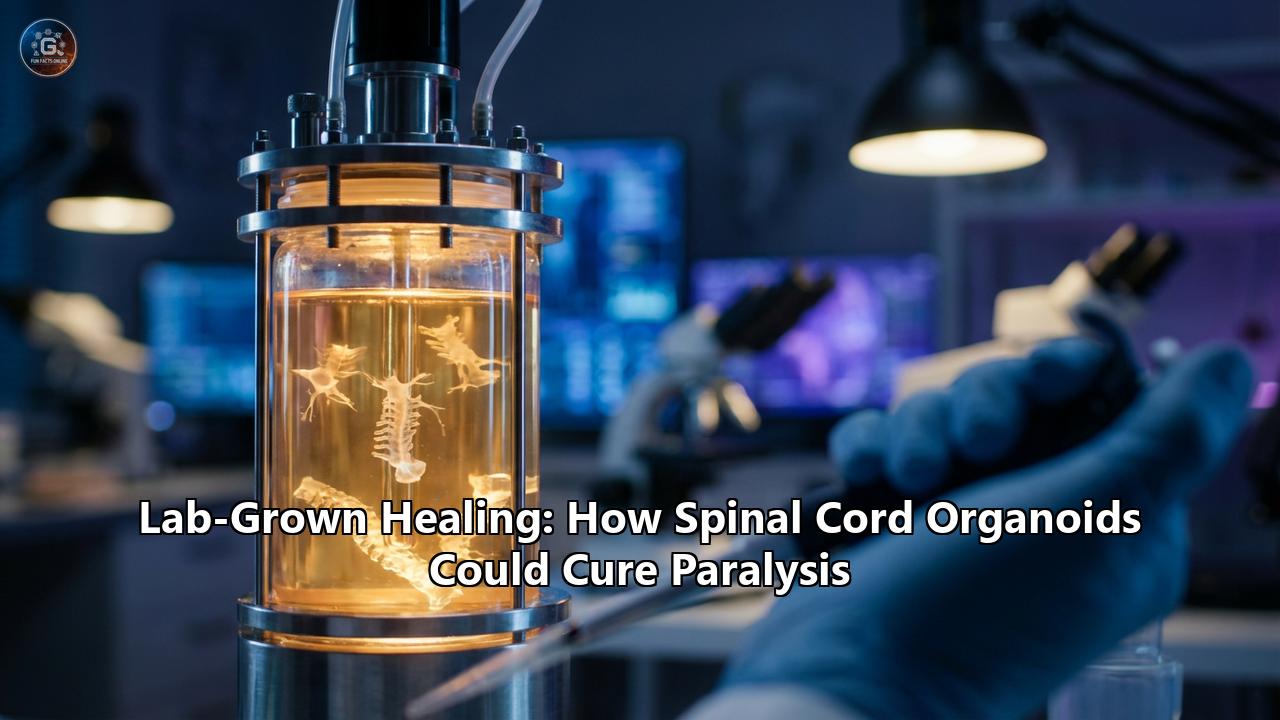
By Day 60, floating in the spinning liquid of a bioreactor, you have a Spinal Cord Organoid (SCO). It is a whitish pearl of tissue, roughly 3 to 4 millimeters wide. Under a microscope, it is a bustling metropolis of electrical activity. Neurons are firing. Glial cells are supporting them. It is, for all intents and purposes, a living piece of human human spinal cord.
But a healthy organoid is not enough. To cure paralysis, you have to break it.
Part III: The February 2026 Breakthrough
On February 11, 2026, a team led by Professor Samuel Stupp at Northwestern University published a paper in Nature Biomedical Engineering that will likely be cited for decades. They didn't just grow these organoids; they subjected them to the brutal reality of trauma.
Using micro-scalpels and compression devices, they created the first accurate lab model of a human spinal cord injury. They replicated the crushing blows of car accidents and the severing cuts of surgical mishaps.
"The reaction was chillingly accurate," Stupp reported. "We saw the immune cells swarm. We saw the neurons die. And most importantly, we saw the formation of the impenetrable glial scar. It was paralysis in a dish."
Then, they introduced the "Dancing Molecules."
The Choreography of Repair
Traditional drugs are static. Imagine trying to open a lock with a key that is frozen in a block of ice. The key is there, but it can't wiggle into the mechanism. The receptors on human cells are vibrating, moving, and shifting constantly. If a drug molecule is stiff and stationary, it often fails to connect with the moving target.
Stupp’s team created a "supramolecular peptide fibril"—a liquid that, when injected into the body (or the organoid), instantly turns into a gel mesh. Inside this mesh are hundreds of thousands of bioactive molecules. But here is the genius: they are not chemically bound to the mesh. They are loose. They vibrate, rotate, and "dance" at incredible speeds.
Because they are moving so fast, they have a massive statistical advantage in hitting the moving cellular receptors. They hit the "repair" button on the neurons thousands of times more effectively than a static drug.
The Results
When injected into the injured human organoids, the results were nothing short of miraculous:
- Scar Dissolution: The density of the glial scar dropped significantly. The physical wall crumbled.
- Neurite Outgrowth: The severed neurons didn't just survive; they grew. Long tendrils (neurites) shot across the gap, reconnecting with the other side.
- Myelination: The protective sheath around the nerves, often lost in injury, began to reform.
"We saw neurites growing in dense, organized bundles," said Stupp. "They were bridging the gap. In the untreated organoids, there was silence. In the treated ones, there was reconnection."
Part IV: The Global Race for Regeneration
While Northwestern has claimed the headlines in 2026, they are not alone. A quiet, intense arms race is underway in laboratories from Tokyo to Geneva, each tackling a different piece of the paralysis puzzle using organoids.
1. The "Assembloids" of Cambridge and RIKEN
At the University of Cambridge and RIKEN in Japan, researchers are solving the "disembodied" problem. A spinal cord organoid in a jar has nowhere to send its signals. It’s like a telephone wire connected to nothing.
These labs are creating "Assembloids." They grow a brain organoid (the command center), a spinal cord organoid (the wire), and a muscle organoid (the actor). They place them next to each other, and the cells reach out. The brain extends axons to the spine; the spine extends axons to the muscle.
In 2025, a team at Cambridge watched as a "mini-brain" sent a signal through a "mini-spine" and made a piece of lab-grown muscle twitch. It was the first time a thought—albeit a chemical one—had translated into movement in a fully synthetic human system. This proves that organoid repair isn't just cosmetic; it's functional.
2. The Swiss Digital Bridge
At EPFL in Switzerland, Grégoire Courtine’s team has long been famous for their "digital bridge"—implanting chips to bypass spinal injuries. Now, they are integrating biology. They are using patient-specific organoids to test gene therapies that "soften" the spinal cord tissue before inserting their electrodes. By combining the "dancing molecules" to heal the biological tissue with their digital implants, they aim to create a "bio-hybrid" cure: biology for the healing, technology for the control.
3. The 3D Scaffolds of Minnesota
At the University of Minnesota, the problem is structure. A real spinal cord is a long cable, not a blob. Organoids tend to grow into spheres. Minnesota researchers are using 3D printing to create silicone guides—microscopic tubes that force the organoid cells to grow in long, straight lines.
"Neurons are like vines," explains lead researcher Dr. Annabel Chen. "If you give them a trellis, they grow tall and straight. If you don't, they grow into a tangle." By printing these "scaffolds" and seeding them with Stupp’s dancing molecules, they are creating spinal cord "patches" that could physically be snapped into a gap in a patient’s spine.
Part V: The Ethical Frontier – Does a Mini-Spine Feel Pain?
As the biology becomes more sophisticated, the philosophy becomes murkier. We are growing human neural tissue. We are making it fire. We are connecting it to muscle. The question must be asked: If you pinch a spinal cord organoid, does it hurt?
The spinal cord is the highway of pain. It is the transmission line for nociception (the processing of noxious stimuli). When researchers at UCSD created organoids that successfully modeled the "pain pathway," bioethicists raised a red flag.
"We are entering the 'Uncanny Valley' of ethics," warns Dr. Sarah Chan of the University of Edinburgh. "These tissues are not conscious in the way a brain is. They have no cerebral cortex to say 'I am me.' But they have the machinery to process suffering. If we injure them to test a drug, are we causing a form of biological distress?"
The consensus for now is "no." Pain requires a brain to interpret the signal. A spinal cord organoid might register the signal of pain (an electrical spike), but without a brain organoid attached and developed enough to possess consciousness, it is like a phone ringing in an empty house. The signal is there, but no one is answering.
However, as "assembloid" research advances and we connect mini-brains to mini-spines, that house may not stay empty for long. Bioethics panels are currently drafting the first "Bill of Rights" for neural organoids, regulating how mature they can become and what stimuli they can be subjected to.
Part VI: The "Valley of Death" and the Clinical Leap
Why does this matter to the person in the wheelchair today?
For decades, spinal cord research has died in the "Valley of Death"—the gap between successful mouse trials and successful human trials.
- The Mouse Problem: Mice are genetically distinct. Their immune systems are different. A drug that dissolves a scar in a mouse often fails in a human because our astrocytes are more aggressive.
- The Organoid Solution: Organoids are the bridge. They are human. If "dancing molecules" work in a human organoid (which they do), the predictive power for a human clinical trial is exponentially higher than data from a rat.
With the validation from the February 2026 study, the FDA has already granted "Orphan Drug Designation" to the dancing molecule therapy.
- 2026-2027: Safety trials in large primates (to ensure the gel doesn't migrate).
- 2028: Phase I Human Clinical Trials. This will likely involve "chronic" patients—those injured years ago. Surgeons will expose the injury site, clean the scar tissue, and inject the supramolecular gel.
- 2030 and beyond: If successful, we could see a world where a spinal cord injury is treated immediately in the ER with an injection that prevents paralysis before it even sets in.
Part VII: The Personalized Patch
The ultimate dream is not just a drug, but a replacement part.
Imagine a patient, John, breaks his back in a motorcycle accident. A 2-centimeter section of his spinal cord is pulped.
In the hospital of 2035:
- Doctors take a blood sample from John.
- They reprogram his blood cells into iPSCs.
- In a bioreactor, they grow a "John-specific" spinal cord organoid on a 3D-printed scaffold, tailored to the exact dimensions of his injury.
- Surgeons implant this living patch into his spine.
Because it is his own DNA, there is no rejection. The "dancing molecules" are used as the glue to fuse the patch to his existing nerves. Rehabilitation begins. Six months later, John walks.
Conclusion: The End of "Permanent"
We are no longer looking at paralysis as a life sentence. We are looking at it as an engineering problem. The "dancing molecules" have provided the tool, and the organoids have provided the testing ground.
For the first time in history, the wiring of the human body is no longer a mystery we must passively accept, but a material we can mold, mend, and regrow. The monitor in the lab is still beeping, but the message has changed. It is no longer counting down the time lost. It is counting down the time until the cure arrives.
Reference:
- https://khanfk.substack.com/p/lab-grown-spinal-cord-organoids-offer
- https://news.feinberg.northwestern.edu/2026/02/11/paralysis-treatment-heals-lab-grown-human-spinal-cord-organoids/
- https://www.sciencealert.com/scientists-grew-mini-human-spinal-cords-then-made-them-repair-after-injury
- https://nrtimes.co.uk/spinal-cord-research-could-accelerate-regenerative-therapies-therapy25/
- https://scantox.com/neural-pathway-organoids-provide-never-before-seen-look-into-pain-signals/
- https://journals.library.columbia.edu/index.php/bioethics/article/view/12504
- https://www.the-scientist.com/spinal-cord-organoids-help-test-paralysis-treatment-74087
- https://www.iflscience.com/lab-grown-mini-human-spinal-cords-healed-in-major-step-towards-paralysis-cure-82531
- https://www.researchgate.net/publication/391726392_Neural_Organoid_Research_Ethics_and_Governance
- https://pmc.ncbi.nlm.nih.gov/articles/PMC12694639/