The Silent Threat: Unraveling the Vascular Science of Brain Aneurysms
Deep within the intricate network of blood vessels that nourish our brain, a silent and potentially deadly threat can emerge. A brain aneurysm, a bulge or ballooning in a blood vessel, can exist for years without a whisper of its presence, only to announce itself with a sudden, catastrophic rupture. This event, a subarachnoid hemorrhage, is a medical emergency of the highest order, often leaving devastation in its wake. Understanding the science behind why these vascular time bombs form, grow, and sometimes burst is a journey into the complex interplay of genetics, lifestyle, and the very mechanics of blood flow.
What is a Brain Aneurysm?
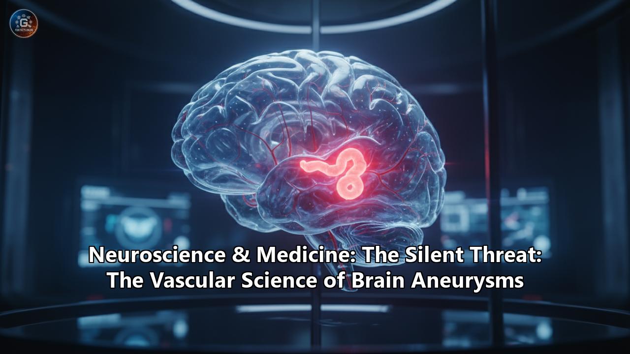
A brain aneurysm, also known as a cerebral or intracranial aneurysm, is a weak, bulging spot on the wall of a brain artery. This abnormal outpouching is likened to a bubble or a thin, worn patch on a tire. The constant pressure of blood flow can cause this weakened area to gradually expand. While many aneurysms remain small and asymptomatic, the primary danger lies in their potential to rupture and bleed into the space surrounding the brain, an event called a subarachnoid hemorrhage. This type of hemorrhagic stroke can lead to severe brain damage, long-term disability, or even death.
It's estimated that unruptured brain aneurysms affect about 3.2% of people worldwide, with approximately 6.7 million people in the United States living with one. However, ruptured aneurysms are far less common, occurring in about 30,000 Americans annually. The average age of rupture is around 50 years old.
The Architecture of a Flaw: Types of Brain Aneurysms
Brain aneurysms are not all created equal. They are classified based on their shape, size, and location, which can influence their risk of rupture and the approach to treatment.
The most common types include:
- Saccular Aneurysms: Often called "berry" aneurysms due to their resemblance to a berry hanging from a vine, these are the most prevalent type. They appear as a rounded, blood-filled sac that protrudes from a blood vessel, usually at a fork or branch in an artery at the base of the brain. These are the aneurysms most likely to rupture.
- Fusiform Aneurysms: This type involves a ballooning or bulging of the artery on all sides, creating a spindle-shaped swelling.
- Mycotic Aneurysms: These are rare aneurysms caused by an infection in the arterial wall. The infection weakens the vessel, leading to the formation of an aneurysm.
Aneurysms are also classified by size, ranging from small (less than 7 millimeters) to giant (larger than 25 millimeters). While larger aneurysms have a greater tendency to rupture, most ruptured aneurysms are actually less than 10 millimeters in diameter.
The Unseen Enemy: Symptoms and Diagnosis
The insidious nature of brain aneurysms lies in their silence. Most unruptured aneurysms produce no symptoms and are often discovered incidentally during imaging tests for other medical conditions.
However, if an unruptured aneurysm grows large enough to press on surrounding brain tissue or nerves, it may cause:
- Pain above and behind the eye
- A dilated pupil in one eye
- Changes in vision or double vision
- Numbness on one side of the face
- A drooping eyelid
- In rare cases, headaches or seizures
In some instances, an aneurysm may leak a small amount of blood, known as a "sentinel bleed." This can cause a sudden, severe headache days or weeks before a major rupture.
The rupture of a brain aneurysm is a dramatic and life-threatening event. The hallmark symptom is a sudden, excruciating headache, often described as "the worst headache of my life" or a "thunderclap headache."
Other symptoms of a ruptured aneurysm include:
- Nausea and vomiting
- Stiff neck
- Blurred or double vision
- Sensitivity to light
- Seizures
- Confusion or loss of consciousness
- Computed Tomography (CT) Scan: This is often the first test performed to detect bleeding in the brain. A CT angiogram (CTA), which involves injecting a contrast dye, can provide detailed 3D images of the blood vessels and help identify the aneurysm's size, shape, and location.
- Magnetic Resonance Imaging (MRI): An MRI uses a magnetic field and radio waves to create detailed images of the brain. A magnetic resonance angiogram (MRA) specifically visualizes the blood vessels.
- Cerebral Angiography: This is considered the gold standard for diagnosing brain aneurysms. It's an invasive procedure where a catheter is threaded from an artery in the groin up to the brain. A contrast dye is then injected to provide highly detailed X-ray images of the arteries, revealing the precise anatomy of the aneurysm.
- Cerebrospinal Fluid (CSF) Analysis: If a rupture is suspected but not confirmed by a CT scan, a lumbar puncture (spinal tap) may be performed. The presence of red blood cells in the cerebrospinal fluid indicates bleeding in the brain.
At the Crossroads of Flow and Weakness: The Science of Aneurysm Formation
The formation of a brain aneurysm is a complex process that begins at the intersection of blood flow dynamics and the structural integrity of the blood vessel wall. Arteries are not rigid pipes; they are living tissues that must withstand the constant pulsatile force of blood flow.
The brain's arterial network, particularly the Circle of Willis at the base of the brain, has numerous branches and bifurcations. These are areas of high hemodynamic stress, where the force of the blood is not evenly distributed. Experts believe that this constant pressure on a weakened area of the vessel wall is what initiates the formation and growth of an aneurysm.
At the cellular and molecular level, the process is now understood to be driven by a chronic inflammatory response. Here's a breakdown of the key events:
- Endothelial Dysfunction: The endothelium is the thin layer of cells lining the inside of blood vessels. Hemodynamic stress can damage these cells, triggering an inflammatory cascade. This dysfunction leads to the expression of inflammatory genes and the recruitment of immune cells, such as macrophages.
- Inflammatory Cell Infiltration: Macrophages and other inflammatory cells are drawn to the site of injury. These cells release a cocktail of chemicals, including cytokines and enzymes called matrix metalloproteinases (MMPs).
- Extracellular Matrix Degradation: The arterial wall's strength and elasticity are provided by a scaffold of proteins called the extracellular matrix (ECM), which includes collagen and elastin. The MMPs released by inflammatory cells begin to break down this matrix.
- Smooth Muscle Cell Apoptosis: Smooth muscle cells are crucial for maintaining the structural integrity of the vessel wall and synthesizing new ECM proteins. The inflammatory environment triggers apoptosis, or programmed cell death, in these cells.
- Wall Remodeling and Weakening: The combination of ECM degradation and the loss of smooth muscle cells leads to a progressive thinning and weakening of the arterial wall. This pathological remodeling creates a susceptible spot that can no longer withstand the pressure of blood flow, causing it to bulge outwards and form an aneurysm.
The Tipping Point: Why Aneurysms Rupture
While the formation of an aneurysm is a gradual process, its rupture is a sudden and catastrophic failure of the weakened vessel wall. The decision of whether an aneurysm will rupture is influenced by a complex interplay of factors.
Hemodynamics and Biomechanics: The forces exerted by blood flow play a critical role. Researchers use computational fluid dynamics (CFD) to model blood flow patterns within aneurysms. These studies have shown that both high and low wall shear stress (WSS) can contribute to rupture, though through different mechanisms.- High Wall Shear Stress: This can lead to the growth and rupture of smaller aneurysms through a direct mechanical response from the vessel wall.
- Low Wall Shear Stress: In larger aneurysms, areas of low and stagnant flow can promote inflammation and thrombus (clot) formation, further destabilizing the aneurysm wall.
The geometry of the aneurysm, including its size, shape, and the angle at which it protrudes from the parent artery, also significantly impacts the hemodynamic forces and the risk of rupture.
Risk Factors for Rupture: Several factors are known to increase the likelihood of an aneurysm rupturing:- Size and Growth: Larger aneurysms and those that are actively growing are at a higher risk of rupture.
- Location: Aneurysms located in the posterior circulation of the brain (the back of the brain) are more prone to rupture.
- Hypertension (High Blood Pressure): Uncontrolled high blood pressure puts increased stress on the aneurysm wall.
- Smoking: Smoking is a major risk factor for both the formation and rupture of aneurysms.
- Family History: A family history of aneurysm rupture may indicate a genetic predisposition and a higher personal risk.
- Previous Rupture: An aneurysm that has already leaked is at a high risk of re-bleeding.
The Aftermath: Consequences of a Ruptured Aneurysm
A ruptured aneurysm leads to a subarachnoid hemorrhage (SAH), where blood floods the space between the brain and the thin tissues that cover it. This bleeding is a devastating event with a high rate of mortality and long-term disability. Nearly 25% of people die within the first 24 hours, and 50% die within 3 months.
The initial hemorrhage is only the beginning of a cascade of potential complications:
- Re-bleeding: The aneurysm is at a high risk of rupturing again, which is often more devastating than the initial bleed. The mortality rate for a re-bleed is about 67%.
- Vasospasm: In the days following a rupture, the blood vessels in the brain can narrow and constrict, a condition known as vasospasm. This can lead to an ischemic stroke by restricting blood flow to parts of the brain, causing further brain damage.
- Hydrocephalus: The leaked blood can block the normal circulation and absorption of cerebrospinal fluid (CSF), causing it to build up within the brain. This condition, called hydrocephalus, increases pressure on the brain and can damage tissue.
- Electrolyte Imbalances: The brain hemorrhage can disrupt the body's sodium balance, leading to swelling of brain cells and permanent damage.
- Seizures: Seizures are a common complication following a ruptured aneurysm.
- Cardiac and Pulmonary Complications: The immense stress of the event can lead to "stress cardiomyopathy" (a form of cardiac failure) and fluid buildup in the lungs (neurogenic pulmonary edema).
Defusing the Time Bomb: Treatment Strategies
The goal of treating a brain aneurysm is to prevent it from rupturing or, if it has already ruptured, to prevent re-bleeding. The decision to treat an unruptured aneurysm depends on a careful assessment of the risks of the procedure versus the risk of rupture. Small, stable aneurysms may be monitored with regular imaging. For ruptured aneurysms, immediate treatment is essential.
There are two main approaches to securing an aneurysm:
1. Surgical Clipping: This is an open-brain surgery where a neurosurgeon removes a small section of the skull to access the aneurysm. Using a microscope, the surgeon places a tiny metal clip across the neck of the aneurysm, cutting off its blood supply. This prevents blood from flowing into the aneurysm sac, thereby eliminating the risk of rupture. Surgical clipping is highly effective, and clipped aneurysms rarely return. However, it is an invasive procedure with risks that include damage to other blood vessels, stroke, and infection. Recovery typically takes 4 to 6 weeks. 2. Endovascular Coiling: This is a less invasive procedure performed from inside the blood vessels. A catheter is inserted into an artery, usually in the groin, and guided up to the brain. Tiny, soft platinum coils are then passed through the catheter and deposited into the aneurysm. The coils pack the aneurysm, blocking blood flow and promoting clot formation, which effectively seals it off from the parent artery.In some cases, other endovascular techniques may be used:
- Stent-Assisted Coiling: A small, flexible mesh tube called a stent is placed in the parent artery across the neck of the aneurysm to help hold the coils in place, particularly for wide-necked aneurysms.
- Flow Diversion: A specialized stent-like device, known as a flow diverter, is placed in the parent artery to redirect blood flow away from the aneurysm. This reduces the pressure on the aneurysm wall, causing it to shrink and eventually heal over time. This technique is particularly useful for large or complex aneurysms that are difficult to treat with clipping or coiling.
Endovascular treatments generally have a shorter recovery time than surgical clipping, but there is a risk of the aneurysm recurring over time, which may require further treatment.
Other Treatments:- Medications: After a rupture, medications such as calcium channel blockers are used to prevent vasospasm. Pain relievers, anti-seizure medications, and drugs to control blood pressure are also part of the management.
- Shunting: If hydrocephalus develops, a shunt (a thin tube) may be surgically placed to drain excess cerebrospinal fluid from the brain to another part of the body.
- Rehabilitation: For those who survive a ruptured aneurysm, physical, speech, and occupational therapy are often necessary to regain lost function and adapt to any permanent disabilities.
The Frontier of Aneurysm Research: A Glimpse into the Future
The field of brain aneurysm research is rapidly evolving, with scientists and clinicians working to better understand, diagnose, and treat this silent threat.
Genetics and Molecular Pathways: Large-scale genetic studies have identified specific gene variants that increase the risk of developing brain aneurysms. This research is paving the way for identifying individuals at high risk who may benefit from screening. Scientists are also delving deeper into the molecular pathways of inflammation, hoping to develop medical therapies that can stabilize or even reverse aneurysm growth, potentially preventing the need for surgery or endovascular procedures. For example, statins and aspirin are being investigated for their potential anti-inflammatory effects on aneurysm progression. Advanced Imaging and Hemodynamic Modeling: The use of advanced imaging techniques and computational fluid dynamics is becoming more sophisticated. This allows for more precise risk stratification of unruptured aneurysms, helping to identify which ones are most likely to rupture and therefore require treatment. Innovations in Endovascular Treatment: The technology for endovascular repair is constantly improving. New generations of coils, stents, and flow diverters are being developed to treat even the most complex aneurysms with greater safety and efficacy. Research is also focused on enhancing the healing process after these procedures, for example, by promoting the growth of a new endothelial layer across the neck of the treated aneurysm. Improving Outcomes after Rupture: Researchers are actively seeking new ways to manage the devastating consequences of a subarachnoid hemorrhage. This includes developing better treatments for vasospasm and finding new strategies to mitigate the inflammatory response that contributes to secondary brain injury.The journey to conquer the silent threat of brain aneurysms is ongoing. Through continued research into the intricate vascular science that governs their formation and rupture, and through the development of innovative diagnostic and therapeutic tools, the medical community is steadily improving its ability to manage this complex and often devastating condition. The goal is to one day transform the narrative of the brain aneurysm from one of a silent threat to one of a manageable and preventable condition.
Reference:
- https://hospital.uillinois.edu/primary-and-specialty-care/neurology-and-neurosurgery/neurological-conditions-we-treat/brain-aneurysm/diagnosis-imaging
- https://www.ahajournals.org/doi/10.1161/strokeaha.113.002390
- https://pmc.ncbi.nlm.nih.gov/articles/PMC4533330/
- https://www.ohsu.edu/brain-institute/brain-aneurysm-diagnosis-and-treatment
- https://www.brighamandwomens.org/neurosurgery/brain-aneurysm
- https://www.ninds.nih.gov/health-information/disorders/cerebral-aneurysms
- https://en.wikipedia.org/wiki/Intracranial_aneurysm
- https://www.mayoclinic.org/diseases-conditions/brain-aneurysm/diagnosis-treatment/drc-20361595
- https://www.mayoclinic.org/diseases-conditions/brain-aneurysm/symptoms-causes/syc-20361483
- https://ouci.dntb.gov.ua/en/works/lReaQGK4/
- https://www.researchgate.net/publication/278731098_Hemodynamic_Differences_in_Intracranial_Aneurysms_before_and_after_Rupture
- https://pubmed.ncbi.nlm.nih.gov/25761423/
- https://www.youtube.com/watch?v=eBp0H7qvYQA
- https://med.miami.edu/labs/starke-lab/research
- https://www.youtube.com/watch?v=01srLFGlty8
- https://www.nhs.uk/conditions/brain-aneurysm/
- https://www.healthline.com/health/brain-aneurysm-repair
- https://www.youtube.com/watch?v=ZRjJp4Jl1QM
- https://matthewlawsonmd.com/great-review-of-new-brain-aneurysm-treatments-endovascular-today/
- https://scispace.com/papers/a-review-of-technological-innovations-leading-to-modern-2s7zphfg
- https://scholars.mssm.edu/en/publications/a-review-of-technological-innovations-leading-to-modern-endovascu/