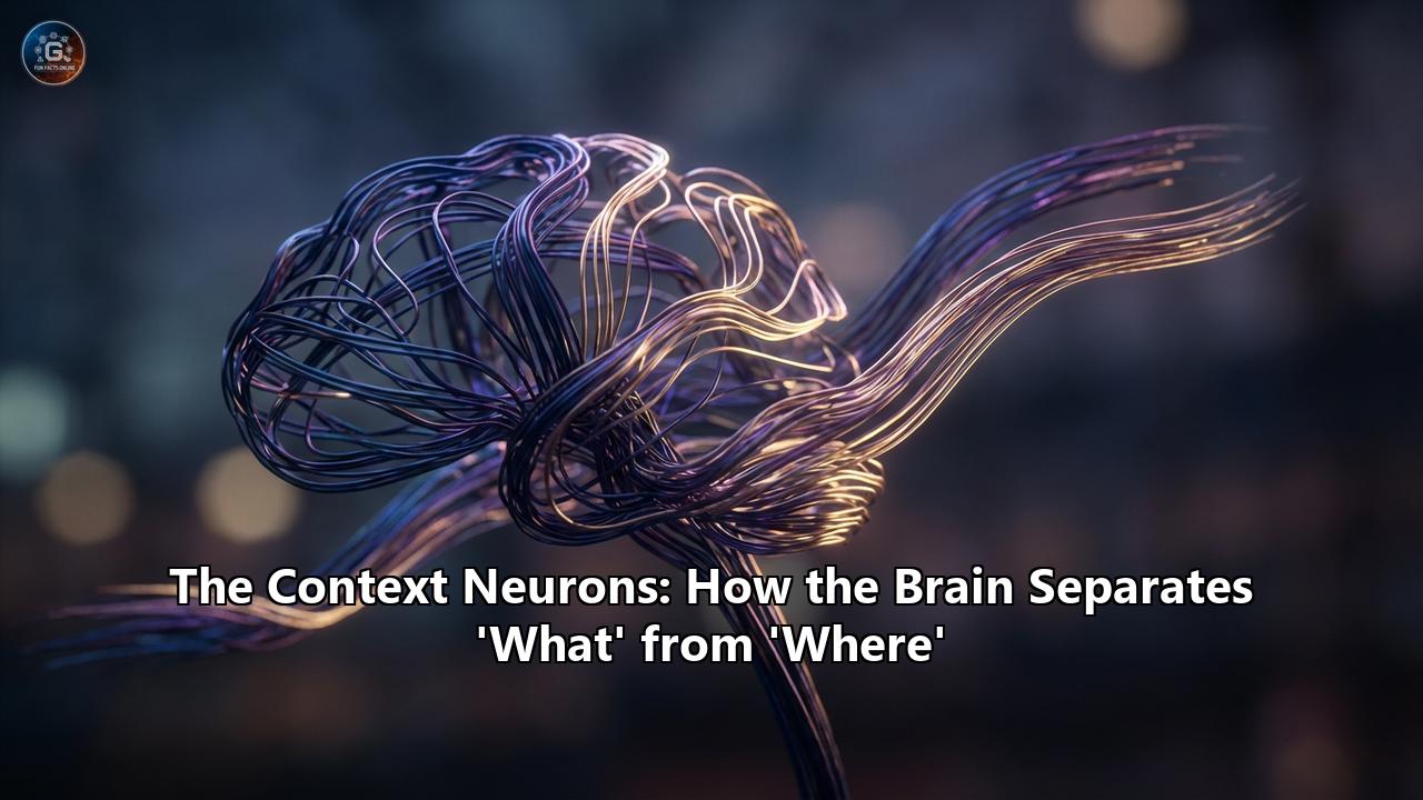
The faint hum of the MRI machine is often the soundtrack to modern neuroscience, but the real music plays silently within the dark, folded matter of the human brain. For decades, scientists have listened to this symphony, trying to decipher how a chaotic storm of electrical impulses transforms into the seamless experience of reality. We open our eyes, and we don't just see shapes and colors; we see things—a coffee cup, a familiar face, a stray cat—anchored precisely in places—on the desk, across the table, under the car.
But perhaps the most remarkable feat of the human mind is not just knowing what something is or where it is, but understanding the invisible web that connects them: the context.
Imagine seeing your boss. If you see her in the boardroom, your brain retrieves a specific set of behavioral protocols: sit up straight, speak professionally, discuss quarterly targets. Now, imagine seeing that same boss at a heavy metal concert, wearing a band t-shirt and moshing in the pit. The "object" (the boss) is identical. The "location" (the venue) is different. But the meaning of the event has shifted entirely. Your brain must instantly decouple the identity of the person from the professional context and re-attach it to a social, recreational one.
For years, this flexibility was a mystery. How does the brain store the concept of "Boss" so that it is stable enough to be recognized anywhere, yet flexible enough to function in completely different situations?
The answer, according to a groundbreaking wave of recent research peaking in 2024 and 2025, lies in a specialized architecture of separation. We have found the "Context Neurons."
This article explores the deep, anatomical and computational mechanisms by which the human brain separates the "What" (identity) from the "Where" (location) and the "Which" (context), and how it miraculously weaves them back together to create the tapestry of episodic memory. We will journey from the early days of the "Two-Stream Hypothesis" to the cutting-edge discovery of specific neuron types in the medial temporal lobe that act as the librarians of our experience, ensuring that while our memories are vast, they never get mixed up.
Part I: The Great Divide — The Origins of What and Where
To understand the sophisticated machinery of context neurons, we must first look at the foundational highways of the visual brain. The separation of information begins the moment light hits the retina, but it becomes dogmatic once signals reach the primary visual cortex (V1) at the back of the brain.
The Two-Stream Hypothesis
In 1982, neuropsychologists Leslie Ungerleider and Mortimer Mishkin proposed a theory that would dominate neuroscience for decades: the Two-Stream Hypothesis. Through lesion studies in macaques, they observed that damage to different parts of the brain resulted in two distinct types of blindness.
- The Ventral Stream ("What"): One pathway traveled ventrally (downward) from the occipital lobe into the temporal lobe. Monkeys with damage here could effectively navigate obstacles and reach for things, but they couldn't distinguish between a triangle and a square. They had lost the ability to identify what an object was.
- The Dorsal Stream ("Where"): The other pathway traveled dorsally (upward) into the parietal lobe. Monkeys with damage here could describe a shape perfectly. They knew it was a square. But they couldn't tell you where it was. They would reach for it and miss. They had lost the representation of where the object existed in space.
This bifurcation suggested that the brain treats identity and location as fundamentally different data types. It makes evolutionary sense. To survive, an organism needs to know what a predator is (is it a lion or a rock?) and where it is (is it coming toward me or moving away?). The processing requirements for these tasks are distinct. Identity requires invariance—a lion is still a lion whether it is seen from the front, side, in light, or shadow. Location requires precision—the exact coordinates relative to the viewer's body are required for flight or fight.
The Anatomy of the Ventral Stream (The "What" Pathway)
The journey of "What" is a process of increasing complexity. It begins in V1, which detects simple edges and orientations. As information flows to V2 and V4, the brain begins to process color and curvature. By the time signals reach the Inferior Temporal (IT) Cortex, neurons have become remarkably specific.
In the IT cortex, we find cells that do not care about spots of light, but rather about complex shapes. This is the home of the famous "Jennifer Aniston Neuron" (or concept cells), a discovery made in the early 2000s where single neurons were found to fire only when a patient saw Jennifer Aniston—whether in a photo, a drawing, or even just her written name.
These Content Neurons are the masters of semantic identity. They embody the abstract concept of an object. Crucially, they tend to be "position invariant." A neuron coding for a banana will fire whether the banana is on the left or right side of your vision. It captures the essence of the object, stripping away the spatial context.
The Anatomy of the Dorsal Stream (The "Where" Pathway)
Meanwhile, the Dorsal stream heads up to the Posterior Parietal Cortex. This system is obsessed with coordinates. It feeds into the motor cortex, guiding our hands and eyes.
Historically, this was called the "Where" pathway, but later researchers like Goodale and Milner refined it to the "Action" pathway. They argued that this stream doesn't just map space for the sake of a map; it maps space to do things. It transforms visual inputs into motor outputs.
However, the rigid separation of these streams posed a problem known as the Binding Problem. If the "banana" neuron is firing in the temporal lobe, and the "kitchen counter" coordinates are firing in the parietal lobe, how does the brain know that the banana is on the counter? Why don't we hallucinate the banana floating in the garage?
The answer lies deeper in the brain, where these two streams finally reunite.
Part II: The Convergence — The Medial Temporal Lobe
The reunion of "What" and "Where" takes place in the Medial Temporal Lobe (MTL), specifically within the structures surrounding the hippocampus. This is the engine room of memory.
For years, textbooks described the hippocampus as a funnel where everything mixes together. But recent high-resolution fMRI and single-unit recording studies have revealed that the separation of "What" and "Where" is maintained right up to the doorstep of memory encoding.
The gatekeeper of the hippocampus is the Entorhinal Cortex (EC). And just like the visual cortex, it is divided.
The Lateral Entorhinal Cortex (LEC): The "What" Gatekeeper
The Lateral Entorhinal Cortex (LEC) receives its primary inputs from the Ventral stream. It is the librarian of Items.
Neurons in the LEC respond to specific objects, odors, and distinct sensory features. If you place a new toy in a rat's cage, the LEC lights up. If you move the toy to a new spot, the LEC doesn't care—it only cares that the toy is there. It is the reservoir of content.
The Medial Entorhinal Cortex (MEC): The "Where" Gatekeeper
The Medial Entorhinal Cortex (MEC) receives inputs from the Dorsal stream and the parietal cortex. It is the librarian of Space.
This is the home of the Nobel Prize-winning Grid Cells. These neurons fire in a perfect hexagonal grid pattern as an organism moves through an environment. They provide an internal GPS, a metric of space that is independent of the objects within it. The MEC tells the brain where you are and the geometry of the room, but it is largely blind to what is in the room.
The Scaffold and the Item
This anatomical setup provides a profound insight into how we build memories. We have the MEC providing a "spatiotemporal scaffold"—a blank grid of space and time. We have the LEC providing the "items" to populate that grid.
Think of it like a theater stage.
- The MEC builds the stage, the lighting, and the backdrop (Context/Space).
- The LEC provides the actors and the props (Content/Items).
- The Hippocampus is the director that places the actors onto the stage to create a Scene (Episodic Memory).
Part III: The Discovery of Context Neurons
Until very recently, the assumption was that once information reached the hippocampus, it was "bound" together inextricably. In rodent studies, researchers often found neurons that had "mixed selectivity." A neuron might fire only for "Cheese" (Item) when it was in the "North Corner" (Context). If you moved the cheese to the South Corner, the neuron went silent. This is a Conjunctive Representation—the What and Where are fused.
However, the human brain appears to have evolved a more sophisticated layer of flexibility, a mechanism that allows us to decouple these elements even deeper in the memory hierarchy.
In January 2026, a landmark study published by researchers at the University of Bonn (led by Prof. Florian Mormann and Dr. Marcel Bausch) fundamentally changed our understanding of this process. They utilized a rare opportunity to record directly from the brains of epilepsy patients who had electrodes implanted for surgical planning.
The Experiment: Biscuits and Questions
The researchers designed a clever task to tease apart "Content" from "Context."
Patients were shown images of various objects (e.g., a biscuit, a dog, a car).
But before each image, they were given a specific task context, represented by a question:
- "Is it bigger?" (referring to the object's size being larger than a shoebox).
- "Is it heavier?" (referring to the object's weight).
This created a matrix of experience. The patient might see a Biscuit in the context of "Bigger?" or a Biscuit in the context of "Heavier?"
If the brain simply "bound" these memories immediately, one would expect to find neurons that fire only for the specific combination: Biscuit-Bigger.
The Findings: Two Distinct Libraries
The results were startlingly clean. The researchers didn't find a soup of mixed neurons. They found two distinct, segregated armies of cells.
- Content Neurons: These cells fired whenever the "Biscuit" appeared, regardless of whether the question was about size or weight. They represented the object identity in its pure form.
- Context Neurons: These cells fired whenever the "Bigger?" task was active, regardless of whether the image was a biscuit or a dog. They represented the abstract cognitive framework of the task.
Unlike rodents, where these representations are often muddied, the human medial temporal lobe maintained a strict division of labor.
The "Gatekeeper" Mechanism
The most fascinating discovery was how these two groups interacted. They didn't just fire in parallel; they communicated.
The researchers observed a temporal delay. The Content Neurons would fire first (identifying the object), and then, milliseconds later, the Context Neurons would adjust their firing patterns. It appeared that the object identity was being "routed" through the context.
The authors likened this to a "Neural Library." By storing Content and Context in separate libraries, the brain achieves infinite combinatorics. If we had to have a separate neuron for every possible combination of object and context (e.g., "Grandma at the beach," "Grandma at the store," "Grandma in a car"), we would run out of neurons.
By separating them, the brain can simply link the "Grandma" neuron (Content) with the "Beach" neuron (Context) on the fly. This is the secret to the flexibility of human memory. It allows us to generalize. We know that "Grandma" is the same person regardless of where she is.
Part IV: The Mechanics of Separation — Why It Matters
Why has the human brain evolved to keep these streams separate for so long? The answer lies in the competing demands of Pattern Separation and Pattern Completion.
Pattern Separation (The "Difference" Engine)
To remember where you parked your car today, you need to distinguish it from where you parked yesterday. The "What" (your car) is identical. The "Context" (Space/Time) is different.
If your brain merged these too early, you would suffer from catastrophic interference. You would walk to yesterday's spot.
The Dentate Gyrus of the hippocampus is famous for this. It takes the inputs from the LEC (Car) and MEC (Location) and orthogonalizes them—it forces them apart into distinct neural patterns. This ensures that similar experiences don't overwrite each other.
Pattern Completion (The "Same" Engine)
Conversely, if you see your car partially obscured by a truck, you need to recognize it as the same car. This requires the "What" pathway to be robust against noise.
By keeping the "What" representation stable (the Content Neuron for the car) while the "Where" representation shifts, the brain allows you to maintain a stable sense of reality despite a constantly changing environment.
The Role of "Theta" and "Gamma" Rhythms
How does the brain synchronize these separate libraries? The answer is in the timing.
Neuroscientists have long observed "Theta oscillations" (4-8 Hz brain waves) in the hippocampus during exploration.
Recent models suggest a Phase Precession mechanism.
- Content information might ride on the trough of the theta wave.
- Context information might ride on the peak of the theta wave.
- Fast Gamma bursts (30-100 Hz) then zip between them, binding the two distinct packets of information into a cohesive conscious moment.
This allows the brain to multiplex: transmitting "Identity" and "Location" on the same wire (the axon) without mixing the signals, simply by using different time slots.
Part V: When Separation Fails — Disorders of Context
The clinical importance of this "What" vs. "Where" separation becomes heartbreakingly clear when we look at diseases that disrupt it.
Alzheimer’s Disease and the Entorhinal Cortex
Alzheimer’s disease does not attack the brain randomly. It almost always begins in the Lateral Entorhinal Cortex (LEC)—the gateway of "What" and "Content."
This explains the specific early symptoms. Patients don't just forget "where" they are (MEC function); they often lose the ability to bind objects to contexts.
They might recognize a face (Content intact) but have zero idea why they know this person or where they met them (Context disconnected).
As the disease spreads to the MEC and Hippocampus, the "Map" crumbles. The scaffold collapses. The patient becomes lost in their own neighborhood because the Grid Cells that provide the spatial context can no longer integrate with the visual landmarks.
Capgras Delusion
In this rare psychiatric disorder, a patient recognizes a loved one’s face (the "What" is correct) but believes they are an imposter.
Some theories suggest this is a disconnection between the visual "What" stream and the emotional context provided by the amygdala. The content neuron fires ("That looks like Dad"), but the emotional context neuron remains silent. The brain resolves this conflict with a delusion: "It looks like Dad, but it doesn't feel like Dad, so it must be a robot/alien."
Schizophrenia and Context
Schizophrenia is often described as a failure of context processing. A healthy brain uses context to inhibit irrelevant associations. If you are at a funeral, you inhibit the urge to laugh.
In schizophrenia, the "Context Neurons" in the Prefrontal Cortex and Hippocampus may fail to fire or fail to gate the "Content Neurons." The result is a flood of inappropriate associations. A random object (a red car) feels intensely significant because the brain has failed to place it in its proper, neutral context.
Part VI: The New Frontier — "Where" is not just Space
One of the most exciting developments in the 2025 research landscape is the realization that the "Where" pathway (Dorsal/MEC) is not just about physical space. It is about Cognitive Space.
The Cognitive Map Theory
The same "Grid Cells" that map a room also appear to map Conceptual Space.
In a study where participants had to navigate a "space" of bird neck lengths and leg lengths (morphing birds on a screen), the MEC fired in a grid pattern.
The brain treats "relationships" as geometry.
- Content: The Bird.
- Context: The abstract variables (Longer neck, shorter legs).
This suggests that the "Context Neurons" discovered in Bonn are part of a universal machinery. Whether the context is "Physical Location" (The kitchen), "Task Rule" (Is it bigger?), or "Social Hierarchy" (Is he my boss?), the brain uses the dorsal/MEC machinery to build a scaffold, and hangs the ventral/LEC "items" upon it.
Part VII: Implications for Artificial Intelligence
The biological separation of "What" and "Where" is currently inspiring a revolution in AI.
Traditional Convolutional Neural Networks (CNNs) often struggle with what is called Out-of-Distribution Generalization. If you train an AI to recognize a cow on green grass, and then show it a cow on a sandy beach, it might fail. It has "overfit" the context. It has bound "Cow" and "Grass" into a single, muddy representation.
The human brain’s architecture—separating Content Neurons from Context Neurons—solves this.
Newer AI architectures, such as Transformers and Neuro-Symbolic AI, are attempting to mimic this. They use "Attention Mechanisms" to dynamically bind separate representations.
- Query (Q): What am I looking for? (Context)
- Key (K): What features are available? (Content)
- Value (V): The result.
By explicitly separating the "Positional Encoding" (Where) from the "Token Embedding" (What), Transformers like GPT-4 function eerily similarly to the Entorhinal Cortex. They treat the "meaning" of a word (Content) as separate from its "position" in the sentence (Context).
However, biological brains are still vastly more efficient. A human needs only one example to learn that a "Smartphone" functions the same way in a bedroom as in a boardroom. AI often needs thousands. The secret likely lies in the physical separation of these neuronal libraries, allowing for instant, zero-shot recomposition of reality.
Part VIII: The Symphony of Experience
As we move through our lives, we feel a sense of seamless unity. We do not feel the "What" pathway struggling to identify the coffee cup while the "Where" pathway calculates the distance to our lips. We do not feel the "Context Neurons" switching libraries when we walk from the office to the bar.
But this seamlessness is an illusion created by the precise, rhythmic coordination of billions of separated components.
The discovery of specialized Context Neurons in the human brain is the final piece of the puzzle that began with Ungerleider and Mishkin. We now know that the brain is a master of modularity. It deconstructs the world into its atomic parts—Identity, Location, Time, Rule—and stores them in separate vaults.
It is only in the fleeting, electrical present—in the gamma bursts of the hippocampus—that these vaults open, and the scattered notes of reality are bound together into the music of a memory.
This architecture of separation is what gives us our freedom. Because our "What" is not shackled to our "Where," we can imagine a castle in the sky. We can remember a lost love without being in the room where we last saw them. We can extract wisdom from one context and apply it to another.
In separating the components of our world, the brain gives us the power to transcend them.
Deep Dive: The Cellular Mechanism of Context Binding
To truly appreciate the elegance of this system, we must zoom in to the synaptic level. How exactly does a "Context Neuron" exert its influence?
The prevailing theory involves Gating via Inhibition.
In the absence of a specific context, the brain is flooded with potential actions and memories. If you hold a hammer, you could throw it, drop it, or hit a nail.
The "Hammer" (Content) activates all these associations.
The "Context Neuron" (e.g., "Fixing the roof") does not activate the correct action directly. Instead, it sends inhibitory signals to suppress the wrong ones (don't throw it, don't drop it). It carves away the noise, leaving only the relevant behavior.
This is why the Prefrontal Cortex (PFC) is so heavily connected to the Hippocampus. The PFC provides the "Rule" or "Goal" (the Context). It projects down to the medial temporal lobe.
Recent 2025 studies utilizing optogenetics in primates have shown that silencing this top-down projection from the PFC causes the "Context Neurons" in the hippocampus to go silent. The result? The animal recognizes the object but cannot perform the correct context-dependent task.
The Evolution of the Contextual Brain
Why did this evolve? We can look to our ancestors.
For a reptile, "Food" is "Food." It eats it whenever it sees it.
For a mammal, "Food" is conditional.
- Food + Territory of Alpha Male = Do Not Eat.
- Food + Safe Den = Eat.
The evolution of the Hippocampus and the Entorhinal Cortex was essentially the evolution of a "Context Engine." It allowed mammals to decouple the stimulus from the response. It introduced the variable of conditionality.
In humans, this exploded into symbolic thought. Language is pure context. The word "Bank" means one thing in the context of "River" and another in the context of "Money."
The "Content Neuron" for the sound Bank is the same. The "Context Neuron" determines the meaning.
Thus, the very neural machinery that allows a rat to navigate a maze without getting lost is the same machinery that allows a poet to use a metaphor, or a programmer to write an "If/Then" statement.
The Future of Memory Research
The identification of these specific neuron types opens the door to futuristic therapies.
- Contextual Prosthetics: Could we stimulate "Context Neurons" to help Alzheimer’s patients retrieve lost memories? If we can artificially trigger the "Kitchen Context," perhaps the "Coffee Cup" memory will naturally resurface via pattern completion.
- Trauma Therapy: PTSD is essentially a failure of context separation. A car backfiring (Content) triggers a war memory because the brain has failed to separate the "War Context" from the "Civilian Context." The content is triggering the wrong file. Targeted inhibition of specific Context/Content bindings could theoretically "unpair" the trauma from the trigger.
Conclusion
The "Context Neurons" are the conductors of our cognitive life. They stand at the crossroads of perception and memory, quietly directing traffic, ensuring that the infinite stream of sensory data is organized into a coherent narrative. They separate the What from the Where, not to keep them apart, but to allow them to dance together in infinite variations. In this separation lies the secret of human creativity, adaptability, and the enduring continuity of the self.
Reference:
- https://en.wikipedia.org/wiki/Two-streams_hypothesis
- https://en.wikipedia.org/wiki/Entorhinal_cortex
- https://www.hopkinsmedicine.org/health/conditions-and-diseases/anatomy-of-the-brain
- https://scitechdaily.com/rewriting-the-rules-scientists-solve-an-episodic-memory-mystery/
- https://www.medfak.uni-bonn.de/en/faculty/news/a-mystery-of-episodic-memory-solved
- https://fgu.cas.cz/en/2025/12/new-research-reveals-close-interaction-between-two-visual-streams-in-the-brain/
- https://www.news-medical.net/news/20260107/Separate-neurons-for-content-and-context-enable-flexible-human-memory.aspx
- https://pmc.ncbi.nlm.nih.gov/articles/PMC3866456/
- https://pmc.ncbi.nlm.nih.gov/articles/PMC12314160/