The ability to map oxygenation non-invasively within the human body represents one of the holy grails of modern medical imaging. For decades, the gold standard for measuring blood oxygen saturation was either the invasive arterial blood gas (ABG) analysis or the ubiquitously used, yet surface-limited, pulse oximeter. However, neither of these tools can answer the critical question often asked in neurology, cardiology, and oncology: Is this specific tissue getting enough oxygen right now?
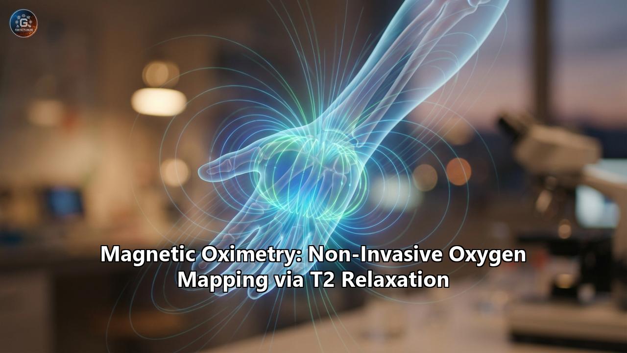
Enter Magnetic Oximetry. This is not a single device, but an emerging paradigm in Magnetic Resonance Imaging (MRI) that leverages the quantum mechanical properties of the hemoglobin molecule to create high-resolution, three-dimensional maps of oxygen delivery and consumption. By exploiting the phenomenon of T2 and T2 relaxation, radiologists and physicists are now able to see the "shadow" of metabolic hunger in a stroke-affected brain, quantify the suffocating fibrosis of a failing liver, and predict the viability of a fetus in a preeclamptic placenta—all without injecting a single drop of contrast dye.
This article explores the biophysical foundations, current technological breakthroughs, and clinical applications of Magnetic Oximetry. It details how a discovery made by Linus Pauling in 1936 has evolved into 2026’s cutting-edge diagnostic tools, transforming our understanding of physiology from the scale of electron spins to the pumping of the human heart.
I. The Physics of Spin: How Hemoglobin Becomes a Magnet
To understand how an MRI scanner can measure oxygen, one must descend into the quantum realm of the hemoglobin molecule. The entire field of magnetic oximetry rests on a happy accident of evolution: the magnetic personality of iron.
The Pauling-Coryell Discovery
In 1936, chemist Linus Pauling and his student Charles Coryell published a landmark paper in the Proceedings of the National Academy of Sciences. Using a Gouy balance—a sensitive device for measuring magnetic susceptibility—they demonstrated that the magnetic state of hemoglobin changes drastically depending on whether it is holding onto an oxygen molecule.
- Deoxyhemoglobin (dHb): When hemoglobin is empty (deoxygenated), the central iron atom (Fe2+) is in a "high-spin" state. It possesses four unpaired electrons in its outer orbital. In the world of quantum mechanics, unpaired electrons act like tiny bar magnets. This makes deoxyhemoglobin paramagnetic—it is attracted to magnetic fields and, crucially, it distorts the magnetic field around it.
- Oxyhemoglobin (HbO2): When oxygen binds to the iron, the electron configuration shifts. The iron atom essentially shares electrons with the oxygen molecule, pairing them up. For decades, this was described simply as a "low-spin" diamagnetic state (having no unpaired electrons). Modern X-ray spectroscopy and quantum chemical calculations have refined this view, suggesting an "intermediate-spin" state with significant delocalization, but for the purposes of MRI, the effect is the same: oxyhemoglobin is diamagnetic. It has almost no magnetic moment and does not distort the surrounding magnetic field.
T2 and T2 Relaxation: The Signal of Silence
An MRI scanner works by aligning the protons (hydrogen nuclei) in water molecules with a massive magnetic field ($B_0$). A radiofrequency pulse tips these protons out of alignment, and as they spin (precess) back to equilibrium, they emit a signal.
The presence of paramagnetic deoxyhemoglobin acts as a magnetic disruptor. Imagine a choir of protons singing a single, clear note (the MRI signal). If you introduce deoxyhemoglobin molecules, they act like individuals shouting out of tune, creating local magnetic field inhomogeneities. This causes the protons in the vicinity to "dephase," or lose their coherence, much faster than they otherwise would.
- T2 Relaxation (Spin-Spin Relaxation): This is the irreversible loss of signal coherence due to interactions between protons. Deoxyhemoglobin shortens T2 relaxation times. Blood that is deoxygenated appears darker on T2-weighted images.
- T2 Relaxation: This parameter captures both the intrinsic T2 effects and the additional dephasing caused by local magnetic field irregularities (inhomogeneities). Because deoxyhemoglobin creates significant field gradients around red blood cells, T2 is exquisitely sensitive to oxygenation levels. This is the basis of the BOLD (Blood Oxygen Level Dependent) effect used in functional MRI (fMRI).
In simple terms: Oxyhemoglobin is magnetically invisible; deoxyhemoglobin is a magnetic contrast agent. By measuring how quickly the MRI signal decays (the relaxation time), we can calculate exactly how much deoxyhemoglobin is present, and conversely, the oxygen saturation ($sO_2$) of the blood or tissue.
II. The Brain: Mapping the Fuel of Thought
The brain is the most metabolically demanding organ, consuming 20% of the body’s oxygen. Consequently, it has been the primary testing ground for magnetic oximetry techniques.
1. TRUST MRI: Weighing the Venous Exhaust
One of the most robust implementations of magnetic oximetry is T2-Relaxation-Under-Spin-Tagging (TRUST) MRI. Developed to measure global cerebral venous oxygenation ($Y_v$), TRUST separates the signal of pure blood from the surrounding tissue.
- How it works: The sequence tags blood spins magnetically and then allows them to flow into the venous sinus (like the superior sagittal sinus). By acquiring a series of images with different T2-weightings, the scanner fits a decay curve to calculate the precise T2 of the blood.
- Calibration: The T2 of blood is dependent not just on oxygen, but also on hematocrit (Hct) and the time between magnetic pulses. TRUST relies on rigorous calibration models (based on bovine and human blood experiments) to convert this T2 value into a percentage of oxygen saturation.
- Clinical Utility: TRUST has become a vital tool in studying conditions like Sickle Cell Anemia (SCA). In SCA, the abnormal shape of red blood cells and the presence of hemoglobin S (HbS) alter the magnetic susceptibility. Recent studies comparing TRUST with catheterization have shown that standard calibration models must be adjusted for SCA patients, but once corrected, TRUST offers a non-invasive window into stroke risk for these children.
2. QSM: The Quantitative Revolution
While TRUST measures global oxygenation in large veins, Quantitative Susceptibility Mapping (QSM) offers a pixel-by-pixel map of magnetic susceptibility across the entire brain.
QSM solves a complex mathematical inverse problem. The MRI scanner measures the phase of the signal (the timing of the proton "choir"). Deoxyhemoglobin shifts this phase. By removing background magnetic fields from the skull and air sinuses, QSM algorithms calculate the magnetic susceptibility ($\chi$) of every voxel.
- Oxygen Extraction Fraction (OEF): By measuring the susceptibility difference between a vein and the surrounding tissue, QSM can calculate the OEF—the percentage of oxygen the brain tissue has removed from the blood.
- Stroke and Penumbra: In ischemic stroke, the core of the infarct is dead, but the surrounding "penumbra" is starving. This tissue extracts every possible molecule of oxygen to survive, leading to a drastically high OEF. QSM can map this "misery perfusion" with high spatial resolution, identifying brain tissue that can still be saved by thrombectomy (clot removal) even hours after stroke onset.
- Tumor Characterization: Glioblastomas (GBM) are aggressive brain tumors characterized by rapid, chaotic growth that outstrips their blood supply, creating hypoxic cores. QSM, combined with quantitative BOLD (qBOLD), allows for the calculation of the Cerebral Metabolic Rate of Oxygen ($CMRO_2$). A 2025 study demonstrated that machine learning models fed with $CMRO_2$ maps could differentiate between Glioblastomas and solitary brain metastases with 93% accuracy—a critical distinction for surgical planning.
3. Artificial Intelligence in Neuro-Oximetry
The marriage of AI and magnetic oximetry is accelerating the field.
- Deep Learning Reconstruction: Calculating a QSM map traditionally required long computation times to solve the inverse dipole problem. New Convolutional Neural Networks (CNNs) and Vision Transformers can now reconstruct high-fidelity QSM maps from raw phase data in seconds, making real-time oxygen mapping feasible in acute stroke settings.
- Synthetic Data Generation: Generative AI models (like GANs) are being used to create synthetic "hypoxic" brain datasets to train diagnostic algorithms, overcoming the scarcity of labeled pathological data.
III. The Heart: Non-Invasive Prognostics
The heart is a pump that feeds itself first. Myocardial oxygenation is the ultimate determinant of heart health, yet assessing it has traditionally required passing a catheter into the coronary sinus—a risky and invasive procedure.
T2 Mapping for Heart Failure (The 2026 Breakthrough)
In January 2026, a groundbreaking study published in JACC: Advances by researchers from the University of East Anglia and Leeds changed the landscape of cardiac diagnostics. They developed a protocol using T2 mapping to estimate the oxygen saturation of the blood in the coronary sinus non-invasively.
- The Problem: In heart failure, the heart struggles to pump. To compensate, the body extracts more oxygen from the blood, leading to lower mixed venous oxygen saturation ($SvO_2$). Measuring this required right heart catheterization.
- The MRI Solution: The researchers found that the T2 relaxation time of the blood in the right ventricle and coronary sinus correlated perfectly with invasive catheter measurements.
- The Impact: In a cohort of 628 heart failure patients followed for three years, those with "healthier" MRI-derived oxygen levels had significantly better survival rates. This suggests that a standard 45-minute cardiac MRI can now provide the same "hard" hemodynamic numbers as an invasive procedure, allowing for frequent, safe monitoring of heart failure progression and medication response.
*T2 vs. T2: Edema vs. Iron*
In cardiac MRI, the distinction between T2 and T2 is vital:
- T2 Mapping: Primarily used to detect edema (water content) in myocarditis or acute myocardial infarction. Water has a long T2.
- T2 Mapping: The gold standard for detecting myocardial iron overload, common in Thalassemia patients receiving blood transfusions. Iron deposits are strongly paramagnetic, causing T2 values to plummet (decay very fast). Monitoring T2 allows clinicians to titrate chelation therapy before irreversible heart failure occurs.
Oxygenation-Sensitive (OS) CMR
Beyond static maps, "Oxygenation-Sensitive" CMR uses T2 sensitivity to perform stress tests without contrast agents. Patients perform breathing maneuvers (like hyperventilation followed by a breath-hold) inside the scanner.
- Vasodilation: A long breath-hold causes CO2 to build up, a potent vasodilator. Healthy coronary arteries dilate, increasing blood volume and oxygenation (increasing T2 signal).
- Steal Phenomenon: In diseased arteries, vessels are already maximally dilated or stiff. The blood "steals" away to healthy areas, causing a paradoxically detectable drop in oxygenation in the ischemic territory. This "breathing stress test" can detect coronary artery disease without the need for pharmacological stressors like adenosine.
IV. The Kidneys: Watching the Filtration Fail
The kidney is a metabolic paradox: the cortex receives huge blood flow for filtration, while the medulla operates on the brink of hypoxia to maintain the osmotic gradients necessary for concentrating urine. This precarious balance makes the kidney uniquely susceptible to hypoxic injury.
Renal BOLD MRI (R2)
The metric of choice here is R2--- (which is simply $1/T2$). A higher R2 means faster decay, more deoxyhemoglobin, and thus hypoxia.
- Chronic Kidney Disease (CKD): As kidney function declines, fibrosis sets in, destroying capillaries. BOLD MRI studies have consistently shown that R2 values in the cortex increase (oxygen drops) as CKD stage advances. This confirms the "chronic hypoxia hypothesis"—that lack of oxygen is a primary driver of kidney failure progression.
- Acute Allograft Rejection: In kidney transplants, distinguishing between Acute Rejection (AR) and Acute Tubular Necrosis (ATN) is difficult but critical. A 2024 prospective study found that medullary R2 levels were significantly lower in Acute Rejection compared to ATN. The inflammation in rejection increases blood flow (hyperemia), paradoxically increasing oxygenation temporarily, whereas necrotic tubules in ATN cannot use oxygen efficiently.
- Drug Response: BOLD MRI is used to test new drugs. For example, giving a patient a diuretic like furosemide shuts down the ion pumps in the medulla. Since these pumps use 80% of the kidney's oxygen, their shutdown should cause a massive "rebound" in oxygenation (drop in R2). If this rebound doesn't happen, it indicates the tubular cells are damaged or non-functional.
V. The Abdomen and the Fetus: New Frontiers
Magnetic oximetry is expanding beyond its traditional strongholds into the abdomen and the womb.
Liver Fibrosis and Inflammation
The liver receives a dual blood supply (portal vein and hepatic artery), making its oxygenation complex.
- Fibrosis: As the liver becomes fibrotic (scarred), resistance to portal flow increases, and the liver becomes more dependent on arterial blood. However, the overall microenvironment becomes hypoxic. BOLD MRI has shown promise in staging liver fibrosis, with R2 values correlating with the histological fibrosis score.
- Iron Overload: The liver is the body's primary iron store. Quantitative Susceptibility Mapping (QSM) has emerged as a superior biomarker for liver iron compared to T2, as it is a fundamental physical property independent of magnetic field strength ($1.5T$ vs $3T$). This allows for precise monitoring of hereditary hemochromatosis.
- Inflammation Probes: Novel "smart" MRI probes are being developed that only become paramagnetic in the presence of reactive oxygen species (ROS) produced by inflammatory cells. While still in preclinical stages, these could allow for the direct mapping of hepatitis activity before fibrosis sets in.
Placental and Fetal Oximetry
The placenta is the fetus's lung. Its failure (placental insufficiency) is a leading cause of stillbirth and growth restriction.
- The "Lungs" of the Fetus: In preeclampsia, the placental vasculature fails to remodel, leading to high resistance and poor oxygen exchange. A landmark observational study showed that placental T2--- is significantly reduced in women with preterm preeclampsia compared to controls. The placenta essentially appears "darker" and more heterogeneous on T2 maps, reflecting its hypoxic, stressed state.
- Fetal Streaming: MRI is fast enough to visualize the "streaming" of blood in the fetal heart. Oxygenated blood from the umbilical vein shoots through the ductus venosus and streams preferentially across the foramen ovale to the brain. T2 mapping can quantify the saturation of this blood in the umbilical vein vs. the fetal descending aorta, providing a direct measurement of oxygen delivery to the fetus. This is currently the only way to measure fetal oxygenation without highly risky fetal blood sampling.
VI. Comparative Technologies: MRI vs. NIRS vs. PET
Magnetic Oximetry does not exist in a vacuum. How does it stack up against its competitors?
| Feature | Magnetic Oximetry (MRI) | Near-Infrared Spectroscopy (NIRS) | PET (Positron Emission Tomography) |
| :--- | :--- | :--- | :--- |
| Mechanism | Paramagnetism of deoxyhemoglobin (T2/T2/QSM) | Optical absorption differences of Hb/HbO2 | Radioisotope decay ($^{15}O$) |
| Invasiveness | Non-invasive (no contrast needed for intrinsic methods) | Non-invasive (optical pads on skin) | Invasive (requires injection of radioactive tracer) |
| Penetration Depth | Unlimited (Whole body, deep organs) | Shallow (1-3 cm). Can reach cortex, but not deep brain or central organs. | Unlimited |
| Spatial Resolution | High (1-3 mm). Can map individual veins and tumor sub-regions. | Low (Diffused signal, cm scale). | Medium (4-6 mm). |
| Temporal Resolution | Seconds to Minutes. (fMRI is sub-second, but quantitative oximetry is slower). | Real-time (Millisecond resolution). Excellent for trend monitoring. | Slow (Minutes). Requires cyclotron on-site for oxygen isotopes. |
| Cost & Access | High ($$$$). Requires MRI scanner. | Low ($). Portable, bedside compatible. | Very High ($$$$). Rare, primarily research. |
| Key Limitation | Motion artifacts; requires patient transport to scanner. | Signal contamination from skin/skull; limited depth. | Radiation exposure; short half-life of tracers. |
The Verdict:- NIRS is the king of the bedside. It is perfect for continuous monitoring of a neonate’s frontal lobe in the NICU or a muscle during exercise. However, it cannot see the thalamus, the fetal heart, or the renal medulla.
- PET is the historical "gold standard" for metabolic rate ($CMRO_2$) but is dying out for this purpose due to the logistical nightmare of requiring an on-site cyclotron to produce $^{15}O$ gas (half-life: 2 minutes) and the radiation burden.
- Magnetic Oximetry is the comprehensive mapper. It is the only modality that combines high-resolution anatomy with deep-tissue oxygen quantification. It is the tool of choice for diagnosis, prognosis, and treatment planning, whereas NIRS is the tool for monitoring.
VII. Future Directions: The Era of Multi-Parametric Oximetry
The future of magnetic oximetry lies in integration.
1. Multi-Parametric Mapping (MPM):Future protocols will not just measure T2. They will simultaneously acquire T1 (fibrosis), T2 (edema), T2 (oxygen/iron), and Diffusion (cellular density) in a single "Fingerprinting" scan. AI algorithms will synthesize these inputs to provide a holistic "biopsy-free" characterization of tissue. A liver scan will report: "Grade 2 Fibrosis, mild inflammation, severe iron overload, moderate hypoxia."
2. 17O-MRI:While proton MRI measures the delivery of oxygen (via hemoglobin), imaging the oxygen isotope $^{17}O$ can measure the consumption of oxygen directly as it is metabolized into water ($H_2^{17}O$). Although 17O is a stable (non-radioactive) isotope, it is expensive and requires specialized hardware. However, recent breakthroughs in 2024-2025 using dynamic 17O-MRI at 3 Tesla have shown it is feasible to map regional $CMRO_2$ in humans, offering a direct metabolic readout that complements the hemodynamic BOLD signal.
3. "Smart" Pulse Oximeters?While not MRI, the principles of magnetic oximetry are inspiring new point-of-care devices. Research is underway to develop miniaturized magnetic resonance sensors (low-field, portable MRI) that could wrap around a limb to measure deep tissue oxygenation, bridging the gap between the NIRS patch and the hospital scanner.
Conclusion
Magnetic Oximetry is a testament to the power of interdisciplinary science. It began with a chemist (Pauling) asking a question about electrons, evolved through the hands of physicists building superconducting magnets, and is now in the hands of AI researchers and clinicians saving lives.
From the "misery perfusion" of a stroke victim to the "fetal streaming" of an unborn child, magnetic oximetry allows us to see the invisible fuel of life. It transforms the MRI scanner from a camera that takes pictures of anatomy into a physiological laboratory, capable of asking and answering the most fundamental question in medicine: Is this tissue alive?* As algorithms improve and scanners become faster, we are rapidly approaching a future where a "comprehensive oxygen map" is a standard part of every medical workup, illuminating the dark corners of human physiology.
Reference:
- https://www.youtube.com/watch?v=dsxL93PrPmY
- https://utoronto.scholaris.ca/items/88aa7eb7-b7e7-4fa3-8171-710e319e52f0
- https://www.researchgate.net/figure/Fetal-streaming-measured-and-visualized-by-T2-MRI-A-differences-between-mean-AAo-and-MPA_fig6_341181294
- https://apm.amegroups.org/article/view/59835/html
- https://www.frontiersin.org/journals/physiology/articles/10.3389/fphys.2022.913443/full
- https://www.ahajournals.org/doi/abs/10.1161/HYPERTENSIONAHA.120.14701
- https://pubmed.ncbi.nlm.nih.gov/32336233/
- https://pmc.ncbi.nlm.nih.gov/articles/PMC10321255/