Have you ever looked at a flock of starlings murmuring in the twilight sky or a school of fish moving as a single, liquid entity, and wondered how thousands of individual minds coordinate such seamless, breathtaking ballets? For decades, biologists sought the answers purely in the realm of biochemistry—signaling molecules, genetic cascades, and receptor pathways. But a silent revolution has taken place at the intersection of physics and biology. We now know that the cells inside your body are performing an equally complex, microscopic dance governed not just by chemical whispers, but by the profound and elegant laws of topology and fluid dynamics.
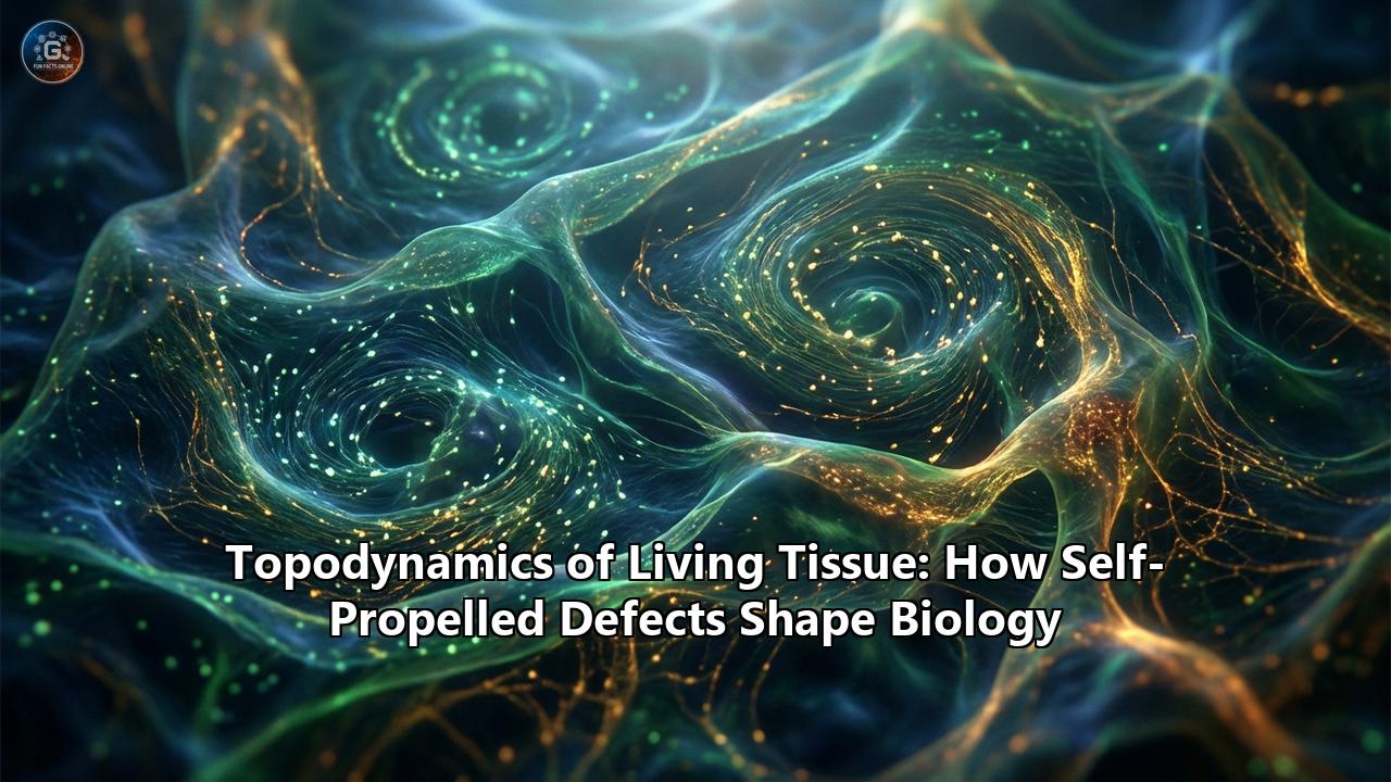
Welcome to the cutting-edge science of topodynamics—the study of how dynamic topological structures emerge in living matter. Far from being a mere bag of chemicals, living tissue behaves as "active matter," a state of material where every individual component consumes energy to move, push, and pull. When millions of these active biological engines pack tightly together, they create spontaneous, mesmerizing patterns of flow and stress. Hidden within these patterns are singular points of profound geometric disruption known as topological defects.
Once thought to be abstract mathematical curiosities or mere manufacturing glitches in smartphone displays, these self-propelled topological defects are now recognized as the master architects of biology. They are the unseen forces that sculpt our organs, heal our wounds, decide which cells live or die, and even dictate the spread of cancer.
The Liquid Crystal in You
To understand topodynamics, we must first look at the technology in your pocket. The screen of your smartphone likely relies on Liquid Crystal Display (LCD) technology. Liquid crystals are a bizarre phase of matter that sits somewhere between the rigid order of a solid crystal and the chaotic freedom of a liquid. The molecules in a liquid crystal can flow freely like water, but they possess an elongated, rod-like shape that forces them to align parallel to their neighbors, creating what physicists call "nematic order".
In a startling twist of biomimicry, human tissues are essentially living liquid crystals. Many of our cells—from the epithelial cells lining our gut and skin to the fibroblasts weaving our connective tissues—are elongated and densely packed. Because they are squished together, they naturally align their long axes with one another to save space, forming a biological nematic liquid crystal.
But there is a crucial difference between the liquid crystals in your phone and the cells in your body. Your phone's display is "passive"; it requires an external electrical voltage to change the alignment of its molecules. Your tissues, however, are active nematics. Every single cell is a microscopic engine, burning ATP (the cellular fuel) to generate internal mechanical forces. Cells constantly crawl, contract, and divide. This continuous injection of energy at the microscopic level pushes the tissue far from thermodynamic equilibrium, generating spontaneous, swirling flows known as "active turbulence".
When living cells continuously push and pull against their neighbors in this state of active turbulence, their perfect parallel alignment inevitably breaks down. In the mathematical landscape of the tissue, regions of geometric frustration emerge. These localized points where the orientational order completely collapses are called topological defects.
The Geometry of Chaos: Comet and Trefoil Defects
If you try to comb a hairy sphere, you will inevitably end up with a cowlick or a bald spot. This is a famous mathematical theorem (the Poincaré-Bhopal theorem, or "hairy ball theorem"), and it beautifully illustrates the concept of a topological defect: a point where the local orientation is mathematically undefined.
In the two-dimensional sheets of cells that make up our epithelial linings, topological defects come primarily in two distinct shapes, characterized by a number called the "topological charge":
1. The +1/2 Defect (The Comet):Imagine the cells aligning in a pattern that looks like a comet or a shooting star. There is a distinct rounded "head" and a splayed "tail." If you walk in a circle around the core of this defect, the orientation of the cells rotates by 180 degrees (or $\pi$ radians), giving it a +1/2 charge. Because of its asymmetrical shape, the +1/2 defect possesses a magical property in active matter: it is self-propelled. The active mechanical forces generated by the cells do not balance out perfectly around the defect's core. In "extensile" tissues (where cells push outward along their long axis), the +1/2 defect acts like a microscopic bulldozer, moving continuously in the direction of its comet head. In "contractile" tissues, it moves backward toward its tail.
2. The -1/2 Defect (The Trefoil):This defect looks like a triangular star or a three-leaf clover. If you trace a circle around it, the cell orientation rotates by 180 degrees in the opposite direction, earning it a -1/2 charge. Unlike the comet, the trefoil is perfectly symmetrical. Therefore, the active forces balance out, and the -1/2 defect is essentially stationary, simply being dragged around by the background flows of the tissue.
For a long time, biophysicists observed these swirling comet and trefoil patterns in petri dishes under microscopes, marveling at their mathematical beauty but assuming they were just a harmless byproduct of cell movement. That assumption was shattered when researchers discovered that these self-propelled defects are biologically functional. They act as intense mechanical hotspots, translating the language of topology into the biochemistry of life and death.
The Extrusion Executioner: How Defects Govern Cell Death
In any healthy tissue, homeostasis must be maintained. If cells divide too rapidly without old cells dying off, the tissue will overcrowd, paving the way for tumors. To prevent this, epithelial tissues undergo a process called cell extrusion, where unwanted, old, or defective cells are squeezed out of the cellular sheet and undergo apoptosis (programmed cell death).
For years, biologists assumed this was a purely biochemical decision made by the individual cell. However, pioneering topodynamic research revealed a universal correlation: the sites where cells are squeezed out and die perfectly match the locations of +1/2 topological defects.
As the self-propelled +1/2 comet defect plows through the epithelial monolayer, it acts like an energetic snowplow. The misalignment of the cells at the head of the comet generates immense mechanical friction and compressive stress. This localized, crushing pressure can be sustained for over an hour. The cells trapped at the core of the defect are subjected to such extreme mechanical deformation that they are squeezed from all sides.
This physical pressure triggers a mechanotransductive alarm bell within the cell. The crushing force causes a critical protein called YAP (Yes-associated protein) to migrate from the cell's nucleus into the cytoplasm. The eviction of YAP from the nucleus signals the activation of Caspase-3, the executioner enzyme of apoptosis. The tissue literally uses the self-propulsion of topological defects to hunt down and squeeze out cells, acting as an active, mechanical immune system to prevent overcrowding. If scientists weaken the junctions between cells (for instance, by removing the protein $\alpha$-catenin), the tissue becomes looser, the size of the defects changes, and extrusion rates skyrocket, further proving that the topodynamics of the tissue dictate its survival.
The Morphogenetic Sculptor: Shaping the Body
If topological defects act as executioners in mature tissues, they act as master sculptors during the dawn of life. Morphogenesis is the process by which a shapeless blob of embryonic cells folds, stretches, and grows into the intricate 3D architecture of organs, limbs, and nervous systems. How does a flat 2D sheet of cells "know" where to bend to form a finger, a tentacle, or a hair follicle?
The answer lies in the accumulation of active stress at topological defects. When the 2D nematic field of cells is constrained, the self-propelled +1/2 defects crash into each other or into tissue boundaries. In certain conditions, they can merge to form higher-order integer defects, like +1 defects, which look like swirling cellular tornadoes (vortices) or bursting fireworks (asters).
These integer defects are absolute powerhouses of topodynamic stress. Recent studies on the regenerative freshwater animal Hydra have shown that the organism's body plan is literally drawn by topological defects in the nematic order of its muscle fibers. When a Hydra regenerates a severed head or foot, the new biological structures do not just emerge at random. They grow precisely at the sites of +1 topological defects. The defect concentrates the tissue's contractile forces to a single point, causing the flat tissue to buckle outward into the third dimension, forming a tentacle. In lab experiments, scientists have manipulated the actin fibers in Hydra to artificially create secondary topological defects, resulting in bizarre, bicephalous (two-headed) organisms.
This phenomenon extends to human development. The spontaneous formation of topological defects acts as a mechanical beacon. The crushing or stretching forces at the defect core physically bend the epithelial sheet, initiating tissue folding. Whether it is the formation of the neural tube (the precursor to the brain and spinal cord) or the budding of limbs, self-propelled defects serve as the physical organizers of morphogenesis, translating 2D planar chaos into organized 3D biological architecture.
The Neural Highways and the Physics of Healing
Topodynamics is not limited to death and folding; it is also the driving force behind mass cellular migration. During the development of the brain, neural progenitor cells must migrate over massive distances to form the complex layers of the cortex. Under a microscope, these neural stem cells behave as highly active nematic particles.
Researchers have found that topological defects dictate the migratory routes of these neural streams. The +1/2 defects act as localized command centers, steering the collective migration of thousands of cells. Because the defect is self-propelled, it carves a path through the tissue, and the surrounding cells, following the nematic order, fall into line behind it like cars on a freshly paved highway.
This identical mechanism is activated when you get a cut. In a healthy, intact layer of skin, the cells are relatively jammed. But when a wound occurs, the sudden empty space alters the boundary conditions of the living liquid crystal. The tissue undergoes an "unjamming transition," shifting from a solid-like state to a fluid-like state. Topological defects immediately form at the wound edge. The self-propelled +1/2 defects orient themselves toward the empty space, generating powerful traction forces that pull the surrounding cells forward to seal the gap. The healing of a scratch on your arm is, fundamentally, an exercise in active nematic topodynamics.
The Dark Side: Cancer and the Brittle-to-Ductile Transition
Like any powerful force of nature, topodynamics has a dark side. The very same mechanisms that allow a tissue to extrude old cells or migrate to heal a wound can be hijacked by cancer.
A tumor is characterized by uncontrolled cell division. This rapid proliferation introduces massive amounts of active stress into the tissue, dramatically increasing the density and speed of topological defects. As a tumor grows, it must eventually break out of its home tissue and invade the bloodstream to spread—a process known as metastasis.
Recent multi-phase-field modeling of biological tissues has uncovered that the extreme stresses generated at topological defects can cause the epithelial barrier to physically rupture. As +1/2 defects self-propel aggressively through the tumor microenvironment, they encounter regions of high friction. If the defect's mechanical stress exceeds the tensile strength of the cell-cell junctions, the tissue undergoes a "brittle-to-ductile transition". The continuous nematic order tears, creating microscopic holes. Cancer cells exploit these topodynamically generated micro-tears, using them as escape hatches to leave the primary tumor and invade surrounding tissues.
Furthermore, the very process of defect-driven extrusion is subverted. While healthy tissue uses the compressive stress at a +1/2 defect to kill and push out a cell (apical extrusion), certain oncogenic (cancer-causing) mutations can reverse the polarity of the extrusion. Instead of popping the cell out into the gut lumen or the skin surface to be shed, the defect forces the live, mutated cell downward (basal extrusion) into the underlying connective tissue, instantly initiating an invasive metastasis. Understanding the topodynamics of these extrusion hotspots gives oncologists a completely new physical paradigm for predicting where and when a tumor will metastasize.
A New Paradigm: From Active Fluids to Active Solids
Up until the mid-2020s, the predominant physical framework for understanding these self-propelled defects was rooted in fluid dynamics. Living tissues were modeled as "active nematic fluids," where the movement of the +1/2 defect was entirely driven by the advection of the fluid flows generated by the cells' active stresses. In simpler terms: the cells push, generating a microscopic fluid current, and the defect surfs on that current.
However, groundbreaking theoretical and experimental research in 2025 and 2026 has introduced a profound paradigm shift: tissues are often better described as active solids. In highly confluent epithelial tissues or dense muscular arrays, cells adhere to one another with incredible strength. The tissue does not flow like water; it resists deformation like a flexible rubber sheet.
How, then, do topological defects self-propel if there is no fluid flow to carry them?
In an active nematic solid, the self-propulsion of the +1/2 defect occurs through a process of local structural remodeling rather than fluid advection. The active stresses generated by the nematic texture couple directly with the elastic strain of the solid medium. As the +1/2 defect sits in the tissue matrix, it actively degrades and reforms the cell-cell adhesions immediately in front of it, while pulling the matrix together behind it.
It moves much like a tracked vehicle or a tank over solid ground, laying down its own path and destroying it in its wake, without the tissue itself actually flowing. This revolutionary discovery explains how profound morphogenetic changes—like the realignment of solid muscle fibers during limb formation, or the stabilization of integer defects in solid tumors—can occur in rigidly packed biological environments where fluid flow is impossible. It reveals that the topodynamics of living tissues possess a staggering versatility, capable of operating in both the liquid and solid states of biological matter.
Engineering the Future: Synthetic Topodynamics
We stand on the precipice of a new era in bioengineering and regenerative medicine. By decoding the topodynamic language of cells, we are no longer limited to biochemical manipulation. We can control the fate of living tissue using pure geometry.
Biophysicists have begun using advanced microcontact printing to create geometrically confined arenas for cell cultures. By etching specific microscopic shapes—like circles, stars, or teardrops—onto a glass slide and allowing cells to grow inside them, scientists can forcibly dictate the nematic alignment of the tissue.
Because the boundary conditions are strictly controlled, scientists can mathematically predict exactly where topological defects will form. By forcing a +1/2 defect to form at a specific coordinate, researchers can artificially trigger cell death and extrusion at that exact microscopic location on demand. Conversely, by engineering a +1 vortex defect, they can command a flat layer of lab-grown cells to spontaneously sprout a 3D protrusion.
The implications for tissue engineering are staggering. Historically, growing a 3D organ in a lab required seeding cells onto an artificial, biodegradable plastic scaffold. But with the mastery of synthetic topodynamics, the scaffold becomes obsolete. By carefully modulating the geometric confinement, the active stresses, and the topological boundaries, we can program a flat sheet of stem cells to undergo "self-assembly." The cells will spontaneously generate the defects necessary to fold themselves into a vascularized liver lobule, a beating heart ventricle, or a segment of the neural tube.
The Symphony of Physics and Biology
For centuries, biology and physics were treated as distinct realms. Physics was the domain of cold, inanimate matter—planets, pendulums, and particles. Biology was the domain of the messy, unpredictable, and animate.
The discovery of the topodynamics of living tissue has dissolved that boundary forever. It has revealed that the chaos of life is underpinned by a profound mathematical elegance. The cells in your body are not just biological entities; they are active topological agents, collectively calculating the geometry of their environment to make decisions about life, death, shape, and movement.
From the quiet extrusion of a dying cell in the lining of your stomach, to the sweeping migration of neurons forming the architecture of your thoughts, to the majestic folding of an embryo into a human being, life relies on the self-propelled anomalies of its own making. The topological defects—the very points where the physical order of the tissue breaks down—are the master keys to its biological function. In the beautiful, swirling turbulence of active matter, the breakdown of order is not a glitch in the system. It is the very engine of life itself.
Reference:
- https://pmc.ncbi.nlm.nih.gov/articles/PMC5439518/
- https://ibecbarcelona.eu/ca/integrative/
- https://elifesciences.org/articles/82435
- https://sites.lsa.umich.edu/shankar-lab/wp-content/uploads/sites/1188/2022/06/TopoActive_NRP2022.pdf
- https://www.frontiersin.org/journals/physics/articles/10.3389/fphy.2020.00184/full
- https://pubmed.ncbi.nlm.nih.gov/28406198/
- https://pmc.ncbi.nlm.nih.gov/articles/PMC7611436/
- https://www.aimspress.com/aimspress-data/mine/2020/2/PDF/mine-02-02-011.pdf
- https://www.researchgate.net/profile/Amin-Doostmohammadi
- https://journals.biologists.com/jcs/article/131/24/jcs218156/77035/Mechanical-forces-in-cell-monolayers
- https://www.mbi.nus.edu.sg/science-features/defects-in-epithelial-tissue-organization/
- https://www.researchgate.net/publication/357612210_Activation_of_Topological_Defects_Induces_a_Brittle-to-Ductile_Transition_in_Epithelial_Monolayers
- https://arxiv.org/html/2509.24905v1
- https://arxiv.org/html/2503.05053v1
- https://arxiv.org/html/2503.05053v2
- https://www.researchgate.net/publication/360705413_Topological_defects_in_biological_matter