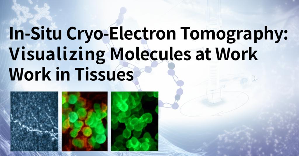
Imagine peering directly into the intricate dance of life, witnessing molecules at their workstations within the very fabric of our tissues. This isn't science fiction; it's the rapidly advancing frontier of in-situ cryo-electron tomography (cryo-ET). This revolutionary technique is transforming our understanding of health and disease by allowing scientists to visualize the molecular machinery of cells in their natural, unaltered environments.
For decades, structural biologists have meticulously studied isolated molecules, providing invaluable insights. However, this often meant taking them out of their complex cellular neighborhoods. In-situ cryo-ET is changing the game. By flash-freezing tissues – a process called vitrification – it preserves them in a near-native, life-like state, preventing the formation of ice crystals that would obliterate delicate structures.
Think of it like this: traditional methods were akin to studying a single worker's tools in a workshop, while in-situ cryo-ET allows us to see the entire factory floor in operation, with all the workers, their tools, and their interactions, all functioning together.
Slicing Through Complexity to Reveal Molecular ChoreographyOne of the major hurdles in imaging tissues is their thickness. Electrons used in microscopes can only penetrate a very short distance. To overcome this, scientists employ a sophisticated technique called cryo-focused ion beam (cryo-FIB) milling. This acts like a molecular scalpel, precisely carving out ultra-thin windows, or "lamellae," from the vitrified tissue. These lamellae, typically only a few hundred nanometers thick (that's thinner than a wavelength of light!), are then imaged from multiple angles using a powerful electron microscope.
Sophisticated computer algorithms then reconstruct these tilted 2D images into a stunning 3D volume – a tomogram. This tomogram is essentially a high-resolution 3D map of the cellular landscape within the tissue, revealing the spatial organization of organelles and, crucially, the macromolecular complexes at work.
Breakthroughs Paving the Way for Unprecedented DiscoveriesRecent advancements are rapidly pushing the boundaries of what's possible with in-situ cryo-ET. Automated workflows for sample preparation, including robotic systems for FIB milling, are increasing throughput, allowing scientists to analyze more samples more quickly. New generations of FIB instruments, some using plasma ion sources instead of gallium, are enabling faster and cleaner milling of these delicate tissue slices.
Furthermore, improvements in electron detectors and image processing software, including the use of artificial intelligence and deep learning for identifying and classifying molecules within the crowded cellular environment, are enhancing the resolution and clarity of the resulting 3D visualizations. Scientists are now able to achieve sub-nanometer resolution for some structures, meaning they can distinguish individual protein complexes and even parts of their structures as they operate within the cell.
From Cellular Architecture to Disease MechanismsThe applications are vast and profoundly impactful. Researchers are using in-situ cryo-ET to:
- Unravel complex cellular processes: Witnessing how different molecular components of a cell interact in real-time provides a deeper understanding of fundamental biological mechanisms, such as cell division, migration, and communication.
- Study pathogen-host interactions: Visualizing how viruses or bacteria invade cells and hijack cellular machinery at the molecular level is crucial for developing new antiviral and antibiotic therapies.
- Investigate the molecular basis of diseases: Scientists can now directly observe structural changes in tissues associated with diseases like neurodegenerative disorders (e.g., Parkinson's, Alzheimer's) or cancer, potentially identifying new targets for treatment. For instance, studies have visualized amyloid fibrils directly within frozen tissue, offering new perspectives on their formation and interaction with the cellular environment.
- Bridge structural biology and clinical research: The ability to study human patient tissues is forging a direct link between high-resolution structural insights and the holistic understanding of human biology in health and disease.
Despite the remarkable progress, challenges remain. Preparing high-quality, ultra-thin lamellae from complex tissues without introducing artifacts is still a highly skilled and time-consuming process. Identifying specific molecules within the densely packed cellular environment, especially those that are rare or lack distinct features, can be difficult. Achieving the atomic-level resolution common in studies of isolated proteins is still a significant hurdle for in-tissue studies.
However, the pace of innovation is relentless. Continued advancements in automation, correlative light and electron microscopy (CLEM) for precisely targeting specific regions of interest, improved data processing algorithms, and new imaging modalities are poised to overcome these limitations.
In-situ cryo-electron tomography is not just providing snapshots; it's offering a dynamic view of life's molecular choreography within its native stage – our tissues. As this technology continues to evolve, it promises to unlock even more secrets of the cellular world, paving the way for groundbreaking discoveries in biology and medicine and ultimately, a healthier future. The era of visualizing molecules truly at work, within us, has decisively begun.

