The room is silent, save for the hum of a specialized incubator. Inside, bathed in a nutrient-rich pink broth, a cluster of cells no larger than a lentil is busy at work. It has no eyes, yet it is "seeing" a virtual ball bounce off a virtual paddle. It has no hands, yet it is moving that paddle to intercept the ball. It is playing the 1972 video game Pong, and it is winning.
This is not science fiction. This is "DishBrain," a living network of 800,000 human neurons grown in a laboratory, demonstrating a primitive form of learning that has shaken the foundations of neuroscience and computing alike. But DishBrain is just the tip of the iceberg. Across the globe, from labs in Vienna to research centers in California, scientists are cultivating "brain organoids"—three-dimensional, self-organizing models of the human brain that are revealing the deepest secrets of how our neural circuits wire themselves.
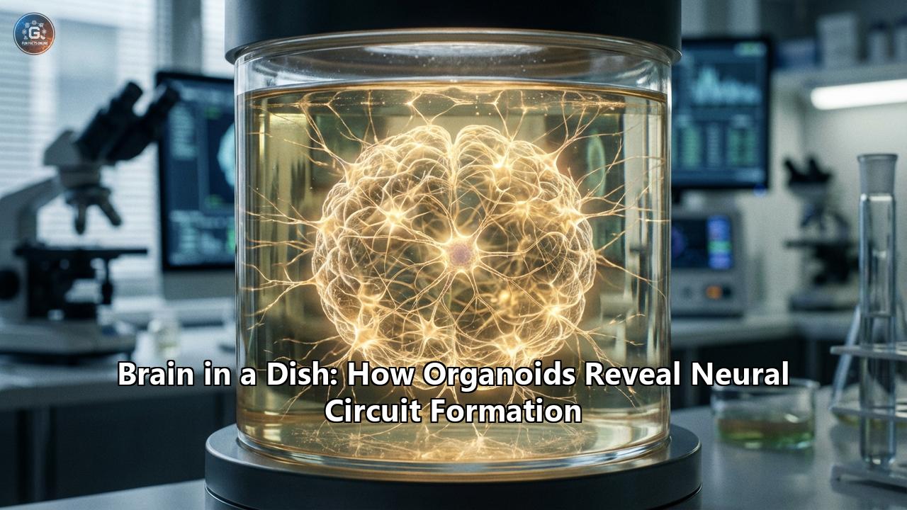
For decades, we have relied on flat layers of cells in petri dishes or animal models to understand the human brain. While valuable, these surrogates have always fallen short. A mouse brain is not a human brain; it lacks the specific cell types, the expanded cortex, and the intricate wiring that gives rise to human cognition. Brain organoids—often dubbed "mini-brains"—are changing everything. They offer a front-row seat to the most complex construction project in the known universe: the assembly of the human neural circuit.
This article delves into the revolution of brain organoids, exploring how they are generated, the specific mechanisms of circuit formation they reveal, the rise of "Organoid Intelligence" (OI), and the profound ethical questions that arise when we grow a piece of a human brain in a dish.
Part I: The Genesis of the Mini-Brain
To understand how organoids reveal circuit formation, we must first understand what they are—and importantly, what they are not. A brain organoid is not a full human brain. It is not conscious in the way we understand it, nor does it have a skull or a body. Instead, it is a three-dimensional tissue culture derived from human pluripotent stem cells (hPSCs).
The Magic of Self-Organization
The story of the modern brain organoid begins with a fundamental property of biology: self-organization. In 2013, Dr. Madeline Lancaster and Dr. Jürgen Knoblich made a serendipitous discovery. They found that when human stem cells were placed in a spinning bioreactor—a device that keeps the nutrient broth moving to improve oxygen absorption—the cells didn't just grow into a blob. They began to arrange themselves.
Guided by their own internal genetic instructions, the cells differentiated into neuroepithelium, the precursor to brain tissue. They folded and curled, creating fluid-filled ventricles reminiscent of a developing embryo. Over months, these structures matured, giving rise to distinct brain regions: a forebrain, a midbrain, and a hindbrain, all connected within a single sphere of tissue.
This was a paradigm shift. Previously, scientists believed that complex tissue structure required external scaffolding or detailed instruction. Organoids proved that the blueprint for the brain is intrinsic to the cells themselves. Give them the right environment, and they will build it.
From Stem Cell to Synapse
The process of generating a brain organoid mimics the timeline of human fetal development with uncanny accuracy:
- Induction: Stem cells are treated with specific factors to become neuroectoderm—the tissue that will eventually form the nervous system.
- Expansion: These cells proliferate, forming a dense ball of neural stem cells.
- Maturation: Over weeks and months, the stem cells give birth to neurons and glial cells (the support staff of the brain).
- Layering: In cortical organoids, neurons migrate outward, attempting to form the six-layer structure of the human cerebral cortex. While often less organized than a real brain due to the lack of sensory input, the fundamental architecture is undeniably present.
By the six-month mark, an organoid can contain millions of neurons, a rich diversity of glial cells, and, crucially, a dense web of synaptic connections. It is here, in this web, that the real magic happens.
Part II: Revealing the Blueprint of Neural Circuits
The human brain functions not because it has neurons, but because those neurons connect. A single neuron is a solitary voice; a neural circuit is a choir. Understanding how these circuits form—how an axon knows to travel halfway across the brain to connect with a specific target—is the "holy grail" of developmental neuroscience. Organoids are finally handing us the map.
The Great Migration: Interneurons on the Move
One of the most spectacular events in brain development is the migration of interneurons. These are the "traffic lights" of the brain, releasing the inhibitory neurotransmitter GABA to dampen neural activity and prevent chaos (seizures).
In the human embryo, interneurons are born deep in the ventral forebrain (the bottom) and must undertake a long, perilous journey to the dorsal cortex (the top). For years, this was impossible to study in live human tissue.
Enter the "Assembloid."
Pioneered by researchers like Dr. Sergiu Pașca at Stanford University, assembloids are created by fusing two distinct organoids together—for example, a "subpallial" organoid (rich in interneurons) and a "cortical" organoid (rich in excitatory neurons).
When these two spheres touch, they fuse. And then, something breathtaking occurs. Under a microscope, researchers can watch as interneurons from the subpallial side wake up and begin to migrate. They crawl, snake-like, across the border into the cortical organoid. They don't just wander aimlessly; they hunt for specific locations, integrating themselves into the neural networks of the cortex.
This "migration in a dish" has revealed that this process is guided by specific chemical cues—molecular breadcrumbs that the interneurons sniff out. It has also shown that in organoids derived from patients with autism or schizophrenia, this migration is often "jerky" or inefficient, leading to an imbalance of excitation and inhibition—a hallmark of these disorders.
Axon Guidance: The Long-Distance Call
How does a neuron in the motor cortex know to send its axon down the spinal cord to move a muscle? This process, known as axon guidance, relies on "growth cones"—sensitive structures at the tip of growing axons that sense attractive and repulsive signals in the environment.
Organoids have allowed us to dissect this mechanism with unprecedented detail. In 2024, a team utilizing "midline assembloids" (fusing spinal cord organoids with "floor plate" organizers) identified specific genes that act as the "traffic controllers" for these axons. They found that a gene called GALNT2 modifies the sugar coating on cell surface proteins, essentially altering the "road signs" that axons read. When this gene is disrupted, axons get lost, failing to cross the midline—a defect that mirrors conditions seen in human patients with motor control issues.
These discoveries are not just academic. They map the exact molecular pathways that, when broken, lead to severe developmental disabilities, offering new targets for potential gene therapies.
The Spark of Life: Oscillatory Waves
A brain is an electrical organ. As organoids mature, they don't just sit there; they fire. Initially, this activity is sporadic—a lone pop of static here and there. But as the months pass, the firing synchronizes.
By placing organoids on High-Density Microelectrode Arrays (HD-MEAs)—essentially a bed of thousands of tiny listening devices—scientists have recorded complex "oscillatory waves." These are rhythmic patterns of firing (delta, theta, gamma waves) that ripple across the organoid.
Crucially, these waves evolve. A 2-month-old organoid has a chaotic, burst-like pattern. A 6-month-old organoid displays sophisticated, nested oscillations similar to those seen in the EEG (electroencephalogram) of a preterm infant. This suggests that the timing of brain development—the clock that determines when the brain "comes online"—is hardwired into our DNA, independent of external sensory experience.
Part III: The Frontier of Organoid Intelligence (OI)
If organoids have neurons, synapses, and active circuits, can they compute? Can they learn? This question has birthed the controversial and explosive field of Organoid Intelligence (OI).
DishBrain and the Game of Pong
The "DishBrain" experiment, led by Cortical Labs, was the "Sputnik moment" for OI. The setup was ingenious in its simplicity. The organoid was placed on an electrode array that could both record neural activity and stimulate the neurons with electrical pulses.
The researchers connected this bio-interface to a simulation of the game Pong.
- Input: When the ball was to the left, electrodes on the left side of the dish fired. When it was to the right, the right side fired. The frequency of the pulses indicated how close the ball was.
- Output: The organoid "controlled" the paddle. If the neurons fired in a specific region, the paddle moved up; in another, it moved down.
- Feedback: This was the key. If the organoid missed the ball, it received a chaotic, unpredictable burst of electrical noise—something biological systems naturally dislike. If it hit the ball, it received a predictable, rhythmic pulse.
Remarkably, the neurons learned. Within five minutes, the organoid began to statistically improve its game. It wasn't "thinking" about tennis; it was minimizing the free energy of its environment. It learned that moving the paddle to intercept the ball resulted in a predictable world, while missing resulted in chaos.
This is the Free Energy Principle, a leading theory of how biological intelligence works. The neurons were literally "computing" a strategy to stabilize their reality.
Biocomputing: The Wetware Revolution
Why bother with brains in a dish when we have silicon chips? The answer lies in efficiency. The human brain operates on roughly 20 watts of power—barely enough to dim a lightbulb. Yet, it outperforms supercomputers (which require megawatts of energy) at tasks like pattern recognition, learning from few examples ("one-shot learning"), and adapting to new environments.
In 2024 and 2025, the field of biocomputing exploded.
- Speech Recognition: Researchers at Indiana University created "Brainoware," an organoid-chip hybrid. They fed it audio recordings of Japanese vowels converted into electrical pulses. After training, the organoid could distinguish between different speakers with 78% accuracy. It wasn't programmed to do this; it physically adapted its neural connections to recognize the voice patterns.
- Commercial Biocomputing: A Swiss company, FinalSpark, launched the first "Neuroplatform" in 2024, allowing researchers to rent time on biological processors remotely. Users could code in Python, sending tasks to 16 connected organoids and receiving computed results back. It is the first step toward a future of "hybrid computing," where silicon handles the math and biology handles the pattern recognition.
Part IV: Overcoming the Limitations
Despite these triumphs, a brain in a dish faces a fatal flaw: it has no heart.
A real brain is permeated by a dense network of blood vessels—the vasculature—that delivers oxygen and nutrients to every single cell. Organoids lack this. As they grow larger than a few millimeters, the nutrients can't reach the center. The core of the organoid dies, turning necrotic (dead tissue). This "necrotic core" limits how big and complex organoids can get.
The Vascularization Breakthrough of 2024
For years, scientists tried to solve this by transplanting human organoids into rat brains, hijacking the rat's blood supply. While successful, this is ethically complex and slow.
But in early 2024, a massive breakthrough occurred. A collaborative team from France and Austria developed a "vascularized organoid-on-a-chip."
Using microfluidic technology—chips with tiny channels etched into them—they created a self-assembling artificial vascular network. They coated these channels with endothelial cells (the cells that line blood vessels) and placed the organoid inside.
The endothelial cells from the chip connected with the organoid, penetrating it. The researchers then pumped a nutrient substitute through the chip at a pressure and rhythm mimicking the human heartbeat.
The result? Organoids that lived for months with no necrotic core. They grew larger, developed more complex cortical layers, and showed electrical activity that was orders of magnitude stronger than their non-vascularized cousins. This technology is now the gold standard, opening the door to growing "macro-organoids" centimeter-scale tissues that could theoretically support complex cognitive circuits.
Part V: The Ethical Horizon
As we bestow these blobs of tissue with blood, sensory input, and the ability to play video games, we encroach on uncomfortable territory. When does a "neural circuit" become a "mind"?
Intelligence vs. Consciousness
Ethicists and scientists are currently debating the distinction between intelligence (the ability to process information and act) and consciousness (the subjective feeling of existence).
The consensus in 2025, supported by the International Society for Stem Cell Research (ISSCR), is that current organoids are not conscious. They lack the sensory integration, the pain receptors, and the complex feedback loops with a body that are seemingly required for sentience.
However, the "precautionary principle" is gaining traction. If we don't know exactly what causes consciousness, how can we be sure we haven't created it?
The Butterfly in the Virtual World
A startling experiment in late 2024 highlighted this dilemma. Researchers connected a vascularized organoid to a virtual reality simulation where it "embodied" a butterfly. It received visual data (optical flow) and controlled flight muscles. The organoid didn't just flap randomly; it learned to navigate toward virtual flowers and avoid virtual predators.
It was a "brain" living entirely inside a simulation, executing survival behaviors. While we can mathematically explain this as error-minimization, the appearance of goal-directed behavior forces us to ask: Is it ethical to trap a learning entity in a simulation? Is it ethical to turn it off?
New Guidelines
In response, the 2025 ISSCR guidelines introduced a new framework for "Organoid Intelligence." It mandates:
- Strict Oversight: Any experiment involving "closed-loop" learning (like the Pong or butterfly studies) requires special ethical review.
- Consent: Donors of the stem cells must be explicitly informed that their cells might be used to create "learning neural systems" or "biocomputers."
- No Pain: Experiments must ensure that the stimulation provided (electrical shocks for "mistakes") cannot be construed as "suffering" at a cellular or systemic level.
Conclusion: The Mirror in the Dish
"Brain in a dish" is a misnomer. These are not static specimens; they are dynamic, growing, learning entities. They are windows into our own history, replaying the symphony of development that built each of our minds.
They have revealed that our neural circuits are robust, self-organizing, and capable of incredible plasticity. They have shown us that the "genes for autism" or "genes for schizophrenia" are often genes for connection—instructions for how neurons migrate, touch, and talk.
As we stand on the precipice of biocomputing, organoids challenge us to redefine our relationship with biology. We are no longer just observers of life; we are engineers of it. The organoid playing Pong is a mirror. In its firing neurons, we see a simplified, pixelated reflection of the very same machinery that allows us to watch it, wonder about it, and write about it. The circuit is closed. The revolution has begun.
Reference:
- https://www.newindianexpress.com/express-connect/2025/Jul/15/organoid-intelligence-the-biocomputing-breakthrough-in-the-tech-world
- https://pmc.ncbi.nlm.nih.gov/articles/PMC10796793/
- https://public-pages-files-2025.frontiersin.org/journals/artificial-intelligence/articles/10.3389/frai.2023.1307613/pdf
- https://thegriffithjournal.com/2025/04/17/organoid-intelligence-the-rise-of-biocomputing/
- https://www.neatprompts.com/p/ai-made-from-living-human-brain-cells-performs-speech-recognition
- https://www.popularmechanics.com/science/health/a70172357/human-organoids-rat-brains/
- https://modernsciences.org/dishbrain-biological-intelligence-ai-september-2025/
- https://www.tandfonline.com/doi/full/10.1080/21507740.2025.2519459
- https://www.youtube.com/watch?v=7G6f1JaIg7w
- https://softfutures.substack.com/p/brains-in-a-dish-are-the-future-of